Korean J Gastroenterol Vol. 62 No. 4, 253-255 http://dx.doi.org/10.4166/kjg.2013.62.4.253 pISSN 1598-9992 eISSN 2233-6869
IMAGE OF THE MONTH
Korean J Gastroenterol, Vol. 62 No. 4, October 2013 www.kjg.or.kr
후복막에 발생한 골외 Ewing 육종
이시형, 장병익
영남대학교 의과대학 내과학교실
Retroperitoneal Extraskeletal Ewing’s Sarcoma
Si Hyung Lee and Byung Ik Jang
Department of Internal Medicine, Yeungnam University College of Medicine, Daegu, Korea
CC This is an open access article distributed under the terms of the Creative Commons Attribution Non-Commercial License (http://creativecommons.org/licenses/
by-nc/3.0) which permits unrestricted non-commercial use, distribution, and reproduction in any medium, provided the original work is properly cited.
교신저자: 장병익, 705-717, 대구시 남구 현충로 170, 영남대학교 의과대학 내과학교실
Correspondence to: Byung Ik Jang, Department of Internal Medicine, Yeungnam University College of Medicine, 170 Hyeonchung-ro, Nam-gu, Daegu 705-717, Korea.
Tel: +82-53-620-3830, Fax: +82-53-654-8386, E-mail: [email protected] Financial support: None. Conflict of interest: None.
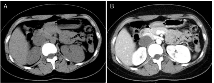
Fig. 1. Initial CT scan. (A) Pre-enhance axial image shows 3.7 cm sized cystic mass without solid component. (B) This mass is not enhanced after contrast administration.
증례: 19세 여자 환자가 3개월 전부터 시작된 복통을 주소 로 외부 병원을 방문하여 시행한 복부 전산화단층촬영에서 후 복막에 종괴가 의심되어 검사 및 치료를 위하여 본원을 방문 하였다. 환자는 비특이적인 복통 외에는 특이증상이 없었으 며, 이학적 검사에서 이상소견은 없었다. 환자의 과거력과 가 족력에서 특이사항은 없었다. 내원시 신체활력징후는 혈압 120/85 mmHg, 맥박 60회/분, 호흡수 18회/분, 체온 36.5oC
였다. 검사실 소견은 말초혈액검사에서 백혈구 6,540/mm3, 혈색소 12.9 g/dL, 헤마토크리트 37%, 혈소판 450,000/mm3, BUN 10.21 mg/dL, 크레아티닌 0.73 mg/dL, AST 21 IU/L, ALT 18 IU/L, ALP 154 U/L, GGT 17 U/L, LDH 332 U/L였 다. 내원 전 외부 병원에서 시행한 복부 전산화단층촬영에서 는 우후복막에 3.7 cm 크기의 낭종이 관찰되었으나 낭종 내 부에 고형성분이나 조영증강 소견은 보이지 않아 단순낭종 또
254
이시형, 장병익. 후복막에 발생한 골외 Ewing 육종The Korean Journal of Gastroenterology
Fig. 2. Follow-up CT scan after 6 months. (A) The mass has enlarged to about 6 cm in size. (B) Contrast-enhanced abdominal CT scan shows 6 cm sized heterogenous enhancing mass with central low attenuation at retrocaval area.
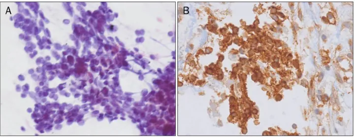
Fig. 3. (A) Microscopic finding of ultrasound guided biopsy shows small round neoplastic cells with irregular nuclei and sparse cytoplasm (H&E,
×400). (B) This tumor cells shows strong diffuse membrane immunohistochemical reactivity with CD99 antibodies (CD 99, ×400).
는 혈종으로 진단하였다(Fig. 1). 보존적 치료 후에도 복통은 지속되었으며, 6개월 후 재시행한 복부 전산화단층촬영에서 종양은 6 cm으로 커졌고 병변의 가장자리에 조영증강되는 불 균질한 종괴로 관찰되었다(Fig. 2). 후복막에서 발생한 악성종 양(부신경절종, 골외 Ewing 육종 등)을 감별하기 위해 내시경 초음파하 미세침생검을 시행하였다. 미세침생검 조직에서 비 전형적인 원형세포(round cell)가 발견되었으나 면역화학염 색에 실패하여 초음파하 조직생검을 시행하였다. 조직생검에 서 발견된 종양세포는 작은 난원형 또는 단방추형의 세포로 구성되어 있었으며, 면역화학염색에서 CD99에 강양성 반응 을 나타냈다(Fig. 3). 그러나, LCA, CD3, CD20, cytokeratic (AE1/AE3), EMA, smooth muscle actin, Desmin, S100 protein, Myogenic, CD56 등의 면역화학염색에는 음성을 나 타내어 골외성 Ewing 육종으로 진단되었다.
진단: 골외 Ewing 육종
Ewing 육종군은 전통적인 골 Ewing 육종, 흉벽에 발생하 는 Askin tumor, 뼈나 연조직에 발생하는 골외 Ewing 육종 으로 구성된다.1 이 중 뼈에 발생하는 Ewing 육종이 가장 흔 하며, 청소년기에서 발생하는 골종양중 두 번째로 흔한 종양 으로 보고되었다. 그러나 골외 Ewing 육종은 다른 종양에 비 해 흔하지 않으며, 신체의 연조직에 발생하는 종양으로 Tefft 등2이 1969년 처음으로 보고하였다. 대개 10대와 20대에서 주로 발생하고 대개의 경우 유전자 변이가 발견되며, t(11;22)(q24;q12)의 유전자 변이가 가장 흔한 것으로 보고되 었다.3 발생부위는 연조직이 있는 어느 곳이나 발생가능하나 주로 신체의 말단부(extremities) 또는 흉부에 발생하며 드물 게 후복막에 발생할 수 있다.1,4 골외 Ewing 육종이 후복막에 발생하는 경우 가장 흔한 증상은 복통으로 보고되었다.5
Lee SH and Jang BI. Retroperitoneal Extraskeletal Ewing’s Sarcoma
255
Vol. 62 No. 4, October 2013
후복막에 종괴가 발견되는 경우 후복막(primary)에서 발생 하는 종양인지, 신장, 부신, 요관에서 발생하는 종양인지 감별 하는 것이 중요하며, 또한 치료계획을 세우기 위해 양성과 악 성의 감별이 중요하다. 이전의 보고에서 후복막에서 발생하는 고형 종양의 대부분은(약 80%) 악성인 것으로 보고되었으며,5 감별해야 할 악성종양으로 lymphoma, soft tissue sarcoma, rhabdomyosarcoma, liposarcoma, congenital neuroblas- toma 등이 보고되었다. 양성종양으로는 leiomyoma, extra- adrenal chromaffinoma, mucinous custadenoma, hae- mangiopericytoma 등이 원인이 될 수 있다. 또한, 동양에서 는 결핵으로 인한 후복막 농양(abscess)과의 감별이 중요하 다.
골외 Ewing 육종의 진단은 전산화단층촬영과 자기공명영 상이 진단에 유용한 것으로 보고되었다.6 특징적인 방사선학 적 특징은 알려져 있지 않으나, 종양은 대부분 가성막 (pseudeocapsule)을 가지고 있으므로 조영 전 전산화단층촬 영에서 경계가 명확하고 저음영의 종괴로 나타나며, 조영 후 전산화단층촬영에서는 불균질하게(heterogenous) 강하게 조 영 증강되는 특징이 있다.6 또한 종괴 내부의 궤사로 인해 저 음영의 부분이 나타날 수 있고 석회화는 흔하지 않은 것으로 보고되었다.6 원격전이 장소는 폐, 뼈 등이 흔하며, 전산화단 층촬영과 자기공명영상을 이용하여 원격전이를 진단할 수 있 다.
골외 Ewing 육종의 조직소견은 불규칙한 핵과 작은 세포 질을 가지는 작은 원형의 종양세포가 판(sheets)을 형성하거 나, 소엽(lobule)을 형성하며, 드물게 rossette을 형성하는 경 우 진단할 수 있으며, CD99 면역화학염색에서 강한 양성을 나타내는 특징이 있다. 골외 Ewing 육종의 가장 효과적인 치 료는 수술 후 항암치료, 방사선치료라고 알려져 있으며, 진단 당시 수술이 어려운 경우에는 수술 전 항암치료 후 수술적 치료를 고려할 수 있다.7,8
이 증례에서는 처음 외부 병원에서 촬영하였던 전산화단층 촬영에서는 조영 전후 변화가 없는 저음영의 종괴로 나타났으 나, 6개월 후 추적 전산화단층촬영에서 골외 Ewing 육종의 전형적인 소견을 나타내었다. 그러므로, 청소년기에 저음영의 종괴가 나타날 경우 단순한 낭종과의 감별진단이 중요하다.
증상이 지속되는 경우, 조영증강되는 부분이 있거나 벽의 비 후가 있는 경우, 추적 검사에서 크기의 증가가 있는 경우에는 악성 종양을 의심하여 반드시 조직검사를 시행하는 것이 좋을 것으로 생각한다.
REFERENCES
1. Maheshwari AV, Cheng EY. Ewing sarcoma family of tumors. J Am Acad Orthop Surg 2010;18:94-107.
2. Tefft M, Vawter GF, Mitus A. Paravertebral "round cell" tumors in children. Radiology 1969;92:1501-1509.
3. Downing JR, Head DR, Parham DM, et al. Detection of the (11;22)(q24;q12) translocation of Ewing's sarcoma and periph- eral neuroectodermal tumor by reverse transcription polymer- ase chain reaction. Am J Pathol 1993;143:1294-1300.
4. Raney RB, Asmar L, Newton WA Jr, et al. Ewing's sarcoma of soft tissues in childhood: a report from the Intergroup Rhabdomy- osarcoma Study, 1972 to 1991. J Clin Oncol 1997;15:574-582.
5. Murtaza B, Saeed S, Khan NA, et al. Retroperitoneal masses: dif- ferent clinical scenarios. J Ayub Med Coll Abbottabad 2008;
20:161-164.
6. Kennedy JG, Eustace S, Caulfield R, Fennelly DJ, Hurson B, O'Rourke KS. Extraskeletal Ewing's sarcoma: a case report and review of the literature. Spine (Phila Pa 1976) 2000;25:1996- 1999.
7. Lee WS, Kim YH, Chee HK, et al. Multimodal treatment of primary extraskeletal Ewing's sarcoma of the chest wall: report of 2 cases. Cancer Res Treat 2009;41:108-112.
8. El Weshi A, Allam A, Ajarim D, et al. Extraskeletal Ewing's sarco- ma family of tumours in adults: analysis of 57 patients from a single institution. Clin Oncol (R Coll Radiol) 2010;22:374-381.