Korean J Gastroenterol Vol. 72 No. 3, 155-158 https://doi.org/10.4166/kjg.2018.72.3.155 pISSN 1598-9992 eISSN 2233-6869
IMAGE OF THE MONTH
Korean J Gastroenterol, Vol. 72 No. 3, September 2018 www.kjg.or.kr
궤양성 대장염에서 병발한 다발성 괴저성 농피증 1예
김승범, 이시형
영남대학교 의과대학 내과학교실
Multiple Pyoderma Gangrenosum in Ulcerative Colitis
Sung Bum Kim and Si Hyung Lee
Department of Internal Medicine, Yeungnam University College of Medicine, Daegu, Korea
CC This is an open access article distributed under the terms of the Creative Commons Attribution Non-Commercial License (http://creativecommons.org/licenses/
by-nc/4.0) which permits unrestricted non-commercial use, distribution, and reproduction in any medium, provided the original work is properly cited.
Copyright © 2018. Korean Society of Gastroenterology.
교신저자: 이시형, 42415, 대구시 남구 현충로 170, 영남대학교 의과대학 내과학교실
Correspondence to: Si Hyung Lee, Department of Internal Medicine, Yeungnam University College of Medicine, 170 Hyeonchung-ro, Nam-gu, Daegu 42415, Korea.
Tel: +82-53-620-3830, Fax: +82-53-654-8386, E-mail: [email protected], ORCID: https://orcid.org/0000-0001-7221-7506 Financial support: This work was supported by the 2016 Yeungnam University Research Grant.
Conflict of interest: None.
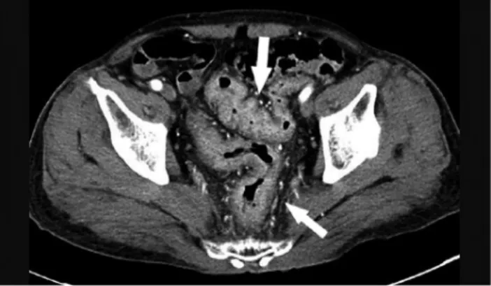
Fig. 1. Abdominal computed tomography scan showed wall thickening of sigmoid colon and rectum (white arrows).
A
A BB
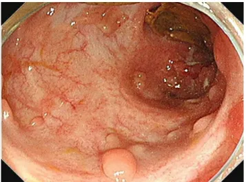
Fig. 2. Colonoscopy showed diffuse hyperemic mucosa with aphthous ulcers and mucosal edema and hemorrhages at sigmoid colon (A) and multiple deep ulcers with exudate at the rectum (B).
Fig. 3. Rectum biopsy showed lymphoplasmacytic infiltration, ulceration, and a few distorted crypts (H&E, ×10).
증례: 66세 남자가 3개월 전부터 간헐적인 혈변이 있어오다 가 4주 전부터 하루 15회 이상으로 악화되는 혈변을 주소로 방문하였다. 환자는 천식 및 전립샘 비대증으로 약물 치료 중이 었다. 방문 당시의 전신양태는 양호하였고, 활력 징후는 정상이 었다. 말초혈액 검사에서 백혈구 11,350/μL, 혈색소 14.7 g/dL 였고 erythrocyte sedimentation rate 43 mm/h와 CRP 7.11 mg/dL였다. 방문하여 시행한 복부 CT 검사에서 에스상 결장 과 직장 벽의 비후 소견이 관찰되었다(Fig. 1). 대장 내시경에서 에스상 결장과 직장에 미만성 점막 발적, 부종, 아프타 궤양이
156
김승범, 이시형. 궤양성 대장염에서 병발한 다발성 괴저성 농피증 1예The Korean Journal of Gastroenterology A
A BB
Fig. 4. On admission day 2, the patient presented painful nodules and erythematous lesions on his right knee (A) and left foot (B) that progressed rapidly to irregular ulcers.
AA BB
Fig. 5. Biopsy specimen from the ulcer of the right knee showed granulation tissue, lymphocyte infiltration, and abscess formation (H&E, ×100) (A) and abundant neutrophilic infiltration, resulting in abscess formation (H&E, ×200) (B).
A
A BB CC
Fig. 6. Incision and drainage of skin lesions of the left thumb (A), foot (B), and right knee (C) was performed.
A
A BB CC
Fig. 7. Healing ulcers with crust and hyperpigmentation on the left thumb (A), foot (B), and right knee (C) after systemic steroid therapy.
Fig. 8. Follow up sigmoidoscopy after treatment shows ulcer scar and pseudo-polyps at sigmoid colon and rectum.
관찰되었고 직장에서는 다발성의 지도상 궤양이 관찰되었다 (Fig. 2). 대장 조직 검사에서 음와 변형과 궤양이 관찰되었다 (Fig. 3). 환자는 중등도의 좌측 궤양성 대장염 진단하에 경구 balsalazide (4.5 g/day)로 치료를 시작하였다. 입원 2일째에
환자는 왼쪽 엄지손가락과 왼쪽 발가락, 오른쪽 무릎에 동통을 동반한 홍반 마디가 발생하였고(Fig. 4), 이는 불규칙한 변연을 동반한 궤양으로 빠르게 진행하였다. 피부 병변의 조직 검사에 서 급성 화농성 염증과 농양 형성 및 육아 조직이 관찰되었다 (Fig. 5). 정형외과와 협진 후 피부 병변에 대한 절개배농을 시행하였다(Fig. 6). 피부 병변에 대한 균 배양 검사에서 세균이 나 진균은 배양되지 않았다. 피부 병변에 대한 병리학적 검사에
Kim SB and Lee SH. Multiple Pyoderma Gangrenosum in Ulcerative Colitis
157
Vol. 72 No. 3, September 2018
서 백혈구파괴혈관염 또는 암의 소견은 관찰되지 않았다. 피부 에 대한 페설지 반응(pathergy reaction) 검사에서 양성 소견 을 보였다. 감염성 원인들이 배제되고 임상 양상들과 병리 소견 을 종합하여 피부 병변은 궤양성 대장염에 동반된 괴저성 농피 증으로 진단하였다. 환자는 경구 prednisolone (30 mg/day)으 로 치료를 시작하였고, 매일 피부 병변에 대한 단순 드레싱을 시행하였다. Prednisolone 치료 시작 후 14일째에 환자의 혈변 과 피부 병변이 점차 호전되었다(Fig. 7). Prednisolone은 3개 월에 걸쳐 감량하였고 혈변과 궤양성 피부 병변의 재발은 관찰 되지 않았다. 외래에서 시행한 구불창자 내시경 검사에서 점막 치유를 시사하는 궤양 반흔과 가폴립이 관찰되었다(Fig. 8).
진단: 괴저성 농피증
괴저성 농피증은 무균의 염증성 및 비종양성 희귀 피부 질 환이다.1괴저성 농피증의 발생률은 백만 명 당 3명에서 10명 으로 알려져 있으며, 30-50대에서 호발하는 것으로 알려져 있 다.2괴저성 농피증은 피부 병변 주위에 자색의 경계를 가지는 구진 또는 농포로 나타나며, 진피의 괴사가 진행되면 심부 궤 양의 형태로 나타난다.3괴저성 농피증의 약 50%에서 염증성 장질환, 류마티스 관절염, 파라단백혈증 또는 혈액암 등의 전 신 질환과 동반되어 나타날 수 있다.1,4 궤양성 대장염에서 괴 저성 농피증의 발생률은 크론병보다 더 높은 것으로 알려져 있다.5 정확한 괴저성 농피증의 발생 기전은 밝혀지지 않았으 나 호중구 화학주성 등의 호중구 기능 장애, 염증복합체의 과 활동, 면역 조절 장애와 유전적 감수성이 중요한 역할을 하는 것으로 알려져 있다.6괴저성 농피증의 진단은 임상적으로 이 루어지며, 다른 피부 질환들을 배제하고 전형적인 임상 소견 들을 바탕으로 진단할 수 있다. 다양한 표현형이 보고되었으 며, 가장 흔한 표현형은 궤양형이다.3 다른 표현형으로는 농 포, 수포와 증식형이 있다. 괴저성 농포증은 전신에 나타날 수 있으며,7염증성 장질환과 관련된 괴저성 농피증은 정강이 와 스토마 주위에 호발하는 것으로 알려져 있다.8괴저성 농피 증의 경과는 염증성 장질환의 활성도와 연관되어 보이기도 한 다.5괴저성 농피증의 치료에 대한 전향적인 무작위 비교 연구 는 없는 실정이나 초기나 단일 병변의 경우 국소 요법으로 nitrogen mustard, aqueous benzoyl peroxide 등을 도포하 거나 triamcinolone 또는 cyclosporine을 병변 내에 주입할 수 있다.6 Steroid, infliximab, mycophenolate, azathioprine, cyclosporine 등이 전신 요법으로 사용될 수 있다.2,9 121명의 괴저성 농피증 환자를 대상으로 경구 steroid와 cyclosporine 의 효과를 비교 분석한 연구에서 steroid와 cyclosporine 모 두에서 궤양의 완전 완화율은 47%로 동일하였고, 재발률도 각각 28%와 32%로 양 군 간에 통계학적으로 의미 있는 차이 를 보이지 않았다.10괴저성 농피증 환자 30명을 대상으로 in-
fliximab의 치료 효과를 분석한 연구의 69%에서 임상적인 반 응을 보였고 6주째에 21%에서 완전 완화를 보고하였다.11 Brooklyn 등11의 연구에서는 괴저성 농피증이 궤양성 대장염 과 동반된 경우 크론병보다 치료가 더 어려울 수 있음을 보고 하였다. 초기 관해 후 재발률은 25% 이상으로 보고되고 있다.
다발혈관염을 동반한 육아종증에서는 초기에 괴저성 농피 증과 같은 피부 병변으로 나타날 수 있어 감별을 필요로 한 다.12 본 증례에서는 anti-neutrophil cytoplasmic antibody 에 대한 혈액학적 검사에서 음성 소견을 보였고, 신장 또는 호흡기로의 침범 소견은 관찰되지 않았으며, 피부 병변에 대 한 조직 검사에서 혈관염 소견은 관찰되지 않아 다발혈관염을 동반한 육아종증은 배제할 수 있었다.
본 연구에서는 경구 steroid로 성공적으로 치료한 궤양성 대장염에 동반된 괴저성 농피증 1예를 경험하여 보고하고 자 한다. 괴저성 농피증에 대한 수술적 치료는 피부 병변을 악화시킬 수 있으므로 염증성 장질환 환자에서 궤양성 피부 병변이 발생한 경우 괴저성 농피증에 대한 감별 진단이 필 요하겠다.
REFERENCES
1. Lee JI, Park HJ, Lee JY, Cho BK. A case of pyoderma gangrenosum with ulcerative colitis treated with mesalazine. Ann Dermatol 2010;22:422-425.
2. Binus AM, Qureshi AA, Li VW, Winterfield LS. Pyoderma gan- grenosum: a retrospective review of patient characteristics, co- morbidities and therapy in 103 patients. Br J Dermatol 2011;
165:1244-1250.
3. Powell FC, Su WP, Perry HO. Pyoderma gangrenosum: classi- fication and management. J Am Acad Dermatol 1996;34:
395-409; quiz 410-412.
4. Mlika RB, Riahi I, Fenniche S, et al. Pyoderma gangrenosum: a report of 21 cases. Int J Dermatol 2002;41:65-68.
5. Greuter T, Navarini A, Vavricka SR. Skin manifestations of in- flammatory bowel disease. Clin Rev Allergy Immunol 2017;53:
413-427.
6. Ahn C, Negus D, Huang W. Pyoderma gangrenosum: a review of pathogenesis and treatment. Expert Rev Clin Immunol 2018;14:
225-233.
7. Byeon YM, Lee J, Lee SJ, et al. Peritonsillar involvement in pyo- derma gangrenosum associated with ulcerative colitis. Intest Res 2014;12:153-156.
8. Vavricka SR, Schoepfer A, Scharl M, Lakatos PL, Navarini A, Rogler G. Extraintestinal manifestations of inflammatory bowel disease. Inflamm Bowel Dis 2015;21:1982-1992.
9. Herberger K, Dissemond J, Hohaus K, Schaller J, Anastasiadou Z, Augustin M. Treatment of pyoderma gangrenosum: retro- spective multicentre analysis of 121 patients. Br J Dermatol 2016;175:1070-1072.
10. Ormerod AD, Thomas KS, Craig FE, et al. Comparison of the two
158
김승범, 이시형. 궤양성 대장염에서 병발한 다발성 괴저성 농피증 1예The Korean Journal of Gastroenterology most commonly used treatments for pyoderma gangrenosum:
results of the STOP GAP randomised controlled trial. BMJ 2015;350:h2958.
11. Brooklyn TN, Dunnill MG, Shetty A, et al. Infliximab for the treat- ment of pyoderma gangrenosum: a randomised, double blind,
placebo controlled trial. Gut 2006;55:505-509.
12. Al Rajabi W, Venturini M, Sala R, Calzavara-Pinton P. Wegener's granulomatosis of the penis: genital presentation of systemic disease. Dermatology 2006;212:370-372.
![외국인 PCR검사 가능 기관 [Referral Laboratories : 11sites]](data:image/gif;base64,R0lGODlhAQABAIAAAP///wAAACH5BAEAAAAALAAAAAABAAEAAAICRAEAOw==)