Korean J Gastroenterol Vol. 73 No. 4, 239-241 https://doi.org/10.4166/kjg.2019.73.4.239 pISSN 1598-9992 eISSN 2233-6869
IMAGE OF THE MONTH
Korean J Gastroenterol, Vol. 73 No. 4, April 2019 www.kjg.or.kr
급성 봉소염성 위식도염
김태훈, 남궁연, 정선영
1, 부선진
제주대학교 의학전문대학원 내과학교실, 영상의학교실1
Acute Phlegmonous Esophagogastritis
Taehoon Kim, Yeon Namgung, Sun Young Jeong1 and Sun-Jin Boo
Departments of Internal Medicine and Radiology1, Jeju National University School of Medicine, Jeju, Korea
CC This is an open access article distributed under the terms of the Creative Commons Attribution Non-Commercial License (http://creativecommons.org/licenses/
by-nc/4.0) which permits unrestricted non-commercial use, distribution, and reproduction in any medium, provided the original work is properly cited.
Copyright © 2019. Korean Society of Gastroenterology.
교신저자: 부선진, 63241, 제주시 아란13길 15, 제주대학교 의학전문대학원 내과학교실
Correspondence to: Sun-jin Boo, Department of Internal Medicine, Jeju National University School of Medicine, 15 Aran 13-gil, Jeju 63241, Korea. Tel: +82-64-754-8122, Fax: +82-64-717-1131, E-mail: [email protected], ORCID: https://orcid.org/0000-0002-9945-6766
Financial support: None. Conflict of interest: None.
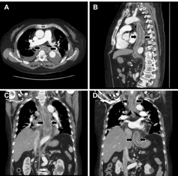
Fig. 1. Initial chest CT. CT scans with axial (A), sagittal (B) and coronal (C, D) images show diffuse wall thickening with intramural low density (arrows) from esophagus to gastric cardia. CT, computed tomography.
증례: 71세 여자가 10일 전부터 발생한 심한 연하 곤란과 연하통, 가슴 통증과 가슴 답답함을 주소로 내원하였다. 증상 발생 초기에는 고형 음식을 삼키기 힘들었으나 내원 시에는 물도 삼키기 어렵다고 하였다. 환자는 관절염 및 반복적인 요 통으로 경구 dexamethasone 2 mg을 4개월 동안 매일 복용 하고 있었다. 이외 과거력에는 특이 소견이 없었으나 내원 시 당뇨가 처음 진단되었다. 음주력이나 흡연력은 없었다.
내원 당시 급성 병색을 보였으며 활력징후는 혈압 130/86 mmHg, 맥박 91회/분, 호흡수 20회/분, 체온 37.4℃였고, 키 165 cm, 체중 83.6 kg으로 체질량지수는 30.7 kg/m2였다. 신체 검사에 서 상복부에 경미한 압통이 있었으며, 간비종대 및 촉지되는 종괴는 없었다. 말초 혈액 검사에서 백혈구 11,900/μL (정상 4,000-10,000), 혈색소 13.6 g/dL (정상 12.0-16.0), 혈소판 170,000/μL (정상 150,000-450,000)였고, 혈액 화학 검사에서 총단백질 5.8 g/dL (정상 6.7-8.3), 알부민 2.8 g/dL (정상 3.8-5.3), 총빌리루빈 2.8 mg/dL (정상 0.2-1.2), 아스파르테이 트아미노전달효소 43 IU/L (정상 8-38), 알라닌아미노전달효소 34 IU/L (정상 4-44), 혈액요소질소 4.3 mg/dL (정상 8.0-20.0), 크레아티닌 0.8 mg/dL (정상 0.6-1.1), C-반응성 단백질 13.88 mg/dL (정상 0.00-0.30)였으며, 혈당 127 mg/dL (정상 70-110), 당화 혈색소 8% (정상 4.0-6.0)였다. 혈액 응고 검사에서 프로트롬빈
C D A B
240
김태훈 등. 급성 봉소염성 위식도염The Korean Journal of Gastroenterology
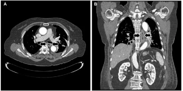
Fig. 2. Follow-up chest CT (3 weeks after medical therapy). CT scans with axial (A) and coronal (B) images reveal the improvement of diffuse wall thickening (arrows) from esophagus to gastric cardia. CT, computed tomography.
Fig. 3. Esophagographic finding (25 days after medical therapy).
Esophagogram shows extraluminal barium collection (arrows) due to mucosal defects in the upper and middle esophagus.
시간 14.7초(정상 9.9-13.5), 활성화부분트롬보플라스틴 시간 32초(정상 20.0-36.0)였다.
흉부 전산화단층촬영에서 식도 전체와 위 분문부의 벽이 전반적으로 두꺼워지고 벽 내부에는 미만성 음영 감소가 관찰 되었으며, 식도 주변의 종격동에는 지방의 침윤과 액체의 저 류가 동반되어 있어 급성 봉소염성 위식도염에 합당한 소견이 었다(Fig. 1). 양측 흉강에 흉수가 저류되어 시행한 진단적 흉 강천자 결과 산도 7.815, 당 113 mg/dL, 단백질 3.0 g/dL, 유수탈수소효소 1,338 U/L로 삼출물로 판단되었다.
급성 봉소염성 위식도염에 대한 치료로 3주 이상의 금식을 유지하고 완전비경구영양법을 시행하였으며, 총 4주 동안 광 범위 항생제(ceftriaxone, metronidazole)를 투여하였다. 치 료하면서 환자가 호소하였던 증상들 및 혈액 검사 결과들은 점차 호전되었으며, 치료 3주째 시행한 흉부 전산화단층촬영 에서 이전에 관찰되었던 전체 식도와 위 분문부의 두꺼워졌던 벽 및 벽 내부의 음영 감소는 거의 호전되었다(Fig. 2). 치료 4주째 시행한 식도조영술에서 상부 및 중부 식도의 세 부위에 점막의 결손으로 인한 조영제의 식도 벽외 저류가 보였으며 (Fig. 3), 상부위장관 내시경에서 동일한 부위에 치유기 궤양 들이 관찰되었다(Fig. 4). 이후 식이를 진행하여 퇴원하였으 며, 외래 추적 관찰 중 특별한 문제는 발생하지 않았다.
진단: 급성 봉소염성 위식도염
위나 식도에 발생하는 봉소염성 염증은 점막은 비교적 잘 보존되지만 점막하층 이상을 광범위하게 침범하는 드물지만 치명적인 화농성 세균 감염 질환이다. 봉소염성 위염은 비교 적 흔하게 보고되지만 봉소염성 식도염은 더 드물고 봉소염성 위식도염은 극히 드물다고 알려져 있다.1-5
급성 봉소염성 위식도염의 병인은 아직까지 명확하지 않 다. 면역 결핍, 영양 부족, 알코올 중독 상태이거나 무위산 증, 조절되지 않는 당뇨, 결합조직 질환, 악성 종양 환자들에 서 더 잘 발생할 수 있지만 기저 질환 없이 건강한 사람에서 도 발생할 수 있다고 알려져 있다.1,2,5 본 증례의 경우 장기 간의 스테로이드 사용으로 인한 면역 억제 상태와 당뇨가 발 병에 영향을 미쳤을 것으로 생각된다. 이 질환에서 일반적으 로 가장 흔히 동정되는 병원균은
Streptococcus
이며, 이외Enterobacter, Escherichia coli, Proteus, Staphylococcus,
Clostridium, Klebsiella pneumoniae
등이 원인균이 될 수 A BKim T, et al. Acute Phlegmonous Esophagogastritis
241
Vol. 73 No. 4, April 2019
Fig. 4. Esophagogastroduodenoscopic finding (26 days after medical therapy). Endoscopy reveals (A) a deep and round healing ulcer (white arrows) in the middle esophagus and (B) a shallow and geographic healing ulcer (black arrows) with inflammatory polyps in the upper esophagus.
있다고 알려져 있다.1,2
급성 봉소염성 위식도염의 증상은 염증의 침범 부위와 정 도에 따라 다를 수 있는데, 위염의 경우 상복부 통증이 발생할 수 있고 식도염의 경우 연하통, 연하 곤란, 가슴 통증, 호흡 곤란 등이 동반될 수 있다. 이외 오심, 구토, 토혈, 딸꾹질 등 의 증상들뿐만 아니라 열을 비롯하여 패혈증에 동반된 전신 증상들이 발생할 수도 있다.5
급성 봉소염성 위염이나 식도염을 진단하는데 전산화단층 촬영 소견은 매우 중요한데, 위나 식도의 벽이 전반적으로 두 꺼워지고 점막하층이나 근육층의 심한 염증 및 농양으로 인하 여 벽내의 음영 감소가 동반되는 것이 전형적인 소견이다.4-6 상부위장관 내시경에서는 침범된 부위의 점막하 부종으로 인 하여 위나 식도의 관강이 좁아지고 잘 팽창되지 않을 수 있으 며, 궤양이 동반될 수도 있다. 궤양은 벽 내의 농양이 배출되 는 통로가 될 수 있기 때문에 치료의 경과나 예후를 판단할 때 매우 중요한 소견일 수 있다.1,7 봉소염성 위염의 경우 악성 병변과의 감별을 위하여 내시경 초음파를 시행하기도 하는데, 내시경 초음파에서는 점막하층과 근육층이 저에코로 두꺼워 져 보일 수 있다.8,9
급성 봉소염성 위식도염은 염증이 국소적인 경우 치사율이 20% 전후이지만 미만성인 경우에는 60%까지 증가하는 치명 적인 질환이기 때문에 신속한 진단과 치료가 매우 중요하 다.2,5 이 질환이 의심되면 금식을 시작하고 즉시 광범위 항생 제를 투여해야 하는데, 장기간 금식이 필요한 경우에는 급식 공장조루술을 통한 영양 공급도 고려해 볼 수 있다.7 한편 코 위관 삽관은 식도 천공 등의 합병증을 유발할 수 있으므로 시행해서는 안 된다.10보존적으로 치료하기 어렵거나 식도 천 공, 종격동염, 농흉, 위 천공, 복막염 등의 심각한 합병증이 동반된다면 수술적 배농 또는 수술적 절제 및 재건술이 필요 할 수 있다.1,3,5 저자들은 경구용 스테로이드제를 장기간 복용
하여 면역 억제 상태에 있던 당뇨 환자에서 발생한 급성 봉소 염성 위식도염이 내과적 치료만으로 호전된 매우 드문 증례를 경험하였기에 보고하는 바이다.
REFERENCES
1. Huang YC, Cheng CY, Liao CY, Hsueh C, Tyan YS, Ho SY. A rare case of acute phlegmonous esophagogastritis complicated with hypo- pharyngeal abscess and esophageal perforation. Am J Case Rep 2017;18:125-130.
2. Kim GY, Ward J, Henessey B, et al. Phlegmonous gastritis: case report and review. Gastrointest Endosc 2005;61:168-174.
3. Hsu CY, Liu JS, Chen DF, Shih CC. Acute diffuse phlegmonous esophagogastritis: report of a survived case. Hepatogastroen- terology 1996;43:1347-1352.
4. Jung C, Choi YW, Jeon SC, Chung WS. Acute diffuse phlegmonous esophagogastritis: radiologic diagnosis. AJR Am J Roentgenol 2003;180:862-863.
5. Kim HS, Hwang JH, Hong SS, et al. Acute diffuse phlegmonous esophagogastritis: a case report. J Korean Med Sci 2010;25:
1532-1535.
6. Yun CH, Cheng SM, Sheu CI, Huang JK. Acute phlegmonous esophagitis: an unusual case (2005: 8b). Eur Radiol 2005;15:
2380-2381.
7. Woo WG, Do YW, Lee GD, Lee SS. Phlegmonous esophagitis treat- ed with internal drainage and feeding jejunostomy. Korean J Thorac Cardiovasc Surg 2017;50:453-455.
8. Hu DC, McGrath KM, Jowell PS, Killenberg PG. Phlegmonous gas- tritis: successful treatment with antibiotics and resolution docu- mented by EUS. Gastrointest Endosc 2000;52:793-795.
9. Kim NY, Park JS, Lee KJ, Yun HK, Kim JS. A case of acute phlegmo- nous gastritis causing gastroparesis and cured with medical treatment alone. Korean J Gastroenterol 2011;57:309-314.
10. Chang PC, Wang WL, Hwang TZ, Cheng YJ. Intramural dissection with mucosal rupture alleviating phlegmonous esophagitis. Eur J Cardiothorac Surg 2012;41:442-444.
A B