www.jkfas.org https://doi.org/10.14193/jkfas.2017.21.3.93
일반적으로 고령 환자의 무지 외반증 수술은 수술 후 통증의 증가, 강직감, 중족 족지 관절의 퇴행성 변화, 취약한 연부조직의 이유로 젊은 환자보다 상대적으로 좋지 않은 결과를 나타낸다는 보고가 있다.4,5) 하지만 평균 수명 및 삶의 질에 대한 요구가 증가함에 따 라 고령에서도 무지 외반증 수술이 늘어가고 있으며, 몇몇 논문에 서는 고령에서 무지 외반증 수술의 좋은 결과를 보고하고 있다.6-8) 무지 외반증 수술은 얼마나 교정각을 얻을 수 있는가, 얼마나 절골 부위의 견고한 고정을 할 수 있는가에 따라 여러 가지 수술법이 소 개되고 있다.9) 절골 부위의 견고한 고정을 위해서는 적절한 수술 방법도 중요하지만 환자의 절골부의 골밀도 정도가 중요한 요소일
서 론
무지 외반증의 유병률은 18세부터 65세 사이에서 23%, 65세 이 상에서 36%로 보고될 만큼 다양한 연령층에서 발생되고 있다.1-3)
Original Article
This is an Open Access article distributed under the terms of the Creative Commons Attribution Non-Commercial License (http://creativecommons.org/licenses/CC
by-nc/4.0) which permits unrestricted non-commercial use, distribution, and reproduction in any medium, provided the original work is properly cited.
Copyright 2017 Korean Foot and Ankle Society. All rights reserved.ⓒ
Purpose: The aim of this study was to evaluate the radiological and clinical outcomes of scarf osteotomy for hallux valgus (HV) defor-
mity in elderly patient with osteoporosis.Materials and Methods: A total of 58 elderly patients (mean age, 72.6 years) underwent scarf osteotomy for HV deformity between 2008
and 2015. The mean follow-up period was 24.4 months. Of the 58 patients, 42 were diagnosed with osteoporosis and 16 were diagnosed as normal. The radiological and clinical outcomes were assessed preoperatively, postoperatively, and at final follow-up, including HV angle, intermetatarsal (IM) angle, American Orthopaedic Foot and Ankle Society (AOFAS) score, patient satisfaction, visual analogue scale (VAS), and complication.Results: There was no significant difference in HV angle and IM angle between the osteoporosis group and normal bone mineral densi-
tometry group at all time points, preoperative, postoperative, and final follow-up. Moreover, there was no statistically significant differ- ence between the two groups with respect to the AOFAS score and VAS evaluations. In the osteoporosis group, the mean HV angle im- proved from 36.7° preoperatively to 11.3° at the time of final follow-up, and the mean IM angle improved from 13.2° to 5.7°. The mean AOFAS score improved from 52.6 preoperatively to 89.1 at the time of final follow-up. With respect to satisfaction, 83.4% of patients were very satisfied or satisfied. There were no serious complications, and all cases showed complete union at the osteotomy site.Conclusion: We believed that scarf osteotomy is a safe, effective procedure for the correction of elderly patients with osteoporosis.
Key Words: Osteoporosis, Hallux valgus, Scarf osteotomy
골다공증이 있는 고령의 환자에서 Scarf 중족골 절골술을 이용한 무지 외반증의 치료
황승현, 이수찬, 남창현, 백지훈, 안혜선
힘찬병원 정형외과 관절의학연구소
The Treatment for Hallux Valgus with Scarf Osteotomy in Elderly Patients with Osteoporosis
Seung Hyun Hwang, Su Chan Lee, Chang Hyun Nam, Ji-Hoon Baek, Hye Sun Ahn
Joint & Arthritis Research, Department of Orthopaedic Surgery, Himchan Hospital, Seoul, Korea
Received April 27, 2017 Revised July 13, 2017 Accepted July 14, 2017 Corresponding Author: Seung Hyun Hwang
Joint & Arthritis Research, Department of Orthopaedic Surgery, Himchan Hospital, 120 Sinmok-ro, Yangcheon-gu, Seoul 07999, Korea
Tel: 82-2-3219-9229, Fax: 82-2-2061-8605, E-mail: [email protected] Financial support: None.
Conflict of interest: None.
간각이 11도 미만인 경우를 경도, 무지 외반각이 20∼40도 이하이 고 제 1∼2중족골간각이 11∼16도 이하인 경우를 중등도, 무지 외 반각이 40도를 초과하거나 제 1∼2중족골간각이 16도를 초과하 는 경우를 중증으로 분류하였다. 중등도 변형은 39예(골다공증군 42예, 정상군 10예), 중증 변형은 19예(골다공증군 13예, 정상군 6 예)였다. 본 연구는 본원 기관생명윤리위원회(Institutional Review Board)의 승인을 받아 진행하였다.
2. 수술방법
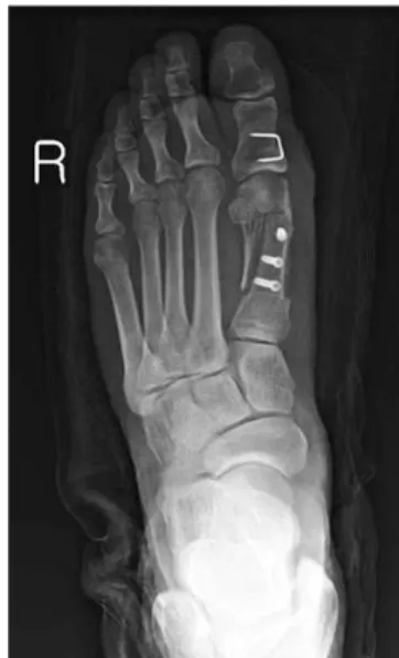
모든 수술은 단일 술자가 동일한 방법으로 수술하였다. 척추 마 취하에 시행하였고 허벅지 중간 부위에 지혈대를 사용하였다. 수 술은 족내측에서 내측 피부절개를 시행한 다음 중족지 지간관절 사이를 통하여 외측 관절낭을 절개하고 무지 내전건을 절개하고, 심부 중족지간 인대를 절개하여 외측 연부조직 유리술을 시행하 였다. Oscillating saw를 사용하여 제 1중족골 골두의 내측 골융기 (medial eminence)를 제거하고 골간단에서 Z-형태의 scarf 절골술 을 시행하고 원위 골편을 towel clip을 사용하여 유지한 상태로 근 위 골편을 외측으로 전위시켰다. 3개의 Kirschner wire를 사용하 여 일시적으로 절골면을 고정한 후 이 강선을 따라서 3개의 소형 유관 양측 피질골 압박나사못(Barouk screw [Johnson & Johnson, DePuy, France]와 Bold screw [Newdeal, Lyon, France])을 사용하 여 고정하였다. 그리고 8 mm staple을 사용하여 Akin 절골술을 같 이 시행하였다(Fig. 1, 2). 수술 후 다음날부터 heel support shoe를 착용하여 부분 체중부하 보행을 시행하였다. 수술 후 6주경부터 일반 신발 착용 및 전체 체중부하를 허용하였다.
3. 방사선학적 및 임상적 평가
방사선학적으로 기립 족부 전후면 방사선 사진에서 수술 전, 수 수 있다.10) 특히 고령일수록 골다공증의 가능성이 높아지고 그로
인한 절골부의 고정 실패 및 골절 등 수술 후 합병증이 발생한다 면 이는 무지 외반증의 결과에 영향을 줄 수 있으므로 더욱더 골밀 도가 중요한 의미를 가질 수 있다.5,10) 하지만 고령의 나이와 관련 한 수술 결과를 보고한 기존의 논문에서도 절골술의 견고한 고정 과 중요하게 연관된 골밀도에 대해서는 언급하고 있지 않다.4,5,9) 이 에 저자들은 65세 이상의 골다공증으로 진단받은 고령의 환자에서 scarf 중족골 절골술을 시행하여 견고한 고정을 시행한다면 양호한 결과를 가져올 것이라고 가설을 세우고 고령의 골다공증 환자와 고령의 골다공증이 아닌 환자를 비교하여 중족골 절골술 시행 후 이에 따른 결과를 방사선학적 및 임상적으로 알아보고자 한다.
대상 및 방법
1. 연구대상
2008년부터 2015년까지 본원에서 중등도 또는 중증 무지 외반 증으로 동일 술자에 의해 중족골 절골술을 받은 65세 이상 환자 중 수술 전 골밀도 검사를 시행하였고, 수술 후 1년 이상 추시 검사가 가능하였던 58예를 대상으로 후향적 연구를 시행하였다.
이 중 골다공증으로 진단받은 환자는 세계보건기구(World Health Organization)에서 T-score에 근거를 둔 정의에 따라 dual energy X-ray absorptiometry (DXA) 장비로 측정한 central bone (대퇴 중 Ward’s triangle 부위를 제외한 요추와 대퇴 부위 측정)의 골밀도 검사수치가 T-score ≤―2.5인 경우로 정의하였고, 58예 중 42예(여성 40예, 남성 2예)는 골다공증으로, 16예(여성 15예, 남성 1예)는 정상으로 진단되었다.
무지 외반의 정도는 Coughlin11) 분류에 따라 경도, 중등도, 중증 으로 분류하였으며 무지 외반각이 20도 미만이고 제 1∼2중족골
Figure 1. Preoperative radiograph. Figure 2. Postoperative radiograph.
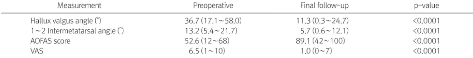
www.jkfas.org 평균 5.7도(0.6∼12.1도)로 제 1∼2중족골간각의 수술 직후 교정각 은 평균 9.2도이며, 최종 추시 시의 교정각 소실률은 18.5%로 나타 났다. 최종 추시 시 무지 외반각의 교정각은 평균 25.4도, 제 1∼2 중족골간각의 교정각은 평균 7.5도로 수술 전에 비해 모두 통계적 으로 유의하게 교정되었다(p<0.05).
AOFAS 전족부 기능 평가는 술 전 평균 52.6점(12∼68점)에서 최 종 추시 시 평균 89.1점(42∼100점), 통증에 대한 VAS 평가는 술 전 평균 6.5점(1∼10점)에서 최종 추시 시 1.0점(0∼7점)으로 모두 유 의하게 향상되었다(p<0.05; Table 2).
최종 추시에서 환자의 수술 후 전체적인 주관적 만족도를 조사 한 결과 골다공증군은 매우 만족 18예(42.9%), 만족 17예(40.5%), 보통 7예(16.7%), 불만족 없음으로, 골밀도 정상군은 매우 만족 8 예(50.0%), 만족 7예(43.8%), 보통 1예(6.3%), 불만족 없음 조사되 어 두 군에서 모두 전체적인 만족도가 높은 것으로 나타났으며 통 계적인 차이는 없었다(Table 3).
전 예에서 술 후 수술 부위의 감염이나 주변 부위의 신경 손상, 불유합이나 중족골 절골의 근위부와 원위부로의 피로 골절 등은 없었으며, 최종 추시 시 합병증이나 술 후 만족도에 따른 재수술 예는 없었다(Fig. 3).
고 찰
무지 외반증 수술은 여러 가지 방법이 소개되어 있지만 일반적 으로 연부조직에 대한 처치와 절골술로 구성되어 있다.12-15) 고령일 수록 골밀도가 감소하게 되고, 특히 골다공증이 있는 경우에는 매 우 불량한 골치유 능력을 가진다. 따라서 고령에서의 무지 외반증 술 직후, 그리고 최종 추시 시의 무지 외반각과 제 1∼2중족골간각
을 측정하였다. 수술 직후의 교정각(교정각=수술 전 각도―수술 직후 각도)과 최종 추시 시의 교정각을 비교하여 교정각 소실 정도 (교정 소실각=최종 추시 시 각도―수술 직후 각도)를 분석하고, 교 정각 소실률(교정 소실률=교정 소실각/수술 직후 교정각)을 계산 하였다.7)
임상적 평가로 American Orthopaedic Foot and Ankle Society (AOFAS) score를 이용하여 전족부 기능 평가를 visual analogue scale (VAS)을 이용하여 통증 평가를 하였다. 또한 환자의 수술에 대한 주관적 만족도를 매우 만족(very satisfied), 만족(satisfied), 보 통(fair), 불만족(poor)의 네 단계로 나누어 조사하였다.
4. 통계 분석
통계에 유의한 증례수를 계산하기 위해서 G* power (version 3.1.4)를 사용하였고, 80% 이상의 검정력을 얻기 위해서는 30증례 가 필요하였다. 각 결과치는 평균과 범위로 나타냈으며, 통계 분 석은 PASW Statistics (18.0 for Windows; IBM Co., Armonk, NY, USA) 통계 프로그램을 이용하여 정규성 검정 후 independent t-test 와 paired t-test로 분석하였으며, 유의성 판정은 p값이 0.05 미만으 로 하였다.
결 과
골다공증으로 진단된 42예의 평균 골밀도(T-score)는 ―2.6 (―2.5
∼―4.8), 평균 연령은 71.6세(65∼82세), 평균 체질량지수는 25.2 kg/m2 (20.3∼37.9 kg/m2), 평균 추시 기간은 23.5개월(12∼59개 월)이었으며, 이들 중 17명은 양측, 8명은 한 측 수술을 시행하였 다. 정상군 16예의 평균 골밀도(T-score)는 ―0.3 (1.9∼―2.4), 평 균 연령은 73.5세(70∼78세), 평균 체질량지수는 26.3 kg/m2 (22.5
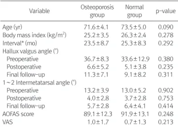
∼30.9 kg/m2), 평균 추시 기간은 25.3개월(12∼42개월)이었으며, 이들 중 4명은 양측, 8명은 한 측 수술을 시행하였다. 골다공증군 과 골밀도 정상군에서 무지 외반각은 각각 술 전 평균 36.7도와 33.6도, 술 후 6.6도와 5.1도, 최종 추시 시 11.3도와 9.1도였으며, 제 1∼2중족골간각은 술 전 평균 13.2도와 13.0도, 술 후 4.0도와 3.7도, 최종 추시 시 5.7도와 6.4도로 모두 두 군 간 유의한 차이는 관찰되지 않았다(Table 1). 또한 최종 추시 시 AOFAS 전족부 기능 평가에서 각각 89.1점과 91.9점, VAS 평가는 각각 1.0점과 0.7점으 로 역시 두 군 간 통계적인 차이는 없었다(Table 1).
골다공증으로 진단된 42예의 무지 외반각은 술 전 평균 36.7도 (17.1∼58.0도)에서 수술 직후 평균 6.6도(0.2∼18.6도), 최종 추 시 시 평균 11.3도(0.3∼24.7도)로 측정되었다. 무지 외반각의 수 술 직후 교정각은 평균 30.1도로 최종 추시 시의 교정각 소실률은 15.6%로 계산되었다. 제 1∼2중족골간각은 술 전 평균 13.2도(5.4
∼21.7도)에서 수술 직후 평균 4.0도(0.1∼11.8도), 최종 추시 시
Table 1. Comparison of Demographic, Radiological and Clinical Out- comes between Two Groups
Variable Osteoporosis group
Normal
group p-value
Age (yr) 71.6±4.1 73.5±5.0 0.090
Body mass index (kg/m2) 25.2±3.5 26.3±2.4 0.278
Interval* (mo) 23.5±8.7 25.3±8.3 0.292
Hallux valgus angle (。) Preoperative Postoperative Final follow-up
36.7±8.3 6.6±5.2 11.3±7.1
33.6±12.9 5.1±3.8 9.1±8.2
0.380 0.235 0.311 1∼2 Intermetatarsal angle (。)
Preoperative Postoperative Final follow-up
13.2±3.9 4.0±2.8 5.7±2.8
13.0±5.2 3.7±2.8 6.4±4.1
0.902 0.753 0.414
AOFAS score 89.1±12.3 91.9±13.1 0.248
VAS 1.0±1.7 0.7±1.3 0.213
Values are presented as mean±standard deviation.
Statistically significant different between the two groups: p<0.05.
AOFAS: American Orthopaedic Foot and Ankle Society, VAS: visual analogue scale.
*1st operation to final follow-up.
에서의 실험이라는 점이다.15) 둘째, 절골술의 선택에 있어서 절골 면의 단면이 넓어 비교적 초기 안정성이 강하고 조기 보행을 할 수 있는 scarf 절골술을 사용하였다는 점이다.16,17) 셋째, scarf 절골 부 위의 고정방식으로 3개의 나사못으로 고정했다는 점인데, scarf 절 골 부위의 고정 나사못의 수에 대한 비교 보고는 없으나 좋은 골질 을 가진 경우에는 1점 고정으로 좋은 결과를 가진다는 보고가 있 다. 골질이 좋지 않은 경우 2개의 나사못을 사용하여 원위부 나사 못을 원위 갈매기형 절골술과 유사하게 중족골 골두로 향하게 하 여 좋은 고정력을 얻을 수 있다는 보고가 있다.18,19) 따라서 저자들 은 더 견고한 고정을 위해 2점 나사못 고정이 아닌 세 개의 나사못 을 사용한 3점 고정을 시행하였고, 이것이 초기의 안정적인 고정에 관여했을 가능성이 있으나 고정 나사못 개수에 대한 절골 부위의 고정력과 임상결과에 대한 추가적인 연구가 필요할 것으로 생각된 다.
본 연구는 몇 가지 제한점이 있다. 첫째 골밀도 검사 시 족부 부 위가 아닌 요추와 대퇴 부위의 골밀도 측정값으로 골다공증을 진 단하여 정확히 중족골의 골밀도를 반영하지 못했다는 점이다. 그 러나 일반적으로 골밀도는 측정 부위(고관절, 척추, 요골, 종골)와 관계없이 골절의 발생빈도를 예측할 수 있다는 보고가 있다.20,21) 수술은 필요한 절골 부위의 나사못 고정의 견고함이 떨어질 수 있
으며, 이를 보완하기 위해 수술 부위의 고정기간이 길어질 수 있 다. Trnka 등5)은 Ludloff 절골술을 이용한 60세 이상의 무지 외반증 수술 환자에서 60세 이하의 환자보다 낮은 임상증상 결과(AOFAS score), 비정상적인 절골 부위의 가골형성 증가를 보고하였고 이런 고령에서의 골감소가 절골 부위의 견고한 고정력을 감소시킬 것이 라고 추론하였다. Hofstaetter 등10)은 사체를 이용한 무지 외반증 수 술의 절골 부위에 대한 생체 공학 연구에서 골밀도가 낮은 군에서 외부 부하에 대한 강성은 감소하고 각 변형이 증가하므로 초기에 절골 부위의 고정과 체중부하에 주의가 필요하다고 보고하였다.
따라서 절골 부위의 견고한 고정이 뼈의 유합 및 골 치유에 있어서 필수적이며, 중요한 골질의 예측에 있어 환자의 나이도 중요하지 만 골밀도가 조금 더 중요한 예측인자가 될 것으로 생각된다. 따라 서 본 연구는 나이를 기준으로 고령과 절골술과의 관계를 보고한 기존의 연구와는 달리 고령에서 골밀도를 기준으로 조사를 하였다 는 데 의의가 있다. 일반적으로 알려진 골다공증과 골과의 상관관 계를 볼 때 골다공증이 있을수록 골절의 가능성도 높아지고 임상 결과 및 합병증이 발생할 빈도가 높을 것을 예상할 수 있으나, 본 연구 결과에서는 골다공증이 있는 환자의 절골술의 결과가 나쁘지 않았다. 이런 일반적인 예측과 상이한 결과의 이유로 몇 가지 가능 성을 생각해 볼 수 있다. 첫째, 앞서 거론한 골밀도와 절골술의 생 체역학 연구논문과 다른 결과가 나타난 이유로, 실제 인체에서는 절골술 후에 시간이 지나면서 가골이 형성되고 골유합이 이루어지 며 절골 부위의 안정성이 증가하고, 주변 근육들에 의한 절골 부위 의 보호효과가 있으나 생체역학 논문은 인체와 다른 통제된 환경 Table 3. Comparison of Overall Satisfaction between Two Groups
Grade Osteoporosis
group Normal group p-value Very satisfied 18 (42.9) 8 (50.0)
0.612
Satisfied 17 (40.5) 7 (43.8)
Fair 7 (16.7) 1 (6.3)
Poor 0 (0) 0 (0)
Values are presented as number (%). The sum of the percentages does not equal 100% because of rounding.
Statistically significant different between the two groups: p<0.05.
Figure 3. Postoperative radiograph at 27-month follow-up.
Table 2. Radiographic and Clinical Outcomes in Osteoporosis Group
Measurement Preoperative Final follow-up p-value
Hallux valgus angle (。) 36.7 (17.1∼58.0) 11.3 (0.3∼24.7) <0.0001
1∼2 Intermetatarsal angle (。) 13.2 (5.4∼21.7) 5.7 (0.6∼12.1) <0.0001
AOFAS score 52.6 (12∼68) 89.1 (42∼100) <0.0001
VAS 6.5 (1∼10) 1.0 (0∼7) <0.0001
Values are presented as mean (range).
Statistically significant different between preoperative and final follow-up examinations: p<0.05.
AOFAS: American Orthopaedic Foot and Ankle Society, VAS: visual analogue scale.
www.jkfas.org 예외적으로 발목골절의 경우 골밀도와 관계가 적다는 보고가 있
으나, 중족골의 골절의 경우 골다공증과 연관이 있다는 보고가 있 다.22) 따라서 골다공증이 있는 경우 중족골의 골절위험도가 증가 하므로, 이는 골다공증이 있는 고령에서의 무지 외반증 수술에서 중족골 교정 절골술 후 내고정 부위 실패나 골절 가능성의 증가를 생각할 수 있다. 하지만 명확한 평가를 위해서 족부 자체의 골밀도 검사가 필요할 것으로 생각된다. 둘째, 임상적 결과평가에 있어 무 지 외반증의 적절한 교정과 절골 부위의 안정적 유지도 중요하지 만, 실제 고령에서 수술 후 동통의 원인으로 중족골―종자골의 진 행된 관절 연골 병변 및 관절염이 중요하므로 이와의 연관성에 대 한 조사가 부족한 점이 있다.23-25) 셋째, 고령이 아닌 다양한 연령에 서 골다공증이 있는 군과 없는 군에 대한 추가적인 비교 연구가 필 요할 것으로 생각된다.
결 론
65세 이상의 골다공증이 있는 고령에서도 scarf 중족골 교정 절 골술을 적절히 사용한다면 좋은 임상적 결과를 얻을 수 있을 것으 로 생각된다.
REFERENCES
11 Cho NH, Kim S, Kwon DJ, Kim HA. The prevalence of hallux valgus and its association with foot pain and function in a rural Korean community1 J Bone Joint Surg Br1 2009;91:494-81 21 Nguyen US, Hillstrom HJ, Li W, Dufour AB, Kiel DP, Procter-
Gray E, et al. Factors associated with hallux valgus in a pop- ulation-based study of older women and men: the MOBILIZE Boston Study1 Osteoarthritis Cartilage1 2010;18:41-61
31 Roddy E, Zhang W, Doherty M. Prevalence and associations of hallux valgus in a primary care population1 Arthritis Rheum1 2008;59:857-621
41 Johnson JE, Clanton TO, Baxter DE, Gottlieb MS. Comparison of Chevron osteotomy and modified McBride bunionectomy for correction of mild to moderate hallux valgus deformity1 Foot Ankle1 1991;12:61-81
51 Trnka HJ, Hofstaetter SG, Hofstaetter JG, Gruber F, Adams SB Jr, Easley ME. Intermediate-term results of the Ludloff oste- otomy in one hundred and eleven feet1 J Bone Joint Surg Am1 2008;90:531-91
61 Jeong BO, Lee SH. Treatment for hallux valgus with chevron metatarsal osteotomy in patients over 60 years old1 J Korean Foot Ankle Soc1 2012;16:223-81
71 Yoo WJ, Chung MS, Baek GH, Yu CH, Moon HJ. Distal chevron osteotomy for moderate to severe hallux valgus deformity in pa- tients aged 50 or older1 J Korean Orthop Assoc1 2008;43:445-501 81 Park HS, Park HT, Lee GS, Kim SH, Lee KT. Operative treatment
for hallux valgus with proximal metatarsal osteotomy in patients
over 55 years old1 J Korean Foot Ankle Soc1 2005;9:69-731 91 Kelikian H. Hallux valgus, allied deformities of the forefoot and
metatarsalgia1 Philadelphia: Saunders; 19651 p11-51
101 Hofstaetter SG, Riedl M, Glisson RR, Trieb K, Easley ME. The influence of patient age and bone mineral density on osteotomy fixation stability after hallux valgus surgery: a biomechanical study1 Clin Biomech (Bristol, Avon)1 2016;32:255-601
111 Coughlin MJ. Hallux valgus1 Instr Course Lect1 1997;46:357-911 121 Schneider W. Influence of different anatomical structures on
distal soft tissue procedure in hallux valgus surgery1 Foot Ankle Int1 2012;33:991-61
131 Choi YR, Lee HS, Jeong JJ, Kim SW, Jeon IH, Lee DH, et al. Hal- lux valgus correction using transarticular lateral release with distal chevron osteotomy1 Foot Ankle Int1 2012;33:838-431 141 Park HW, Lee KB, Chung JY, Kim MS. Comparison of outcomes
between proximal and distal chevron osteotomy, both with supplementary lateral soft-tissue release, for severe hallux val- gus deformity: a prospective randomised controlled trial1 Bone Joint J1 2013;95:510-61
151 Potenza V, Caterini R, Farsetti P, Forconi F, Savarese E, Nicoletti S, et al. Chevron osteotomy with lateral release and adductor tenotomy for hallux valgus1 Foot Ankle Int1 2009;30:512-61 161 Barouk LS. Scarf osteotomy for hallux valgus correction1 Local
anatomy, surgical technique, and combination with other fore- foot procedures1 Foot Ankle Clin1 2000;5:525-581
171 Robinson AH, Bhatia M, Eaton C, Bishop L. Prospective compar- ative study of the scarf and Ludloff osteotomies in the treatment of hallux valgus1 Foot Ankle Int1 2009;30:955-631
181 Perugia D, Basile A, Gensini A, Stopponi M, Simeonibus AU.
The scarf osteotomy for severe hallux valgus1 Int Orthop1 2003;27:103-61
191 Wagner A, Fuhrmann R, Abramowski I. Early results of scarf osteotomies using differentiated therapy of hallux valgus1 Foot Ankle Surg1 2000;6:105-121
201 Cummings SR, Black DM, Nevitt MC, Browner W, Cauley J, En- srud K, et al. Bone density at various sites for prediction of hip fractures1 The Study of Osteoporotic Fractures Research Group1 Lancet1 1993;341:72-51
211 Yates AJ, Ross PD, Lydick E, Epstein RS. Radiographic absorpti- ometry in the diagnosis of osteoporosis1 Am J Med1 1995;98:41S- 7S1
221 Hasselman CT, Vogt MT, Stone KL, Cauley JA, Conti SF. Foot and ankle fractures in elderly white women1 Incidence and risk fac- tors1 J Bone Joint Surg Am1 2003;85:820-41
231 Bock P, Kristen KH, Kröner A, Engel A. Hallux valgus and carti- lage degeneration in the first metatarsophalangeal joint1 J Bone Joint Surg Br1 2004;86:669-731
241 Roukis TS, Weil LS, Weil LS, Landsman AS. Predicting articular erosion in hallux valgus: clinical, radiographic, and intraopera- tive analysis1 J Foot Ankle Surg1 2005;44:13-211
251 Doty JF, Coughlin MJ, Schutt S, Hirose C, Kennedy M, Grebing B, et al. Articular chondral damage of the first metatarsal head and sesamoids: analysis of cadaver hallux valgus1 Foot Ankle Int1 2013;34:1090-61