Portal vein arterialization following
a radical left extended hepatectomy for Klatskin tumor:
A case report
Celeste Del Basso, Roberto Luca Meniconi, Sofia Usai, Nicola Guglielmo, Marco Colasanti, Stefano Ferretti, Giovanni Battista Levi Sandri, Giuseppe Maria Ettorre
Division of General Surgery and Liver Transplantation, San Camillo Hospital, Rome, Italy
Case Report
Portal vein arterialization (PVA) has been attracting attention for its role as a salvage inflow technique in various clinical applications.
Initially performed in shunt surgery for portal hypertension, with the aim of preventing a decreased hepatic inflow, it is largely used in case of hepatic artery thrombosis in the transplantation domain or in the enlarged radical operations in case of hilar cancer invading the hepatic artery. A 62-year-old man underwent a left extended hepatectomy with hepatic bile duct resection and right Roux-en-Y he- paticojejunostomy for hilar cholangiocarcinoma. Computed tomography scan on postoperative day (POD) 5 revealed right hepatic ar- tery pseudo-aneurysm, which was confirmed by an angiography. Stent placement was infeasible. Coiling of the pseudoaneurysm was associated with a risk of complete occlusion inducing critical liver failure. Since his general conditions were deteriorated, the patient underwent an emergency laparotomy. Hepatic artery reconstruction was impossible. Thus, a PVA was performed by anastomosing the ileocecal artery and vein. The intraoperative ultrasound showed satisfactory patency of the PVA with good portal flow in the absence of arterial flow. Doppler ultrasound on POD 15 showed that the cross-sectional area and blood flow of the portal vein were increased.
The patient was discharged on POD 54 in good general condition. Hepatic artery disruption represents potentially lethal complica- tions of hepatic, biliary, and pancreatic surgery. PVA may be a feasible therapeutic strategy to guarantee arterial inflow to the remnant liver. Although PVA is a salvage surgical procedure, increased portal flow should be controlled to avoid portal hypertension and liver fibrosis.
Key Words: Portal vein arterialization; Hilar cholangiocarcinoma; Hepatic artery disruption; Hepatic artery pseudoaneurysm;
Hepatectomy
INTRODUCTION
Since 1960, portal vein arterialization (PVA) has been at- tracting attention due to its role as a salvage inflow technique in various clinical applications. It was initially performed in
shunt surgery for portal hypertension to prevent a decreased hepatic inflow. Later, this indication was abandoned because of improvement of radiological procedures. PVA has been largely used in the transplantation domain or in the enlarged radical operations in case of hilar cancer invading the hepatic artery (HA). Moreover, it has been used in the treatment of fulminant hepatic failure.
Strongly promoted by enthusiasts, it is a surgical technique that must be used as a salvage surgery for HA disruption when surgical reconstruction is technically impossible [1] and when all other possible strategies have been ruled out [2].
We report a case of HA disruption complicating a left ex- tended hepatectomy successfully treated with PVA.
Received: October 15, 2020, Revised: January 4, 2021, Accepted: January 6, 2021
Corresponding author: Celeste Del Basso
Division of General Surgery and Liver Transplantation, San Camillo Hospital, Via Portuense 292, Rome 00149, Italy
Tel: +39-6-55170-245, E-mail: [email protected] ORCID: https://orcid.org/0000-0003-3606-9512
Copyright Ⓒ The Korean Association of Hepato-Biliary-Pancreatic Surgery
This is an Open Access article distributed under the terms of the Creative Commons Attri- bution Non-Commercial License (http://creativecommons.org/licenses/by-nc/4.0) which permits unrestricted non-commercial use, distribution, and reproduction in any medium, provided the original work is properly cited.
CASE
A 62-year-old man without previous medical history was re- ferred to our institution with a diagnosis of hilar cholangiocar- cinoma involving the bile duct confluence and the left hepatic duct (Klatskin type IIIB). Percutaneous biopsy showed a poorly differentiated solid pattern that was CK 7 positive and CD10 negative.
Laboratory data showed severe anaemia, slight elevation of alkaline phosphatase and gamma-glutamyl transpeptidase (g- GT), increased blood level of total bilirubin (9.5 mg/dL) and elevated level of tumour markers: CA 19/9 of 3,061 U/mL and CEA of 8.5 ng/mL.
Prior to admission, he was treated for positioning of a metal- lic endoscopic biliary stent (10 mm × 60 mm) and two percuta- neous transhepatic bilateral biliary drainages (8-Fr). The right drainage subsequently caused an intrahepatic haemorrhage because of the presence of a hepatic angioma. An angiographic procedure with super selective embolization was required.
At the admission, his general conditions were poor. He pre- sented jaundice and itching. His nutritional assessment indi- cated poor conditioning.
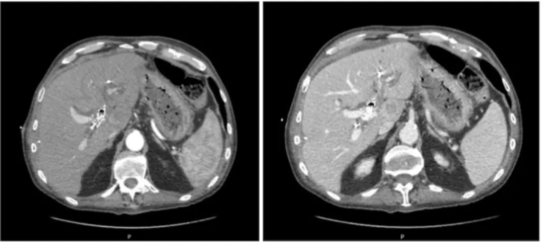
Preoperative total body computed tomography scan (CT) (Fig. 1) confirmed the presence of an infiltrating lesion orig- inating from the biliary bifurcation extended to the second order left bile ducts as well as the caudate segment. The lesion had a diameter of 30 mm × 46 mm, showing contrast enhance- ment and proximal dilatation of the biliary tree. The left HA appeared thread. It was partially involved.
The patient underwent a radical left extended hepatectomy (including segment I) with extra-hepatic bile duct resection and right Roux-en-Y double duct hepaticojejunostomy through the suture of both the right anterior and posterior sectorial bile ducts. Since it was impossible to remove the metallic biliary stent, its distal part was left in the main bile duct stump, close to the head of the pancreas. During surgery, resected margins were assessed by frozen-section histology. Results were nega- tive.
Hepatic hilum dissection was found to be difficult due to tu- mour location and size. In addition, there was an inflammato- ry reaction to the prosthetic device. To achieve a better control, the liver was completely mobilized.
The patient was transferred to our intensive care unit (ICU).
On postoperative day (POD) 3, he was moved to the surgical ward. He was haemodynamically stable with the following blood results: GOT of 49 UI/L, GPT of 145 UI/L, total bilirubin of 2.01 mg/dL (direct bilirubin 1.49 mg/dL), g-GT of 161 UI/
L, and INR of 1.3. He started a full liquid diet which was well tolerated.
Postoperative course was uneventful until POD 5 when the patient complained vomiting. At the examination, his abdo- men was distended and a pancreatic fistula was revealed by the abdominal surgical drains content, with increased level of amylases and lipases (amylases >7,500 U/L; lipases >3,500 U/L).
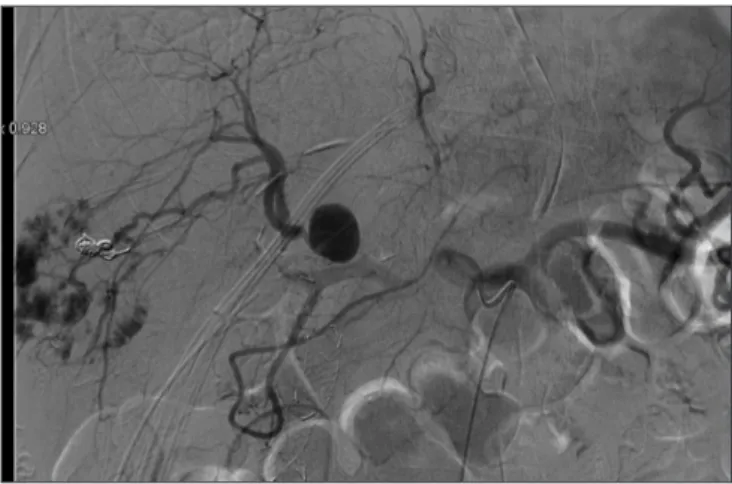
An abdominal contrast CT-scan showed a small collection in gallbladder bed (3 cm × 1.5 cm), abdominal effusion, and right HA branch pseudo-aneurysm (18 mm) whose presence was confirmed through an angiography (Fig. 2). No active bleeding was revealed. Stent placement in the right HA was infeasible.
Coiling of the pseudoaneurysm was associated with a risk of complete occlusion inducing critical liver failure. After the procedure, his general conditions suddenly deteriorated and an emergency laparotomy was performed.
The liver was pale and ischemic. Intraoperative ultrasound showed no arterial flow. The right branch of the HA was dis- rupted in the area of the pseudo-aneurysm. Considering the local inflammatory situation, a HA reconstruction was im- possible. Thus, PVA was used to provide arterial inflow to the remnant liver. A side-to-side arteriovenous shunt was created between the ileocolic artery and the ileocolic vein using two Prolene Suture 7–0 (Fig. 3).
At the end of the procedure, the liver was reddish-brown in colour. It felt rubbery to the touch. The intraoperative ul- trasound showed satisfactory patency of the PVA with good portal flow in the absence of arterial flow. The patient was transferred to the ICU. On POD 3, blood tests revealed total
Fig. 1. Preoperative total body computed tomography scan (arterial and portal phase) showing the presence of metallic biliary stent and bilateral percutaneous drainages.
bilirubin level of 4.88 mg/dL (direct bilirubin, 2.89 mg/dL), platelets of 123,000/µL, and INR of 2.23. An asymptomatic biliary fistula occurred. It was conservatively managed. He was extubated on POD 10. Doppler ultrasound on POD 15 showed that the cross-sectional area and blood flow of the portal vein were increased (main portal vein, 18 cm/sec). The patient was discharged on POD 54 in good general condition. The patient is alive and doing well at six-month follow-up without evidence of recurrence or systemic disease. At three months follow up, the patient was admitted for ascites (2 L/day) and portal hy- pertension was suspected as the major cause. The ascites was rapidly resolved with medical treatment and a single paracen- tesis. The patient was then discharged. No evidence of portal hypertension was found in Doppler ultrasound.
Written informed consent was obtained from the patient for publication of this case report and accompanying images. A copy of the written consent is available for review by the Edi- tor-in-Chief of this journal upon request.
DISCUSSION
We report a rescue case of PVA following major complica- tions after a left extended hepatectomy.
As a matter of fact, HA disruption represents potentially le- thal complications of hepatic, biliary, and pancreatic surgeries [3]. It often leads to biliary ischemia (bile leakage, bile infarcts, biloma formation) and partial liver necrosis.
In the case described here, previous arterial embolization and the challenging hepatic pedicle dissection due to inflammation and biliary stump fistula might have fragilized the HA wall, causing the HA pseudo-aneurysm. On the other side, complete liver mobilization hindered the development of arterial collat- erals.
PVA was historically used in portal hypertension surgery in association with portocaval shunting. It seemed to attenuate the ischemic effect on cholangiocytes caused by a HA disrup- tion, reducing cholestasis and proinflammatory cytokine pro- duction, ductular reaction, and fibrosis [4] in animal models.
Because of anastomotic arterioportal thrombosis and compli- cations related to an excessive liver arterialization with the de- velopment of other therapeutic options, this surgical approach was abandoned in favor of radiological shunting therapies.
PVA is largely used in the field of liver transplantation [5]. It was first described in 1989 by Sheil et al. [6]. After a suprahe- patic inferior vena cava anastomosis, they attached a cannula inserted in the recipient iliac artery to a donor portal vein can- nula, resulting in early liver revascularization.
According to Bonnet et al. [7], it could be particularly useful in case of pre-existing diffuse portal vein thrombosis, notably a contraindication for orthotopic liver transplantation (OLT) or more rarely in case of portal vein thrombosis complicating a recent OLT.
In a patient with HA thrombosis after a liver transplantation, PVA presents the disadvantage of leaving the portal hyper- tension unchanged. Thus, an over arterialization of the liver can eventually lead to fibrosis. Some authors have reported an aneurysmal dilatation of the PV and its intrahepatic branches revealed by CT scan and various complications (most notably right-sided heart failure and portal hypertension) following PVA [8].
Current indications for PVA in the field of liver transplan- tation domain include post-OLT hepatic artery thrombosis (HAT), extended splanchnic vein thrombosis pre-OLT or post- OLT, and low portal flow pre-OLT according to anatomic vari- ations such as the absence of portal and mesenteric veins.
Moreover, according to the literature, PVA can also be used in case of acute liver failure as a life-saving measure in order to provide temporary life support [9,10].
Fig. 2. Angiography showing a hepatic artery pseudoaneurysm.
The picture shows results of a previous super selective angioma em- bolization with coils and the residual metallic stent in the bile duct stump.
Fig. 3. Side to side arteriovenous shunt between the ileocolic artery and the ileocolic vein using two Prolene 7/0 running sutures.
Beside OLT, PVA is also used to prevent HAT after cancer sur- gery, representing an alternative strategy when HA reconstruc- tion after its accidental resection is impossible. In 2004, Kondo et al. [11] described 10 cases of biliary cancer treated with rad- ical surgery performed through an en-bloc resection including HA and an end-to-side arterioportal reconstruction between the common hepatic or gastroduodenal artery and the PV.
Since then, only Young et al. [12] have reported the use of PVA in a cholangiocarcinoma surgery, describing 2 cases of hi- lar cholangiocarcinoma treated with PVA as a salvage strategy.
Finally, PVA can be a promising emergency intervention al- lowing the entire liver cells (hepatocytes and, specifically, chol- angiocytes) to mitigate the hypoxic-ischemic damage by devel- oping arterial collaterals. Results emerging from a Paris group [1], both in case of liver transplant or hepatic resection, have shown an overall survival rate of 63% at a median follow-up of 13 months.
In our case, PVA was performed in an emergency setting to avoid ischemic injury after HA pseudoaneurysm and disrup- tion, thus decreasing serum transaminase levels and limiting the parenchymal ischemia.
Despite this enthusiasm, PVA is a procedure associated with high morbidity rates even if the natural course of a completely de-arterialized liver is death. Complications related to arterio- venous shunt thrombosis can lead to emergency liver trans- plantation as only a rescue option [13].
The aim of PVA is to create a time frame to allow collater- al arterial vessels onset or a definitive surgery performance.
Early thrombosis can be a catastrophic event due to acute liver ischemia, although a delayed occlusion is often well tolerated.
Surveillance is recommended because an embolization of the arterioportal fistula could be necessary to treat or prevent the onset of portal hypertension.
In conclusion, complete arterial hepatic deprivation is known to be associated with severe ischemic damage, including bile infarcts, bilomas, and eventually liver abscesses after infection.
PVA has a beneficial effect on ischemia-hypoxia-induced bile duct injury. However, increased portal blood represents a cause of portal hypertension and liver fibrosis. Thus, measures are needed to control blood flow and patients who have undergone PVA should be followed up.
CONFLICT OF INTEREST
No potential conflict of interest relevant to this article was reported.
ORCID
Celeste Del Basso, https://orcid.org/0000-0003-3606-9512 Roberto Luca Meniconi,
https://orcid.org/0000-0003-3324-8128
Sofia Usai, https://orcid.org/0000-0002-3789-2108
Nicola Guglielmo, https://orcid.org/0000-0003-1052-5006 Marco Colasanti, https://orcid.org/0000-0002-5697-7086 Stefano Ferretti, https://orcid.org/0000-0002-6275-7813 Giovanni Battista Levi Sandri,
https://orcid.org/0000-0002-7893-5047 Giuseppe Maria Ettorre,
https://orcid.org/0000-0002-7501-5472
AUTHOR CONTRIBUTIONS
Conceptualization: CDB, RLM. Data curation: CDB, RLM, SU. Methodology. CDB, RLM, SU. Visualization: CDB, RLM, SU. Writing - original draft preparation. CDB, RLM, SU, GME.
Writing - review and editing: All authors.
REFERENCES
1. Bhangui P, Salloum C, Lim C, Andreani P, Ariche A, Adam R, et al.
Portal vein arterialization: a salvage procedure for a totally de-arte- rialized liver. The Paul Brousse Hospital experience. HPB (Oxford) 2014;16:723-738.
2. Iseki J, Touyama K, Noie T, Nakagami K, Takagi M, Hakamada K, et al. Partial portal arterialization for the prevention of massive liver necrosis following extended pancreatobiliary surgery: experience of two cases. Surg Today 1992;22:568-571.
3. Tessier DJ, Fowl RJ, Stone WM, McKusick MA, Abbas MA, Sarr MG, et al. Iatrogenic hepatic artery pseudoaneurysms: an uncommon complication after hepatic, biliary, and pancreatic procedures. Ann Vasc Surg 2003;17:663-669.
4. Jiang J, Wei J, Wu J, Gao W, Li Q, Jiang K, et al. Partial portal vein arterialization attenuates acute bile duct injury induced by hepatic dearterialization in a rat model. Biomed Res Int 2016;2016:7427246.
5. Melandro F, Lai Q, Levi Sandri GB, Guglielmo N, Di Laudo M, Mo- rabito V, et al. A case of portal vein arterialization after a liver trans- plant. Exp Clin Transplant 2013;11:287-289.
6. Sheil AG, Thompson JF, Stephen MS, Eyers AA, Bookallil M, Mc- Caughan GW, et al. Donor portal vein arterialization during liver transplantation. Transplant Proc 1989;21(1 Pt 2):2343-2344.
7. Bonnet S, Sauvanet A, Bruno O, Sommacale D, Francoz C, Dondero F, et al. Long-term survival after portal vein arterialization for portal vein thrombosis in orthotopic liver transplantation. Gastroenterol Clin Biol 2010;34:23-28.
8. Paloyo S, Nishida S, Fan J, Tekin A, Selvaggi G, Levi D, et al. Portal vein arterialization using an accessory right hepatic artery in liver transplantation. Liver Transpl 2013;19:773-775.
9. Erhard J, Lange R, Rauen U, Scherer R, Friedrich J, Pietsch M, et al.
Auxiliary liver transplantation with arterialization of the portal vein for acute hepatic failure. Transpl Int 1998;11:266-271.
10. Charco R, Margarit C, López-Talavera JC, Hidalgo E, Castells L, Allende H, et al. Outcome and hepatic hemodynamics in liver trans- plant patients with portal vein arterialization. Am J Transplant 2001;1:146-151.
11. Kondo S, Hirano S, Ambo Y, Tanaka E, Kubota T, Katoh H. Arterio-
portal shunting as an alternative to microvascular reconstruction after hepatic artery resection. Br J Surg 2004;91:248-251.
12. Young AL, Prasad KR, Adair R, Abu Hilal M, Guthrie JA, Lodge JP.
Portal vein arterialization as a salvage procedure during left hepat-
ic trisectionectomy for hilar cholangiocarcinoma. J Am Coll Surg 2008;207:e1-e6.
13. Hidalgo E. Portal vein arterialization: ‘enjoy’ it responsibly. HPB (Oxford) 2014;16:739.