Korean J Gastroenterol Vol. 68 No. 3, 166-168 http://dx.doi.org/10.4166/kjg.2016.68.3.166 pISSN 1598-9992 eISSN 2233-6869
IMAGE OF THE MONTH
Korean J Gastroenterol, Vol. 68 No. 3, September 2016 www.kjg.or.kr
게실염으로 오인된 대장암
구훈섭
건양대학교 의과대학 내과학교실
Colon Cancer Misdiagnosed as Diverticulitis
Hoon Sup Koo
Department of Internal Medicine, Konyang University College of Medicine, Daejeon, Korea
CC This is an open access article distributed under the terms of the Creative Commons Attribution Non-Commercial License (http://creativecommons.org/licenses/
by-nc/4.0) which permits unrestricted non-commercial use, distribution, and reproduction in any medium, provided the original work is properly cited.
Copyright © 2016. Korean Society of Gastroenterology.
교신저자: 구훈섭, 35365, 대전시 서구 관저동로 158, 건양대학교 의과대학 내과학교실
Correspondence to: Hoon Sup Koo, Department of Internal Medicine, Konyang University College of Medicine, 158 Gwanjeodong-ro, Seo-gu, Daejeon 35365, Korea.
Tel: +82-42-600-9127, Fax: +82-42-600-9058, E-mail: [email protected] Financial support: None. Conflict of interest: None.
증례: 62세 남자 환자가 전날부터 발생한 복통과 발열을 주소로 방문하였다. 우상복부에 당기는 듯한 복통이 주기적으 로 호전과 악화를 반복하면서 그 강도가 점점 심해지는 양상 이었고, 급성 병색을 보였다. 그러나 혈변, 설사, 체중 감소, 소화불량, 배변 형태의 변화 등은 없었다. 당뇨, 고혈압, 간염, 폐결핵의 병력은 없었고, 암의 가족력과 과거력 또한 없었다.
방문 당시 활력징후는 혈압 100/60 mmHg, 맥박수 70회/분, 체온 37.9oC로 측정되었다. 신체검사에서 결막이 창백하였고, 복부 촉진에서 우상복부 압통이 관찰되었으나 반발 압통은 없 었으며, 종물은 촉지되지 않았다. 말초혈액검사에서 백혈구 16,900/L, 혈색소 10.1 g/dL, 혈소판 244,000/L로 측정되었 고, 일반화학검사에서 혈액요소질소 12.1 mg/dL, 크레아티닌 1.05 mg/dL, 총 단백 7.0 g/dL, 알부민 4.3 g/dL, AST/ALT 17/13 IU/L, 총 빌리루빈 0.4 mg/dL, ALP 97 IU/L로 측정되 었다. CEA는 1.64 ng/mL로 정상 소견을 보였다. 발열 및 복 통의 원인 감별을 위해 복부 전산화단층촬영 검사를 시행하였 다. 원위부 상행결장 주위의 지방 침윤, 염증 및 장벽 비후를 보이는 상행 결장의 게실염이 강하게 의심되었다(Fig. 1).
발열과 복통을 보이는 상행 결장의 게실염으로 진단하고 식이 조절, 수액 공급 및 경정맥 항생제(ciprofloxacin)로 7일 간 입원 치료를 하였다. 치료 후 발열, 복통 증상이 호전되었 으며, 퇴원 시 경구 항생제(ciprofloxacin)를 처방하였다. 퇴원 일주일 후 외래진료 방문 당시 복통, 발열 증상은 모두 호전되
었으며, 체중 감소, 흑색변, 소화불량 등의 이상 소견이 없어 급성 게실염이 잘 치료되었다고 생각하였다. 복부 전산화단층 촬영으로 진단받은 우측 대장 게실염 환자로 치료 후 6주 뒤 대장내시경 검사를 계획하였다. 6주 후 대장내시경 검사에서 원위부 상행결장에 관강 둘레를 100% 차지하고 내시경 통과 는 가능한, 자발성 출혈을 보이는 원주형 궤양 침윤성 종괴가 관찰되었으며, 이 병변에 대해 대장내시경으로 생검을 시행하 였고, 조직검사에서 분화도가 좋은 선암으로 확진되었다(Fig.
2). 추적 복부 전산화단층촬영에서 원위부 상행결장 주변의 염증은 호전되었으나 5 cm 길이의 장벽 윤상 비후 소견이 관찰되었고, 양전자 방출 단층촬영에서는 원위부 상행결장 및 주변 림프절에 fluorodeoxyglucose (FDG) 섭취 증가 소견이 관찰되었다(Fig. 3). 환자는 원위부 상행 결장암 진단 후 우반 결장 절제술을 시행받았다.
진단: 게실염으로 오인된 대장암
대장 게실(colon diverticulum)은 대장벽의 일부가 비정상 적으로 탈출된 소낭이고, 게실증(diverticulosis)은 염증이나 출혈의 합병증이 없는 상태이며, 합병증이나 통증이 동반되면 게실 질환(diverticular disease)이라 한다. 서구의 경우 대부 분 좌측 대장 게실염인데 반해 동양인의 경우에는 55-75%가 우측 대장 게실염이다.1 대장 게실염의 임상 양상은 다양한데, 경도의 복통 및 열부터 15-20%는 농양, 누공, 폐색, 천공으로
Koo HS. Colon Cancer Misdiagnosed as Diverticulitis
167
Vol. 68 No. 3, September 2016
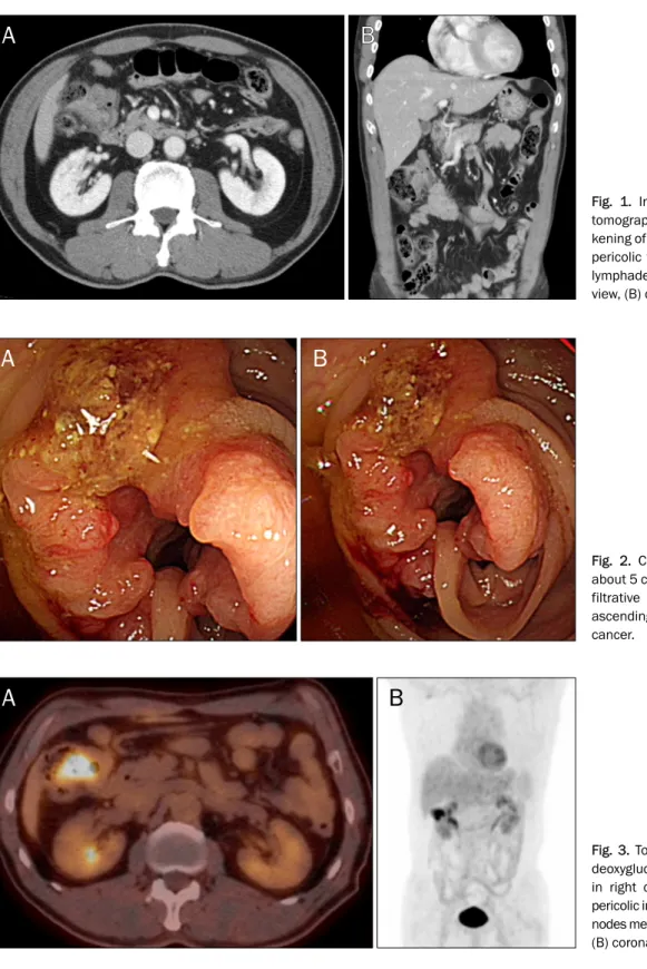
Fig. 1. Initial abdominal computed tomography shows segmental thic- kening of distal ascending colon with pericolic fat infiltration and regional lymphadenopathy. (A) Transverse view, (B) coronal view.
Fig. 2. Colonoscopic findings show about 5 cm, circumferential ulceroin- filtrative mass lesion in the distal ascending colon, suggesting colon cancer.
Fig. 3. Torso PET CT showed fluoro- deoxyglucose (FDG)-avid malignancy in right colon, hepatic flexure with pericolic infiltration and regional lymph nodes metastases. (A) Transverse view, (B) coronal view.
진행할 수 있고 천공으로 진단된 게실염 환자의 12-36%는 사 망할 수 있기 때문에 정확하고 빠른 진단이 매우 중요하다 할 수 있다.2,3 주된 치료는 항생제 사용, 장 휴식(bowel rest), 복통 조절이다. 항생제 치료는 그람 음성 및 혐기성 균을 모두 포함할 수 있는 조합이 필요한데, 외래 환자들의 경우 cipro-
floxacin과 metronidazole 또는 amoxicillin/clavulanate를 사용한다. 입원 환자들의 경우는 정주용 광범위 항생제를 투여 하는데, ceftriaxone과 metronidazole, 단일 제제로는 beta- lactam/beta-lactamase 억제제(piperacillin/tazobactam) 또 는 meropenem을 사용하며, 사용 기간은 7-10일이다.4,5
168
구훈섭. 게실염으로 오인된 대장암The Korean Journal of Gastroenterology
현재 임상에서 급성 대장 게실 질환의 진단에 가장 널리 이용되는 검사법은 복부 전산화단층촬영인데, 이는 급성 게실 염 진단시 검사의 비침습성, 대장 주위 염증 평가의 유용성, 게실염의 범위와 합병증에 대한 평가가 가능한 장점을 가지기 때문에 가장 먼저 시행되는 검사이다.6 복부 전산화단층촬영 에서 게실염은 장벽의 비후, 결장 주변의 염증 침윤, 조영증 강, 농양, 장 폐쇄, 장 주위 림프절의 크기 증가 소견을 보일 수 있어 악성 종양과의 감별이 힘들 수 있다.7 최근에 고해상 전산화단층촬영의 정확도가 80-100%까지 높아졌다는 연구도 있으나, 여전히 전산화단층촬영으로 게실염 환자에서 악성 종 양을 감별하는 것은 어려운 문제이다.8 따라서, 게실염 환자의 악성 종양 감별을 위해 일반적으로 대장내시경 시행이 권고되 고 있다.9
2015년 미국 소화기학회에서는 전산화단층촬영에 의해 확 진된 급성 게실염에 대해 치료 후 대장내시경 검사의 유용성 에 대해 문헌고찰을 통해 평가하였다. 분석에서 치료 4-8주 후 시행된 대장내시경 검사에서 1,000명의 환자당 15건의 대 장암, 38건의 진행성 선종을 발견할 수 있었다.10
급성 게실염의 진단 및 치료에 조기 대장내시경의 유용성 에 대한 연구가 진행되고 있다.11 조기 대장내시경이란 급성 게실염으로 입원 치료 중 내시경을 시행하는 것을 말한다. 일 반적으로 조기 대장내시경은 대장 천공의 가능성이 높다고 알 려져 있으며, 통증 및 게실염에 의한 염증성 협착으로 인해 맹장 삽입률이 75-82%로 낮아 앞으로 조기 대장내시경 검사 에 대한 더 많은 연구가 이루어져야 할 것으로 생각된다. 급성 게실염 치료 후 약 6주경에 대장내시경을 시행할 것을 권고하 고 있다.12-15
이번 증례는 입원할 당시 시행했던 전산화단층촬영에서 장 벽 비후가 동반된 급성 게실염이 의심되었고 임상적으로 게실 염에 합당한 소견이 관찰되었으며 항생제 및 수액 치료 후 증상이 호전되어 입원 중 대장내시경을 시행하지 않았다. 추 적 관찰 중 대장내시경 검사를 시행하지 않았더라면 진단을 계속해서 놓칠 수 있었던 증례로, 게실염으로 강하게 의심되 는 경우라도 악성 종양과의 감별 진단을 위해 치료 후 약 6주 경에 대장내시경 검사가 반드시 필요할 것으로 생각된다.
REFERENCES
1. Hildebrand P, Kropp M, Stellmacher F, Roblick UJ, Bruch HP, Schwandner O. Surgery for right-sided colonic diverticulitis: re- sults of a 10-year-observation period. Langenbecks Arch Surg 2007;392:143-147.
2. Morris CR, Harvey IM, Stebbings WS, Speakman CT, Kennedy HJ, Hart AR. Epidemiology of perforated colonic diverticular disease. Postgrad Med J 2002;78:654-658.
3. Stollman N, Raskin JB. Diverticular disease of the colon. Lancet 2004;363:631-639.
4. Schechter S, Mulvey J, Eisenstat TE. Management of un- complicated acute diverticulitis: results of a survey. Dis Colon Rectum 1999;42:470-475.
5. Weizman AV, Nguyen GC. Diverticular disease: epidemiology and management. Can J Gastroenterol 2011;25:385-389.
6. Birnbaum BA, Balthazar EJ. CT of appendicitis and diverticulitis.
Radiol Clin North Am 1994;32:885-898.
7. Shen SH, Chen JD, Tiu CM, et al. Differentiating colonic divertic- ulitis from colon cancer: the value of computed tomography in the emergency setting. J Chin Med Assoc 2005;68:411-418.
8. Balthazar EJ, Megibow A, Schinella RA, Gordon R. Limitations in the CT diagnosis of acute diverticulitis: comparison of CT, con- trast enema, and pathologic findings in 16 patients. AJR Am J Roentgenol 1990;154:281-285.
9. Hale WB; NDSG. Colonoscopy in the diagnosis and management of diverticular disease. J Clin Gastroenterol 2008;42:1142-1144.
10. Strate LL, Peery AF, Neumann I. American Gastroenterological Association Institute technical review on the management of acute diverticulitis. Gastroenterology 2015;149:1950-1976.e12.
11. Lahat A, Yanai H, Menachem Y, Avidan B, Bar-Meir S. The feasi- bility and risk of early colonoscopy in acute diverticulitis: a pro- spective controlled study. Endoscopy 2007;39:521-524.
12. Almy TP, Howell DA. Medical progress. Diverticular disease of the colon. N Engl J Med 1980;302:324-331.
13. Penfold JC. Perforation of the colon complicating colonoscopy:
report of a case. Dis Colon Rectum 1975;18:626-627.
14. Dean AC, Newell JP. Colonoscopy in the differential diagnosis of carcinoma from diverticulitis of the sigmoid colon. Br J Surg 1973;60:633-635.
15. Panish JF. Limitations and complications of colonoscopy.
Gastrointest Endosc 1980;26(2 Suppl):20S-21S.