교합성 골수강내 금속정술후 발생한 대퇴골 간부 골절의 불유합의 치료
이근배・문은선・송은규・최 진・정성택 전남대학교 의과대학 정형외과학교실
<국문초록>
목 적 : 교합성 골수강내 금속정술 후 발생한 대퇴골 간부 골절의 불유합의 치료 결과를 알아 보고자 하였다.
대상 및 방법 : 1990년 5월부터 2 0 0 0년 7월까지 대퇴골 간부 골절에 대한 교합성 골수강내 금속정술후 발생한 불유합에 대해 치료받은 환자중 1년 이상 추시 가능하였던 3 3명을 대상으로 후향적으로 조사 하였다. 수술 당시 평균 연령은 4 0세( 1 9 - 6 8 )였으며, 남자가 2 7례, 여자가 6례였다. 불유합의 형태는 W e b e r와 B r u n n e r의 분류 방법에 따르면 과혈관형 1 0예(30%), 무혈관형 2 1예(64%), 혼합형이 2예(6%), 무 혈관형중에서 감염성이 5예( 2 3 % )였다. 결과는 수술 방법에 따른 골 유합 기간 및 불유합, 부정 유합, 감염 등의 합병증의 유무로 평가하였다.
결 과 : 수술 방법은 불유합의 원인과 형태에 따라 결정하였으며, 5가지 방법으로 시행하였다. 골수강 내 금속정 교환술을 시행한 경우가 7예, 골수강내 금속정 교환술 및 골 이식술을 함께 시행한 경우 1 1 예, 외고정술 5예, 금속판 고정 및 골 이식술 3예, 해면골 이식술만을 시행한 경우가 7예였다. 골유합 기간은 각각 평균 1 9주, 17주, 20주, 16주, 15주로서 치료 방법 간에 통계학적인 의의는 없었다. 전례에 서 술 후 감염이나 불유합, 부정 유합 등은 관찰되지 않았다.
결 론 : 교합성 골수강내 금속정술후 발생한 대퇴골 간부 골절의 불유합의 치료에 있어서는 불유합의 원인과 형태를 잘 분석하여 그 원인에 맞는 적절한 치료 방법을 선택하는 것이 골유합을 얻는데 매우 중요하다고 생각된다.
색인 단어 : 대퇴골 간부 골절, 불유합, 교합성 골수강내 금속정술
※ 통신저자 : 정 성 택
501-757 광주시 동구 학동 8번지 전남대학교 의과대학정형외과학교실 TEL : 062-227-1640
FAX : 062-225-7794
E-mail : [email protected]
* 본 논문의 요지는 2001년도 대한골절학회 추계학술대회에서발표되었음 .
서 론
교합성 골수강내 금속정술후 발생한 대퇴골 간부 골절의불유합은 분쇄골절, 개방성골절, 골절부위 의 혈액순환 장애, 골절편의 부적절한 정복이나 고 정, 골절부위의신연, 골유합전의과도한운동, 감염 등의복합적인요인에의하여발생할수있기때문에 이와같은원인들을잘분석하여치료계획을세워야
한다3 , 4 , 7 , 8 , 1 2 , 1 6 ). 특히 원인에따른적절한수술방법의
선택이 조기에 골 유합을 성취할 수 있는 가장 중요 한 인자라 생각된다. 이에 본 연구는 교합성 골수강 내금속정술후발생한대퇴골간부골절의불유합에 대해치료한 3 3명의환자를대상으로불유합의원인 에따른치료방법들의결과를분석하고자하였다.
연구 대상 및 방법
1 9 9 0년 5월부터 2 0 0 0년 7월까지본원및타병원에 서교합성골수강내금속정술후대퇴골 간부골절의 불유합이발생하여본원에서 치료받은 3 3명을 대상 으로 하였다. 연구는 의무 기록지와 추시방사선 사
진을기초로하여후향적으로조사하였으며, 추시기 간은 평균1 7개월( 1 2 - 4 6 )이었다. 환자의 연령은 평균 4 0세( 1 9 - 6 8 )이었고, 남자 2 7명, 여자 6명이었다. 최초 의 수상 원인은 교통 사고가 2 5예( 7 5 % )로 가장 많았 고, 그외넘어진 경우 5예(15%), 추락 3예( 9 % )이었다.
1차 수술 후 불유합 판정까지의 기간은 평균 1 7주( 6 - 7 6주)이었다.
초기골절의분류는 Winquist-Hansen 분류1 9 )에따르 면 제 1형이 2예(6%), 제 2형이 8예(24%), 제 3형이 6 예(18%), 제 4형이 1 5예( 4 5 % )였으며, 타병원에서 1차 수술한환자중 2예는골절의형태를분류할수 없었 다. 불유합의형태는 W e b e r와 B r u n n e r의분류1 6 )방법 에 의하면 과혈관형 1 0예(30%), 무혈관형 2 1예( 6 4 % ) , 혼합형이 2예(6%), 무혈관형중에서 감염성이 5예 ( 2 3 % )였다. 기계적 부전과 동반된 경우는 금속정 파 단이 7예, 나사못의이완이1예였다. 1차수술후본원 에서불유합에대한수술을하기전까지추가적인수 술을 받았던 환자는 모두 1 1명, 19회로 골 유합을 얻 기위해역동화, 골이식, 금속판보강술등을시행받 았으며, 수술횟수는 5명에서1회, 4명에서2회, 2명에 서 3회이었다. 결과는 수술 방법에 따른 골 유합 기
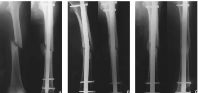
Fig. 1-A : The preoperative roentgenogram of 58-year-old man shows Winguist-Hansen type II femoral shaft fracture (Lt), and the postoperative roentgenogram shows medial fracture gap with displaced fracture fragment(Rt).
Fig.1-B : The 12 months follow-up radiograph shows breakage of nail with varus angulation of fracture(Lt), and the nonunion was managed with larger nail exchange without bone graft(Rt).
Fig.1-C : The 6 months(Lt) and 12 months(Rt) follow up radiograph after larger nail exchange shows complete union.
간및불유합 , 부정유합, 감염등의합병증의유무로 평가하였다.
결 과
불유합에 대한 치료 방법으로는 골수강내 금속정 교환 및 골 이식술이 1 1예(33%), 골수강내 금속정교 환만시행한경우가 7예(21%), 외고정이5예(15%), 금 속판고정술 및골이식술이 3예(9%), 해면골이식술 의경우가 7예( 2 1 % )였다.
치료방법은불유합의원인과형태에 따라결정하 였으며금속정의안정성이 부족하다고 생각되는경 우엔 더 굵은 금속정으로의 교환술을 시행하였고 (Fig. 1), 금속정의안정성은유지되나골결손이심하 여 골유합의진행이어렵다고생각되는경우나무혈 관형 불유합에서는골이식술을시행하였으며, 감염 성불유합으로내고정이어렵거나골단축이있어골 연장술이 필요한 경우에는 외고정을 시행하였다 (Fig. 2). 또한골절부위의금속정의안정성이부족하 고, 골절간격이 넓어 골절편의 직접적인 압박이필 요하다고 생각되는 경우엔 압박 금속판 고정 및 골 이식술을시행하였다(Fig. 3).
골 유합 기간은 골수강내 금속정 교환 및 골 이식 술을시행한경우에서평균 1 7주( 1 2 - 2 8주), 골수강내
금속정 교환술만 시행한경우는평균 1 9주( 1 4 - 3 1주) , 외고정술의경우평균 2 0주( 1 7 - 2 6주), 금속판 고정및 골이식술의경우평균 1 6주(14 -24주), 해면골이식술 을 시행한 경우에서는 평균 1 5주( 1 1 - 2 0주)로서 치료 방법간에어느정도의 차이는보였으나통계학적인 의의는없었다.
합병증으로는전례에서불유합이나 부정유합, 감 염 등은 없었으며, 동측 슬 관절 부분 강직이 3예 있 었다.
고 찰
골수강내 금속정은 대부분의 대퇴골 간부 골절의 최적의 치료 방법으로 널리 사용되고 있으며, 수술 기법의발달과금속정의디자인의개발에힘입어우 수한 결과와 매우 높은 유합율이 보고되고 있다
1 , 2 , 5 , 6 , 1 1 , 1 4 , 1 6 , 1 9 ). 대퇴골 간부 골절에 대한 골수강내 금
속정술 후 불유합율은 매우 낮아서 1 9 8 4년 W i n q u i s t
등1 8 )에 의하면 0 . 9 %의 불유합을 보인다고 하였고,
1 9 8 8년 Brumback 등2 )은 2 %에서 불유합이 발생하였 다고보고하였다. 근래에들어서는골수강내금속정 의수술적응증이복잡골절에까지더욱확대되면서 불유합율은 점차 증가되고 있으며, Kempf 등6 )은 대 퇴골의 심한 분쇄 골절에서 1 0 %의 불유합을 보고하
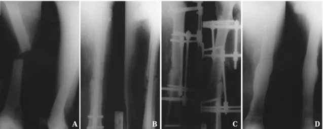
Fig. 2-A : The preoperative radiograph of 41-year-old man shows transverse femoral shaft fracture.
Fig. 2-B : The 11 months follow up radiograph after interlocking IM nailing shows diffuse periosteal reaction through the entire shaft and bony resorption at fracture end.
Fig. 2-C : The postoperative radiograph shows removal of nail and change to external fixation with Ilizarov.
Fig. 2-D : The 18 months follow-up radiograph after external fixation shows complete union without any evidence of infection.
A B C D
였다.
불유합이 발생하는 원인은 대부분 분쇄 골절이나 개방성골절, 부적절한고정, 골절부위의신연, 골절 부위의 혈액 순환 장애, 감염 등과 같은 복합적인 요 인에 의해 발생되는 것으로 보고되어 있으며
3 , 4 , 7 , 8 , 9 , 1 2 , 1 6 ), 본 연구에서의 불유합의 원인도 모두 이
들 중 하나에 해당되었다. 이를 극복하기 위해서는 골절 초기에분쇄 정도나감염 등골절의 양상을 잘 파악해가능한정확한해부학적정복을 이룸과동시 에 골절 간격이 최소화 되도록 노력해야 하며, 알맞 은크기의금속정을사용하여견고한 내고정을도모 하고, 골절 부위의 혈행 장애를 최소화하기 위한 주 의깊은연부조직의조작이필요하며 , 술 후 재 활 강 도나 완전 체중 부하의 시점은 추시 방사선 소견에 따라 개별적인 적용을하는것이중요하다고생각된 다. 또한치료 방법의 선택은 이와 같은 불유합의 원 인을해결하는 방향으로결정되어야하는데, 불유합 의원인이부적절한내고정인경우에는더굵은금속 정으로 교환하고, 무혈관형 불유합에서는자가골이 식을추가하며, 원인이골절부위의신연인경우에는 정적고정을동적고정으로바꿔신연부위에압박이 가도록 하거나 골 이식 또는 확공형의 더 굵은 금속
정 교환술로치료할 수있다2 0 , 2 1 ). 이외에원위 골 간 단 부위의 불유합으로 금속정으로는 안정성이 부족 하다고판단되는경우엔 Poller 나사못을삽입하여불 안정성을줄이거나1 2 ), 추가로금속판을고정하는방
법8,15) 또는압박 금속판과골이식방법등을 고려할
수있으며, 감염성불유합으로내고정이어렵거나골 단축이있어골연장술이필요한경우에는외고정으 로치료할수있다 . 이와같은여러 가지치료 방법들 중 확공형의더굵은금속정으로의교환술은이환율 이낮고, 추가적인골 이식이필요치않으며, 조기재 활이가능하다는장점등으로골수강내금속정술후 발생한대퇴골골절의불유합의치료에가장흔히사 용되는방법으로서골유합율은높게는 K l e m m9 ), Oh
등1 3 )에의해 1 0 0 %의 성공률이보고되었으며, Weresh
등1 8 )은 5 3 %의낮은골유합율을 보고함으로써보고
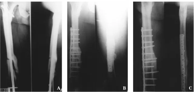
자에따라결과의차이가 매우크다고할수있다 . 저 자들의경우는확공후골수강내금속정교환술로치 료한경우가 7례, 골수강내금속정교환및골이식술 을함께시행했던경우가 1 1예로각각평균 1 9주와 1 7 주에 골유합이 모두관찰되었다. 따라서확공후 골 수강내금속정교환술은대부분의 대퇴골간부골절 의불유합에대해여전히최적의치료방법이라생각 Fig. 3-A : The preoperative radiograph of 48 year-old man with nonunion of femoral shaft fracture shows
incompletely united fracture fragment and large fracture gap.
Fig.3-B : The postoperative radiograph after osteosynthesis with compression plate and bone grafting shows obliteration of fracture gap.
Fig.3-C : The 18 months follow up radiograph shows complete union.
A B C
되나, 성공적인 골 유합율을 이루기 위해서는 모든 형태의 불유합에서 금속정 교환술만을 시행하기보 다는역동화나압박 금속판의사용을고려하거나무 혈관형불유합의경우엔골이식술을동시에시행하 는것이보다높은골 유합율을 얻을수 있는방법이 라 생각된다. 또 다른 치료 방법으로서는 외고정 5 례, 압박금속판고정및 골이식술 3예, 자가 해면골 이식술 7예를 시행하였으며, 전례에서 평균 1 5주에 서 2 0주에골유합을얻을수있었다 . 골유합 기간은 치료 방법 간에 어느 정도 차이를 보여 외고정술과 골수강내 금속정교환술을시행했던 경우에서각각 2 0주, 19주로 가장 많은 치료 기간이 소요되었으나 시행된 예가 적어 통계적인 의의는 없었다. 골유합 율에 있어서 저자들의 경우는 3 3예 전례에서 술 후 감염이나불유합없이우수한골유합소견을보였는 데 이는불유합의원인을분석하여가장중요하다고 생각되는 원인 인자를 해결하는 방향으로 적극적인 치료 방법을 선택하였기 때문이라고 생각하며 향후 더많은증례를 통한치료 방법간의 비교연구가필 요하리라생각된다.
결 론
교합성 골수강내 금속정술후 발생한 대퇴골 간부 골절의불유합의치료에있어서는 불유합의원인및 형태를 잘분석하여환자별로개별화된분석결과에 맞는적절한방법을선택하는것이골유합을얻는데 매우중요하다고생각된다.
R E F E R E N C E S
1. Brumback RJ, Reilly JP, Poka A, Lakatos RP, Bathon H and Burgess AR : Intramedullary nailing of femoral shaft fractures. Part I. Decision-making errors with interlocking fixation. J Bone Joint Surg, 70A:1441-1452, 1988.
2. Brumback RJ, Uwagie-Ero S, Lakatos RP, Poka A, Bathon GH and Burgess AR : Intramedullary nailing of femoral shaft fractures. Part II. Fracture healing with static interlocking fixation. J Bone Joint Surg, 70A:1453-1462, 1988.
3. Cove JA, Lhowe DW, Jupiter LB and Siliski JM : The management of femoral diaphyseal nonunion. J Orthop Trauma, 11:513-520, 1997.
4. Hak DJ, Lee SS and Goulet JA : Success of exchange reamed intramedullary nailing for femoral shaft nonunion or delayed union. J Orthop Trauma, 14:178-182, 2000.
5. Hanks GA, Foster WC and Cardea JA : Treatment of femoral shaft fractures with the Brooker-Wills interlocking intramedullary nail. Clin Orthop, 226:226-218, 1988.
6. Kempf I, Grosse A and Beck GF : Closed locked intramedullary nailing. Its application to comminuted fractures of the femur. J Bone Joint Surg, 67A:709- 720, 1985.
7. Kempf I, Grosse A and Rigaut P : The treatment of noninfected pseudoarthrosis of the femur and tibia with locked intramedullary nailing. Clin Orthop, 212:142-154, 1986.
8. Kim KS, Kim JO, Jung HG and Jung BO : Plate augmentation for the nonunion of femur shaft fractures after interlocking intramedullary nail fixation. J Korean Society of Fractures, 12:21-27, 1999.
9. Klemm KW : Treatment of infected pseudoarthrosis of the femur and tibia with an interlocking nail. Clin Orthop, 212:174-181, 1986.
10. Kuntscher G : Intramedullary surgical technique and its place in orthopaedic surgery. J Bone Joint Surg, 47A: 809-818, 1965.
11. Lee KB, Jung ST, Moon ES, Song EK and Jeong KC : Usefulness of interlocking compression nail in treatment of femoral shaft stable fracture. J Korean Society of Fractures, 14:601-608, 2001.
12. Lee SC and Song MH : Nonunion of the fracture of distal one-third of femoral shaft treated by interlocking intramedullary nailing. J Korean Society of Fractures, 12:259-266, 1999.
13. Oh I, Nahigian SH, Rascher JJ and Farkall JP : Closed intramedullary nailing for ununited femoral shaft fractures. Clin Orthop, 106:206-215, 1975.
14. Rothwell AG and Fitxpatrick CB : Closed Kuntscher nailing of femoral shaft fractures: a series of 100 consecutive patients. J Bone Joint Surg, 60B:504-509, 1978.
15. Ueng SWN, Chao EK, Lee SS and Shih CH : Augmentative plate fixation for the management of femoral nonunion after intramedullary nailing. J Trauma, 43:640-644, 1995.
16. Weber BG and Brunner C : The treatment of nonunion without electrical stimulation. Clin Orthop, 161:24-32, 1981.
17. Webb LX, Winquist RA and Hansen ST : Intramedullary nailing and reaming for delayed union or nonunion of the femoral shaft. Clin Orthop, 212:133-141, 1986.
18. Weresh MJ, Hakanson R, Stover M, Sims SH, Kellam JF and Bosse MJ : Failure of exchange reamed intramedullary nails for ununited femoral shaft fractures. J Orthop Trauma, 14:335-338, 2000.
19. Winquist RA, Hansen ST and Clawson DK : Closed intramedullary nailing of femoral fractures. J Bone Joint Surg, 66A:529-539, 1984
20. Wu CC : The effect of dynamization on slowing the healing of femur shaft fractures after interlocking nailing. J Trauma, 43:263-267, 1997.
21. Wu CC and Chen WJ : The treatment of femoral shaft aseptic nonunion: comparison between closed and open bone-grafting techniques. J Trauma, 43:112-116, 1997.
. .
Treatment of the Nonunion of Femur Shaft Fractures after Interlocking Intramedullary Nailing
Keun-Bae Lee, M.D., Eun-Sun Moon, M.D., Eun-Kyoo Song, M.D., Jin Choi, M.D., Sung-Taek Jung, M.D.
Department of Orthopedics, Chonnam National University Hospital, Gwangju, Korea
Purpose : We analyzed the results of treatment for the nonunion of femur shaft fractures after interlocking intramedullary(IM) nail fixation.
Materials & Methods : Thirty-three patients who underwent interlocking IM nailing due to femur shaft fractures from May, 1990 to July, 2000 and followed up for more than one year were evaluated retrospectively. Mean age at the time of operation was 40 years(Range, 19-68). 27 cases were men and 6 cases were women.
By Weber and Brunner classification of the nonunion, hypervascular type were 10 cases(30%), avascular type 21cases(64%), mixed type 2 cases(6%). Infected type among the avascular type of nonunion were 5 cases(23%). Results were evaluated with bone union by treatment methods and complications.
Results : According to the causes and types of nonunion, we performed IM nail exchange in seven cases, IM nail exchange and bone grafting in eleven cases, external fixation in five cases, compression plating and bone grafting in three cases, and only cancellous bone grafting in seven cases. Radiographical union was achieved in 19 weeks, 17 weeks, 20 weeks, 16 weeks and 15 weeks respectively. There’s no statistically significant difference between treatment methods. There are no cases of nonunion, malunion and infection.
Conclusion : The selection of appropriate treatment method by the cause and type of each nonunion is very important to achieve the bony union in the treatment for the nonunion of femur shaft fractures after interlocking intramedullary nailing.
Key Words : Femur shaft fracture, Nonunion, Interlocking intramedullary nailing Abstract