Trochanteric Fixation Nail-Advanced (TFNA) 근위 대퇴골 골수정을 이용한 근위 대퇴골 골절의 치료
윤재연ㆍ김지완*
동국대학교 일산병원 정형외과학교실, 울산대학교 의과대학 서울아산병원 정형외과학교실*
Treatment of Proximal Femur Fracture with a Newly Designed Nail: Trochanteric Fixation Nail-Advanced (TFNA)
Jae Youn Yoon, M.D., Ji Wan Kim, M.D., Ph.D.*
Department of Orthopaedic Surgery, Dongguk University Ilsan Hospital, Goyang,
Department of Orthopaedic Surgery, Asan Medical Center, University of Ulsan College of Medicine*, Seoul, Korea
Received May 11, 2020 Revised June 10, 2020 Accepted August 4, 2020 Correspondence to:
Ji Wan Kim, M.D., Ph.D.
Department of Orthopaedic Surgery, Asan Medical Center, University of Ulsan College of Medicine, 88 Olympic-ro 43-gil, Songpa-gu, Seoul 05505, Korea
Tel: +82-2-3010-3530 Fax: +82-2-3010-1158 E-mail: [email protected] Financial support: None.
Conflict of interests: None.
Purpose: This study evaluated the clinical results and implant safety of a newly developed implant, Tro- chanteric Fixation Nail-Advanced (TFNA; DePuy Synthes), in the treatment of proximal femur fractures.
Materials and Methods: This was a retrospective cohort study of 26 patients diagnosed with proximal femur fracture and treated surgically with TFNA. The patients’ demographic data, surgical data, radio- logic findings, and functional outcomes, including complications, were evaluated.
Results: The mean age of the patients was 71.2 years (95% confidence interval [CI], 68.2-74.2); 65.4%
were female. The mean Carlson comorbidity index score was 5.4, and the mean Koval grade before fracture was 2.1. Fracture classification included four cases of AO/OTA 31.A1, nine cases of A2, six cas- es of A3, and seven cases of 32A including six cases of atypical femoral fractures. The mean operating time was 53.3 minutes (95% CI, 43.6-63.1). There were no early postoperative complications, such as postoperative infection, deep vein thrombosis, pulmonary embolism, or in-hospital death, except one case of pneumonia. The mean Koval score at the postoperative six-month follow-up was 2.9. Euro- Qol-5 Dimension (EQ-5D) increased from 0.05 to 0.54 after three months and 0.72 at six months post- operatively. Bone union was observed in all cases with a mean union time of 12.9 weeks. No implant failure occurred, and no cases required secondary revision surgery.
Conclusion: A new intramedullary nail system, TFNA, showed excellent outcomes and safety in the sur- gical treatment of proximal femur fractures.
Key Words: Hip fractures, Femoral fractures, Fracture fixation, Treatment outcome, Prosthesis failure
Copyright © 2020 The Korean Fracture Society. All rights reserved.
This is an Open Access article distributed under the terms of the Creative Commons Attribution Non-Commercial License (http://creativecommons.org/licenses/by-nc/4.0) which permits unrestricted non-commercial use, distribution, and reproduction in any medium, provided the original work is properly cited.
Introduction
Hip fractures frequently occur in elderly patients with osteoporosis and are rapidly increasing in prevalence owing to an increase in the elderly population and social activi-
ties.1) Hip fractures are associated with high morbidity and mortality in patients and a high economic burden to soci- ety.2) The basic principle for treatment of intertrochanteric fractures and subtrochanteric fractures is osteosynthesis.
Various surgical implants can be used according to fracture
type, condition of the patient, and the surgeon’s preference.
Among the available implants, intramedullary devices have shown successful clinical outcome with better biomechani- cal stability because they have a short lever arm and require a relatively small skin incision compared with conventional extramedullary devices.3)
Trochanteric fixation nail-advanced (TFNA; DePuy Synthes, Warsaw, IN, USA) was first introduced in 2015 as the result of several design modifications from the existing Trochanteric Fixation Nail (TFN; DePuy Synthes) and was used first in South Korea in 2018.4) The design character- istics of the new device include decreased proximal nail di- ameter (from 17 to 15.66 mm) and decreased radius of cur- vature (ROC, from 1.5 to 1.0 m). The LATERAL RELIEF CUTTM design provides reduced impingement on the lateral cortex and the BUMP CUTTM design, which is a small tu- bercle at the mid-portion of the lateral proximal aperture, is said to allow improved fatigue strength compared to previ- ous nails of similar size (Fig. 1).4-6)
Despite the many advantages of the new design charac- teristics of TFNA, Lamber et al.7) recently published a wor- risome report of 16 cases of early implant breakage in the treatment of hip fracture patients. According to the data, implant failure mostly occurred in AO/OTA 31A3 fractures (reverse oblique type), and almost all implant fractures oc- curred at the proximal aperture.7) The purpose of this study
is to report clinical outcomes and share our experiences re- garding implant safety in hip fracture patients, with a mini- mum follow-up of six months.
Materials and Methods
1. Study design & data collection
This is a retrospective cohort study of patients who were surgically treated with a TFNA device due to proximal femoral fracture from December 2018 to March 2019 at a single university hospital (Asan Medical Center). All patients provided informed consent, and the study was approved by Institutional Review Board of Asan Medial Center (S2020- 2580-0001). High-energy trauma patients, revision osteo- synthesis, and pathologic fractures were excluded from the study. All patients fulfilled the minimum follow-up period requirement of six months.
We evaluated the patients’ demographic factors, such as age, sex, body mass index, bone mineral density (BMD), Koval grade, and Carlson comorbidity index. All patients were screened for deep vein thrombosis (DVT) using Dop- pler sonography or indirect computed tomography venog- raphy and started on a routine thromboprophylaxis using an intermittent pneumatic compression (IPC) device and/
or subcutaneous injection of enoxaparin or fondaparinux from admission to discharge. IPC was indicated for the patients without history or presence of DVT, thrombophle- bitis, congestive heart failure, pulmonary embolism, and other peripheral vascular diseases that may worsen lower limb ischemia. Chemical prophylaxis was also indicated for the patients without high risk for bleeding and those with- out coagulopathy (international normalized ratio >1.5) or thrombocytopenia (platelet count <80,000). Patients were encouraged to start using a wheelchair and to begin am- bulation training as soon as possible if their general condi- tion allowed. We allowed tolerable weight-bearing using a double-crutch or walker according to each patient’s general functioning and preference.
Clinically, operation time, time to rehabilitation, length of stay, and any reports of postoperative complications were
A B
Fig. 1. Photograph showing the design characteristics of the Trochan- teric Fixation Nail-Advanced (TFNA). The LATERAL RELIEF CUTTM (dotted lines) and BUMP CUTTM (arrows) are marked in the pictures.
(A) True lateral view of the proximal aperture of the TNFA. (B) Antero- lateral view of the proximal aperture.
assessed. Mean Harris hip score (HHS) was used to mea- sure the patients’ clinical outcomes after surgery. We evalu- ated functional outcomes using the patients’ quality of life by serially assessing the Euro-Qol-5 Dimension (EQ-5D) after surgery. After discharge, the patients were followed up at regular intervals (postoperative six weeks, three months, six months, and annually thereafter). Both clinical scoring and radiologic assessment were performed at each visit.
Radiographically, we classified fracture type using the AO/OTA classification and observed the fracture heal- ing process. Assessment of fracture healing was done using serial follow-up hip radiographs in anteroposterior and translateral (cross-table) view. Bone union was confirmed when the bridging callus or cortical bone was observed in the cortical bone in at least three directions along the proxi- mal femoral fracture line (anterior, posterior, medial, and lateral cortices). The time to bone union or occurrence of complications, such as nonunion, fixation failure, or implant cut-out, was confirmed as well.
2. Surgical technique
A radiolucent traction table was used for surgery, and all procedures were performed by a single surgeon. Closed reduction of fracture was done with the aid of a fluoro- scopic device. When a closed reduction was not satisfactory, percutaneous long Kelly-Rankin hemostatic forceps or a small Cobb’s elevator was used to assist the reduction of the fragments. We aimed to achieve anatomical or valgus (ex- tramedullary) reduction, and the intraoperative evaluation of the reduction status was satisfactory in all patients. The entry point of the nail was located just medial to the tip of the greater trochanter, and the nail axis was in line with the medullary canal. All patients with subtrochanteric fractures underwent surgical fixation using long nails, depending on fracture pattern, femur bowing, and length of the bones.
For relatively young patients (age <50 years) and those with good bone quality, the proximal fragment was fixed using a lag screw. For senile osteoporotic patients (≥50 years old) or those with rotational instability after fracture reduction, a helical blade device was preferred. Set screws were firmly
tightened and released with counterclockwise 1/4 turns to allow sliding of the proximal segment without rotation.
3. Statistical analysis
Kolmogorov–Smirnov test and Shapiro–Wilk test were conducted to prove the normality of the clinical outcome data, and both HHS (p=0.213 and p=0.385, respectively) and EQ-5D score (p=0.200 and p=0.649, respectively) fol- lowed the normal distribution. The paired t-test was used to compare the patients’ preoperative versus postoperative clinical outcome scores. All statistical analyses were per- formed using IBM SPSS Statistics statistical software (ver.
21; IBM, Aramonk, NY, USA), and a probability level of 0.05 was used for all tests.
Results
A total of 26 patients with an mean age of 71.2 years (95% confidence interval [CI], 68.2-74.2) enrolled in this study. The mean follow-up duration was 6.5 months (95%
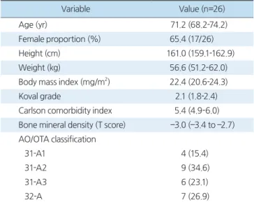
CI, 6.1-6.9). The patients’ demographic factors are present- ed in Table 1. BMD was performed in patients ≥50 years old. The mean T-score for patients who underwent the BMD test was –3.0, with 73.9% (17/23) of patients having
Table 1. Patients’ Demographic Factors
Variable Value (n=26)
Age (yr) 71.2 (68.2-74.2)
Female proportion (%) 65.4 (17/26)
Height (cm) 161.0 (159.1-162.9)
Weight (kg) 56.6 (51.2-62.0)
Body mass index (mg/m2) 22.4 (20.6-24.3)
Koval grade 2.1 (1.8-2.4)
Carlson comorbidity index 5.4 (4.9-6.0) Bone mineral density (T score) ––3.0 (––3.4 to ––2.7) AO/OTA classification
31-A1 4 (15.4)
31-A2 9 (34.6)
31-A3 6 (23.1)
32-A 7 (26.9)
Values are presented as mean (95% confidence interval), mean (num- ber), or number (%).
a BMD score of less than –2.5. There were 19 intertro- chanteric fractures (AO/OTA A31) and 7 subtrochanteric fractures (AO/OTA A32). Six out of 7 subtrochanteric fracture patients were diagnosed with an atypical femur
fracture.
The mean time to start using a wheelchair and ambula- tion was 2.2 days and 2.7 days, respectively, with a mean hospital stay of 8.9 days. There were no early postoperative complications such as infection, deep vein thrombosis, pul- monary embolism, or in-hospital death. There was 1 case of pneumonia that improved within two weeks after sur- gery. Fourteen patients were discharged home, and the other 12 patients were transferred to another hospital for further rehabilitation and recovery. Perioperative patient variables are presented in Table 2.
The radiographically measured mean tip-apex distance (TAD) was 19.4 mm (95% CI, 18.4-20.4). Most cases, except for two cases of subtrochanteric atypical femoral fracture, satisfied the mean TAD of less than 25 mm. Bone union was observed in all cases, and the mean union time was 12.9 weeks (95% CI, 11.6-14.2) (Fig. 2, 3).
Table 2. Perioperative Patient Variables
Variable Value (n=26)
Operation time (min) 53.3 (43.6-63.1)
TFNA nail length
Short nail (170, 200, 235 mm) 19 (73.1) Long nail (320, 340, 360, 380 mm) 7 (26.9) Type of cephalomedullary fixation
Lag screw 9 (34.6)
Helical blade 17 (65.4)
Time to start wheelchair (d) 2.2 (1.3-3.0) Time to start ambulation (d) 2.7 (2.1-3.2) Total hospital stays (d) 8.9 (7.0-10.8) Values are presented as mean (95% confidence interval) or number (%). TFNA: Trochanteric Fixation Nail-Advanced.
A B C D
R R
Fig. 2. (A) Total hip anteroposterior (AP) radiograph of 71-year-old female diagnosed with an intertrochanteric fracture of the right hip (AO/OTA 31A1.3). (B) Immediate postoperative total hip AP and cross-table lateral x-ray. The fractured segment was reduced in a slight valgus position (ex- tramedullary). (C) Postoperative three months x-ray shows callus formation and bone bridging at the medial fracture gap. (D) Postoperative six months x-ray shows a complete union of the fracture.
R R R R
A B C D
Fig. 3. (A) Total hip anteroposterior (AP) radiograph of 90-year-old female diagnosed with an intertrochanteric (reverse obliquity) fracture of the right hip (AO/OTA 31A3.1). (B) Immediate postoperative total hip AP and cross-table lateral x-ray. Cerclage wire before nailing was used to reduce the fragment in an anatomical position. (C) Fracture remained stable, and callus formation was visible around the medial calcar area in the postop- erative three-month x-ray. (D) Complete bone union was achieved at the six-month follow-up.
The mean Koval score at the postoperative six months follow-up was 2.8 (95% CI, 2.2-3.3). The mean HHS score improved from 64.9 (95% CI, 56.4-67.4) at discharge to 76.3 (95% CI, 70.2-82.4) at the six-month follow-up, and the improvement was statistically significant (p<0.001) (Fig. 4). The mean EQ-5D score increased from 0.25 (95%
CI, 0.18-0.31) at discharge to 0.72 (95% CI, 0.63-0.83) at the six-month follow-up, and the increase in score was statistically significant as well (p<0.001) (Fig. 5).
Discussion
TFNA for proximal femoral fractures showed a suc- cessful union rate (100%) with a mean union time of 12.9 weeks. Considering that all 6 atypical subtrochanteric frac- tures achieved bone union within six months, the outcome is satisfactory and is in line with the clinical results of previ- ous studies using other implants.8-13) Both patient functional outcome measures, HHS and EQ-5D score, also showed statistically significant improvement on serial follow-up (p<0.001) and had similar results compared to other previ- ous studies using different implant systems in hip fracture patients.13,14)
In the current study, there were no implant failures or implant fractures. Meanwhile, Lambers et al.7) reported on 16 cases (13 patients) of implant fractures that occurred between 2016 and 2018. Most of the implant fracture pa-
tients in their study had reverse oblique type fractures cor- responding to AO/OTA classification 31A3 (75%), and the implant fracture occurred at the proximal aperture in almost all patients (94%). Either delayed union (69%) or nonunion (31%) were confirmed in all cases, and implant breakage was thought to be attributable to the new design of TFNA, which has a LATERAL RELIEF CUTTM and BUMP CUTTM design around the proximal aperture (Fig. 1).
Both the nail width (17 mm in TFN vs 15.6 mm in TFNA) and wall thickness in the proximal area are reduced in the newly designed nail compared to its predecessor. Design characteristics of the implant, as well as abnormal stress concentration in specific fracture types, may have affected the vulnerability of the nail. Although the percentage of AO/OTA 31A3 (reverse oblique type) patients in our study (23.1%) was lower than that in the study by Lamber et al., all six cases showed successful bone union during follow- up, and no implant fracture occurred during or after the six-month radiological assessment.7)
In seven cases of long nail use, there was no iatrogenic fracture or perforation of the distal anterior cortex at the distal femur, which results from a mismatch between femur and nail. The ROC of the newly designed TFNA is 1.0 m, and the curvature is increased compared to the conventional TFN, which has a ROC with a radius of 1.5 m.4,5) Asian patients usually have more femoral bowing than do Western patients.15,16) In these patients, the distal tip of long straight
Fig. 4. Change in the mean Harris hip score (HHS) following hip sur- gery. A serial survey of the patients was conducted at admission (before discharge) and in the out-patient clinic. The mean HHS score showed gradual improvement, and the increase was statistically significant (p<0.001). Postop.: postoperative.
Fig. 5. Change in the Euro-Qol-5 Dimension (EQ-5D) score following hip surgery. A serial survey of the patients was conducted at admis- sion (before discharge) and in the out-patient clinic. The EQ-5D score showed improvement, and the increase was statistically significant (p<0.001). Postop.: postoperative.
nails can collide with the anterior cortex of the distal femur during implant insertion and cause implant breakthrough or iatrogenic fracture. For this reason, the new design of the TFNA can be a good alternative for patients with greater femoral bowing. By selecting an implant with the proper design, we were able to prevent implant-femur mismatch and the occurrence of iatrogenic fractures.
The free selectability of the cephalomedullary fixation system between a conventional screw and a helical blade is another advantage of this instrument. Although there is controversy in the clinical aspects, the helical blade has ad- vantages in gaining firm bone compaction and additional bone purchase for fracture site compression in osteoporotic patients, preventing implant cut-out or -through.17,18) The mean BMD of our enrolled patients was –3.0, showing a severe degree of osteoporosis in the majority of the patients.
We, therefore, used the blade type in two-thirds of the pa- tients (17 cases, 65.4%), and no cut-out or -through was observed in any patient.
There are several limitations to this study. First, this is a retrospective cohort study, and the level of evidence is relatively low. To compensate, we only included patients who were surgically treated by a single surgeon at a single institution. We also encouraged all patients to visit our out- patient clinic postoperatively to minimize loss to follow-up.
Second, the number of enrolled patients is relatively small, which makes it difficult to derive meaningful conclusions.
The purpose of our study was to briefly report on clinical outcomes and personal experiences of using TFNA in our patient group for the first time. We expect to carry out a prospective case-control study with more enrolled patients soon. Finally, the clinical observation period may be too short to evaluate the safety of the newly developed implant.
In the data of Lamber et al.,7) however, the mean time to implant failure was 5.0±2.2 months, and our follow-up period is not shorter than that of the other study.
Conclusion
Our clinical outcome demonstrated that a new nail, TFNA, was safe and produced satisfactory outcomes.
TFNA may be a useful option for fixation of simple in- tertrochanteric or subtrochanteric fractures with increased femoral bowing. However, we advise clinicians to be aware of the issues related to implant failure and pay careful atten- tion to long-term outcomes and safety.
요 약
목적:
근위 대퇴골 골절 환자에서 사용한 TFNA 금속정의중ㆍ단기 임상 결과 및 안정성을 알아보고자 한다.
대상 및 방법:
본 연구는 후향적 코호트 연구로, 2018년 12월 부터 2019년 3월까지 TFNA를 이용해 근위 대퇴골 골절 수 술을 받은 환자들의 기본 정보를 조사하였으며, 기능적으로 는 수술 전후 6개월간 HHS 및 EQ-5D 점수의 변화를 분석 하였다. 그 외 방사선학적 골유합의 진행, 기구 파손 발생 여 부 등을 확인하였다.결과:
해당 기간 중 총 26명의 환자가 포함되었으며, 1예의 폐 렴 환자를 제외하고 술 후 합병증은 없었다. 내고정물 파쇄 역시 확인되지 않았다. HHS는 64.9점에서 76.3점으로 호전 되었으며, EQ-5D 역시 0.25점에서 0.72점으로 호전되었다.모든 예에서 골유합을 확인했으며, 골절의 평균 유합 시간은 12.9주였다.
결론:
최소 6개월간의 추시 결과 TFNA는 근위 대퇴골 골절환자의 수술적 치료에 있어 효과적이면서도 안전한 기구임을 확인할 수 있었다.
색인 단어:
고관절 골절, 대퇴골 골절, 골절 고정술, 치료 결과, 삽입 파손ORCID
윤재연, https://orcid.org/0000-0003-4449-7314 김지완, https://orcid.org/0000-0002-3524-8706
References
1. Lenich A, Fierlbeck J, Al-Munajjed A, et al: First clinical and biomechanical results of the Trochanteric Fixation Nail (TFN).
Technol Health Care, 14: 403-409, 2006.
2. Williamson S, Landeiro F, McConnell T, et al: Costs of fragility hip fractures globally: a systematic review and meta-regression analysis. Osteoporos Int, 28: 2791-2800, 2017.
3. Bellabarba C, Herscovici D Jr, Ricci WM: Percutaneous treat-
ment of peritrochanteric fractures using the Gamma nail. Clin Orthop Relat Res, (375): 30-42, 2000.
4. DePuy Synthes: TFN-Advanced Proximal Femoral Nailing Sys- tem: surgical technique [Internet]. West Chester (PA), Monument (CO), Depuy Synthes: 2017 [cited 2020 Apr 1]. Available from:
http://synthes.vo.llnwd.net/o16/LLNWMB8/US%20Mobile/
Synthes%20North%20America/Product%20Support%20Ma- terials/Brochures/3936_DSUSTRM06140109-7_TFNA_Core_
rev1.pdf.
5. DePuy Synthes: TFN-Advanced Proximal Femoral Nailing System: value analysis brief [Internet]. Oberdorf, DePuy Synthes:
2016 [cited 2020 Apr 1]. Available from: http://synthes.vo.llnwd.
net/o16/LLNWMB8/INT%20Mobile/Synthes%20Interna- tional/Product%20Support%20Material/legacy_Synthes_PDF/
DSEM-TRM-0515-0375-1_LR.pdf.
6. DePuy Synthes: Titanium Trochanteric Fixation Nail System:
surgical technique [Internet]. Oberdorf, DePuy Synthes: 2016 [cited 2020 Apr 1]. Available from: http://synthes.vo.llnwd.net/
o16/LLNWMB8/INT%20Mobile/Synthes%20International/
Product%20Support%20Material/legacy_Synthes_PDF/DSEM- TRM-0714-0116-3_LR.pdf.
7. Lambers A, Rieger B, Kop A, D’Alessandro P, Yates P: Implant fracture analysis of the TFNA proximal femoral nail. J Bone Joint Surg Am, 101: 804-811, 2019.
8. Pu JS, Liu L, Wang GL, Fang Y, Yang TF: Results of the proxi- mal femoral nail anti-rotation (PFNA) in elderly Chinese pa- tients. Int Orthop, 33: 1441-1444, 2009.
9. Halder SC: The Gamma nail for peritrochanteric fractures. J Bone Joint Surg Br, 74: 340-344, 1992.
10. Barquet A, Mayora G, Fregeiro J, López L, Rienzi D, Fran- cescoli L: The treatment of subtrochanteric nonunions with the
long gamma nail: twenty-six patients with a minimum 2-year follow-up. J Orthop Trauma, 18: 346-353, 2004.
11. Yeh WL, Su CY, Chang CW, et al: Surgical outcome of atypical subtrochanteric and femoral fracture related to bisphosphonates use in osteoporotic patients with or without teriparatide treat- ment. BMC Musculoskelet Disord, 18: 527, 2017.
12. Lee KJ, Yoo JJ, Oh KJ, et al: Surgical outcome of intramedul- lary nailing in patients with complete atypical femoral fracture: a multicenter retrospective study. Injury, 48: 941-945, 2017.
13. Roh YH, Rho J, Nam KW: Treatment of the proximal femoral fracture using the new design cephalomedullary nail: prospective outcomes study. J Korean Fract Soc, 32: 35-42, 2019.
14. Vaquero J, Munoz J, Prat S, et al: Proximal Femoral Nail Anti- rotation versus Gamma3 nail for intramedullary nailing of un- stable trochanteric fractures. A randomised comparative study.
Injury, 43 Suppl 2: S47-S54, 2012.
15. Abdelaal AH, Yamamoto N, Hayashi K, et al: Radiological as- sessment of the femoral bowing in Japanese population. SICOT J, 2: 2, 2016.
16. Egol KA, Chang EY, Cvitkovic J, Kummer FJ, Koval KJ: Mis- match of current intramedullary nails with the anterior bow of the femur. J Orthop Trauma, 18: 410-415, 2004.
17. Goffin JM, Pankaj P, Simpson AH, Seil R, Gerich TG: Does bone compaction around the helical blade of a proximal femo- ral nail anti-rotation (PFNA) decrease the risk of cut-out?: a subject-specific computational study. Bone Joint Res, 2: 79-83, 2013.
18. Chapman T, Zmistowski B, Krieg J, Stake S, Jones CM, Levicoff E: Helical blade versus screw fixation in the treatment of hip fractures with cephalomedullary devices: incidence of failure and atypical “medial cutout”. J Orthop Trauma, 32: 397-402, 2018.