PGHN
Original Article
Clinical Features and Extraintestinal Manifestations of Crohn Disease in Children
Young Ah Lee, Peter Chun*, Eun Ha Hwang*, Sang Wook Mun*, Yeoun Joo Lee*, and Jae Hong Park*
Good Gang-An Hospital, *Department of Pediatrics, Pusan National University School of Medicine, Busan, Korea
Purpose: The aim of this study was to investigate the clinical features and extraintestinal manifestations (EIMs) of Crohn disease (CD) in Korean pediatric patients.
Methods: The medical records of 73 children diagnosed with CD were retrospectively reviewed. Data regarding base- line demographic and clinical characteristics, including CD phenotype at diagnosis based on the Montreal classi- fication, and clinical features and course of EIMs were investigated.
Results: Fifty-two (71.2%) of the patients were males. The mean age of the patients was 12.5 years. The mean fol- low-up period was 3.4 years. The disease location was ileal in 3 (4.1%) of the patients, colonic in 13 (17.8%), ileoco- lonic in 56 (76.7%). The clinical behavior was inflammatory in 62 (84.9%) of the patients, stricturing in 8 (11.0%), and penetrating in 3 (4.1%). Perianal abscesses or fistulas were found in 37 (50.7%) of the patients. EIMs observed during the study period were anal skin tag in 25 patients (34.2%), hypertransaminasemia in 20 (27.4%), peripheral arthritis in 2 (2.7%), erythema nodosum in 2 (2.7%), vulvitis in 1 (1.4%), uveitis in 1 (1.4%), and pulmonary throm- boembolism in 1 (1.4%).
Conclusion: Perianal diseases and manifestations were present in more than half of Korean pediatric CD patients at diagnosis. Inspection of the anus should be mandatory in Korean children with suspicious CD, as perianal fistulas, abscesses, and anal skin tags may be the first clue to the diagnosis of CD.
Key Words: Crohn disease, Extraintestinal, Children
Received:August 28, 2016, Revised:October 24, 2016, Accepted:November 12, 2016
Corresponding author: Jae Hong Park, Department of Pediatrics, Pusan National University Yangsan Hospital, 20 Geumo-ro, Mulgeum-eup, Yangsan 50612, Korea. Tel: +82-55-360-2180, Fax: +82-55-360-2181, E-mail: [email protected]
Copyright ⓒ 2016 by The Korean Society of Pediatric Gastroenterology, Hepatology and Nutrition
This is an openaccess article distributed under the terms of the Creative Commons Attribution NonCommercial License (http://creativecommons.org/licenses/by-nc/4.0/) which permits unrestricted noncommercial use, distribution, and reproduction in any medium, provided the original work is properly cited.
INTRODUCTION
The incidence of inflammatory bowel disease (IBD) in pediatric and adolescent patients has increased. It is estimated that 15% to 25% of patients experience the onset of symptoms before 20 years of
age [1]. In particular, reports have shown an increas- ing incidence of pediatric Crohn disease (CD) in the last decades [2,3]. The epidemiologic and clinical characteristics of CD seem to differ among ethnic groups, but data on the clinical course of pediatric CD are scarce.
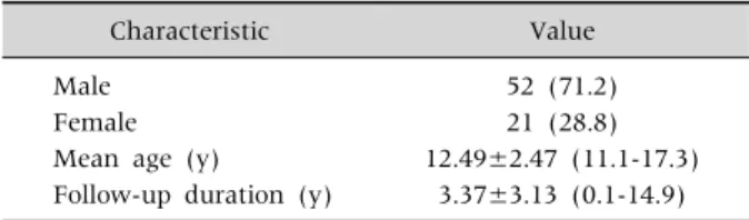
Table 1. Baseline Characteristics of Patients with Crohn Disease (n=73)
Characteristic Value
Male 52 (71.2)
Female 21 (28.8)
Mean age (y) 12.49±2.47 (11.1-17.3) Follow-up duration (y) 3.37±3.13 (0.1-14.9) Values are presented as number (%) or mean±standard deviation (range).
Patients with CD frequently manifest a spectrum of clinical symptoms and signs due to a combination of intestinal and extraintestinal lesions. The patho- genesis of extraintestinal manifestations (EIMs) with CD is poorly understood. An immune mecha- nism has been suspected because of some of the le- sions that accompany CD, such as those involving the skin, joints, eye, mouth, and hepatobiliary tract [4]. The EIMs of CD have been reported to involve al- most every organ system. Sometimes it can be diffi- cult to differentiate true EIMs from secondary extra- intestinal complications. Some EIMs may not corre- late with disease activity but in general EIMs tend to follow the clinical course of IBD [5]. The incidence of the EIMs of IBD in previous studies has ranged from 21-41%, depending on the patient population and criteria used [6]. EIMs were more common in CD than ulcerative colitis (UC) and the clinical spectrum of EIMs varied from mild transitory to very severe le- sions [6].
Although about 25% of Crohn patients are diag- nosed as children and adolescents, and their clinical features differ from those of adults [7], only a few Korean studies involving a small number of patients have been published to date [8-10]. Data on the dif- ferences between the EIMs of IBD in children and adults are also lacking. Understanding the preva- lence and clinical course of the EIMs in children with CD is necessary to provide guidelines for the appro- priate management of CD.
Therefore we aimed to investigate the clinical fea- tures and EIMs of CD in Korean pediatric patients.
MATERIALS AND METHODS
The medical records of 73 children diagnosed with CD between July 1995 and June 2011 at Pusan National University Hospital and Pusan National University Children’s Hospital were reviewed retros- pectively. We evaluated the baseline demographic and clinical characteristics including sex, age, fol- low-up duration, CD phenotype at diagnosis, clinical features and course of EIMs, and the drugs used for treatment.
The diagnosis of CD was based on the clinical fea- tures in combination with endoscopic and radiologic findings, and in most cases it was supplemented by results from histologic examinations. CD phenotype at diagnosis was assessed using the Montreal Classification [11]. The CD location and the disease behavior were determined at the time of diagnosis.
The EIMs were determined at diagnosis and during follow-up.
Arthritis was defined as pain, swelling, redness, and warmth in one or more joints. Erythema nodo- sum was diagnosed when tender, red nodules, main- ly on the extensor surface of the legs, were observed.
The skin lesions were diagnosed by dermatologists.
Patients with suspicious iritis or painful uveitis un- derwent ocular examinations by ophthalmologists.
Hepatobiliary involvement was suspected when ab- normal liver biochemistry results had been detected.
For the diagnosis of pulmonary thromboembolism (TE) in one patient, a ventilation-perfusion scan and angiography were performed.
RESULTS
Characteristics of patients with CD
Among the 73 study patients, 52 (71.2%) of the pa- tients were male and the male/female ratio was 2.5:1. The mean age at diagnosis was 12.49±2.47 (range, 11.1-17.3) years. The duration of follow-up after diagnosis was 3.37±3.13 (range, 0.1-14.9) years (Table 1).
At diagnosis, 56 patients (76.7%) had ileocolonic disease, 13 (17.8%) had isolated colonic disease, and 3 (4.1%) had ileal disease. There was one case of iso-
Table 3.Extraintestinal Manifestations of Patients with Crohn Disease (n=73)
Manifestation n (%)
Perianal skin tag 25 (34.2)
Hypertransaminasemia 20 (27.4)
Peripheral arthritis 2 (2.7)
Erythema nodosum 2 (2.7)
Granulomatous vulvitis 1 (1.4)
Pulmonary thromboembolism 1 (1.4)
Table 2. Montreal Classification of Patients with Crohn Disease (n=73)
Classification n (%)
Location
L1 (ileal) 3 (4.1)
L2 (colonic) 13 (17.8)
L3 (ileocolonic) 56 (76.7)
L4 only (isolated small bowel disease)* 1 (1.4) Behavior
B1 (non-stricturing, non-penetrating) 62 (84.9)
B2 (structuring) 8 (11.0)
B3 (penetrating) 3 (4.1)
P (perianal disease modifier) 37 (50.7)
*Crohn disease was diagnosed with wireless capsule endoscopy.
lated upper disease confirmed by capsule endoscopy (Table 2). With respect to the behavior of the disease, non-stricturing, non-penetrating type was present in 62 patients (84.9%), stricturing in 8 (11.0%), and penetrating in 3 (4.1%). Perianal abscesses or fistulas were found in 37 patients (50.7%) (Table 2).
Treatment of patients with CD
Systemic corticosteroids were administered to all patients (100.0%) for remission induction. Oral 5-aminosalicylic acids (5-ASA) only or 5-ASA plus azathioprine was used for maintenance therapy in 43 (58.9%) and 30 (41.1%) patients, respectively. A to- tal of 17 patients (23.3%) failed remission induction or maintenance and were successfully treated with infliximab.
EIMs of patients with CD
The EIMs of the patients with CD included anal skin tags in 25 patients (34.2%), hypertransami- nasemia in 20 (27.4%), peripheral arthritis in 2 (2.7%), erythema nodosum in 2 (2.7%), vulvitis in 1 (1.4%), uveitis in 1 (1.4%), and pulmonary TE in 1 (1.4%) (Table 3).
Anal skin tags developed in 25 patients (34.2%) (19 male, 6 female). The location of the disease was the small bowel and colon in 15 (60.0%) patients, isolated colon in 8 (32.0%), and isolated small bowel in 2 patients (8.0%).
Hypertransaminasemia developed in 20 (27.4%) patients. The alanine aminotransferase (ALT) level was 116.4±86.1 IU/L (range, 40-346 IU/L). The dura- tion of ALT elevation was 2.4±1.7 months (range, 10 days-6 months). Hepatitis developed at the time CD was diagnosed in one patient (5.0%), during the in- duction period in 11 patients (55.0%), and during the maintenance period in 8 (40.0%) patients. The hep- atitis resolved in all of the patients without specific treatment.
One of the two patients with peripheral arthritis was a 14-year-old boy who had pain in both knees.
The joint symptom developed during the main- tenance therapy, and the patient developed hepatitis at the same time. The other patient was a 14-year-old girl who had right knee pain. Arthritis developed when she elected to stop medical treatment. The dis- ease was located in the ileum and colon in both patients. The symptoms disappeared several months after they resumed medical treatment.
Erythema nodosum developed in two patients, an 11-year-old and 14-year-old boy, whose lesion sites were the colon only and the ileum and colon, respectively. The skin lesions developed before they were diagnosed with gastrointestinal CD and dis- appeared after they underwent treatment with corticosteroids.
Vulvitis developed in a 10-year-old girl and was her presenting symptom of CD. Crohn vulvitis was diagnosed by skin biopsy of the lesion. The location of the disease in this patient was the ileum. The skin lesion did not resolve during treatment of the disease.
Uveitis developed in a 14-year-old boy whose le-
sion site was the colon only, and the condition re- solved 1 month after treatment.
A pulmonary TE developed in an 11-year-old boy whose presenting symptom was severe hema- tochezia probably due to the TE. The initial abdomi- nal computed tomography (CT) revealed enlarge- ment of the right ileocolic branch. Despite proper medical treatment, intermittent severe hema- tochezia continued. The patient eventually received right hemicolectomy and segmental resection of the ileum to control the bleeding. The patient developed repetitive hemoptysis 2 years later. The follow-up CT scan demonstrated dilatation of the main pulmo- nary artery and the right pulmonary artery, and ob- literation of the pulmonary vein. Pulmonary hyper- tension was detected by cardiac angiography. The in- itial location of this patient was the ileum.
Most of the EIMs developed prior to the diagnosis of CD or during relapse of the intestinal lesions in our study. All of the EIMs, except the perianal skin tags, vulvitis and pulmonary TE, resolved during treat- ment of the intestinal disease of CD.
DISCUSSION
This is the first study in Korean pediatric CD pa- tients to investigate the EIMs associated with CD.
Despite the limitations related with the small num- ber of patients included in this study, we were capa- ble of finding that diseases or manifestations involv- ing the anus or the perianal region occurred in more than half of Korean children with Crohn’s disease at diagnosis.
In our study, the incidence of CD showed a male predominance (2.5:1), as was also reported in Korean pediatric (2.2:1)[10] and adult (2.2:1) [12]
studies. Male predominance was also reported in Western pediatric studies [13-15]. However, recent large-scale studies in Western pediatric CD patients have shown a lower male predominance of a male-to-female sex ratio of 1.43-1.45:1 [3,16] com- pared to our study, suggesting a gender difference between Caucasian and Korean ethnicity in the pe- diatric population of CD. Further large-scale pro-
spective studies are required in order to clarify this difference, considering the small-scale and retro- spective design of our study. Meanwhile, the mean diagnosis age of patients in our study was similar to the results of other Korean and Western pediatric studies which ranged from 11.4 to 13.2 years [3,10,16].
Most of the patients with CD in our study had dis- ease in both the small bowel and colon (76.7%). and non-stricturing and non-penetrating behavior (84.9%), results similar to other reports on Korean children [10] and adults [12]. Kim et al. [10] re- ported that 75% of children with CD presented with both small bowel and colonic involvement, and 85%
of patients had inflammatory behavior. Ye et al. [12]
reported that 67.3% of the adults with CD in their sample had disease in both the small bowel and colon. However, the number of patients with stric- turing and penetrating behavior increased from 31.3% at the time of diagnosis to 50.7% at the final evaluation. This discrepancy between our study’s and Ye et al.’s findings [12] may have resulted from the differences in the follow-up periods and the con- traction of disease. The Western study of Müller et al.
[3] reported that 58.7% of children with CD had ileo- colonic disease and 84.4% had inflammatory behavior. Another recent European study reported that ileocolonic disease was seen at presentation in 53% of patients, and nonstricturing and non- penetrating disease in 82% [16].
The rate of perianal abscesses or fistulas in our study was high (50.7%), and these findings were similar to those reported in other Korean pediatric (63.0%) [10] and adult (46.8%) [12] studies.
However, Western studies by Müller et al. [3] and Palder et al. [17] reported perianal disease rates of 14.5% and 28.0% in children with CD, respectively.
Another European study reported that perianal dis- ease (presence of fistula and/or abscess) at diagnosis was seen in 9% of pediatric CD patients [16].
The proportion of patients with anal skin tags in our study was relatively higher compared to previous studies which ranged from 21% to 29% in CD pa- tients [10,18]. Anal skin tags are one of the anorectal
malformations of CD; they are flesh colored and waxy in appearance, painless, can be raised and firm, narrow or broad, and patients are more likely to de- velop multiple tags than isolated tags [18]. Taylor et al. [19] reported that granulomas were more fre- quently found in anal skin tags than in rectal biop- sies (34.6% vs. 30.8%) of patients with CD. Studies on anal skin tags as a potential predictor of CD may be an interesting topic for investigation in the future.
Although there is limited data regarding the oc- currence of EIM in pediatric IBD patients, it seems that one quarter to almost half of them experience at least 1 EIM at the time of diagnosis [20]. Veloso [6]
reported that the inflammatory EIMs (peripheral ar- thritis, erythema nodosum, pyoderma gangrenosum, iritis/uveitis, and aphthous stomatitis) occurred at least once in 25.8% of pediatric and adult patients with IBD, and these manifestations were more com- mon in CD than in UC. They were also more common in patients with colonic involvement than in patients with ileal disease. The cumulative probability of a second manifestation was about 70% after 10 years of follow-up [21].
The proportion of EIMs observed in our study was similar to this study in which a single EIM occurred in 60.5% of patients, whereas multiple manifes- tations occurred in 39.5%. Müller et al. [3], however, reported that 12.5% of children with CD had EIMs.
This big discrepancy regarding the incidence of EIMs between these reports may have resulted from dif- ferent inclusion criteria. The overall rate of EIM will vary based on which conditions are included as an EIM, as well as the period of follow-up. When EIMs are more broadly defined to include other systemic effects such as growth delay, nutritional deficiency, anemia, decreased bone mineral density, and fa- tigue, the rate of EIMs could approach 100% [20].
Elevations in transaminases were reported in up to 17% of the patients with IBD [22]. Primary scleros- ing cholangitis and autoimmune hepatitis are im- portant causes of elevated liver enzymes in these patients. Medications used to treat IBD such as sul- fasalazine, thiopurine, and methotrexate can also have adverse effects on the liver [23]. There are con-
flicting opinions about etiology of transient hyper- transaminasemia in pediatric CD patients under- going their initial treatment with enteral nutrition [24,25]. In our study, modest and transient hyper- transaminasemia developed in 27.4% of the patients.
Several studies have suggested that there is an in- creased risk of venous and arterial TE in adults with IBD. Sonoda et al. [26] reported that a venous TE was detected in 8 of 47 consecutive Japanese patients with active IBD (17.0%). Guerra Montero et al. [27]
reported that 15 of 515 IBD patients (2.9%) had thromboembolic events, and they were more com- mon in patients with extensive disease, colonic in- volvement, and other EIMs. Lazzerini et al. [28] re- ported that TE in children with IBD occurred most often during active disease (82.8%) and more fre- quently in UC. The sites of the TE were the cerebral vessels (54.3%), the vessels in the limbs (26.0%), the abdominal vessels (13.0%), and the vessels in the retina and lungs (6.7%). Nylund et al. [29] reported that hospitalized children and adolescents with IBD are at increased risk for TE. Conservative methods such as hydration, mobilization, and pneumatic de- vices should be considered to prevent TE in hospi- talized children with IBD.
There are some limitations in this study. Since this is a single-center study, common EIMs of CD such as pyoderma gangrenosum, primary sclerosing chol- angitis and psoriasis were not observed. The small number of patients and the short follow-up period may have resulted in observational bias. A further prospective population-based study in pediatric CD is needed to accurately estimate the incidence of EIMs and their natural course.
In conclusion, perianal diseases and manifes- tations occur in more than half of Korean pediatric CD patients, which may be the first clue in diagnos- ing CD. Therefore, inspection of the anus should be mandatory in Korean children with suspicious CD.
ACKNOWLEDGEMENTS
This work was supported by a 2-year research grant of Pusan National University.
REFERENCES
1. Goodhand J, Hedin CR, Croft NM, Lindsay JO.
Adolescents with IBD: the importance of structured transition care. J Crohns Colitis 2011;5:509-19.
2. Benchimol EI, Fortinsky KJ, Gozdyra P, Van den Heuvel M, Van Limbergen J, Griffiths AM. Epidemiolo- gy of pediatric inflammatory bowel disease: a system- atic review of international trends. Inflamm Bowel Dis 2011;17:423-39.
3. Müller KE, Lakatos PL, Arató A, Kovács JB, Várkonyi Á, Szűcs D, et al. Incidence, Paris classification, and fol- low-up in a nationwide incident cohort of pediatric pa- tients with inflammatory bowel disease. J Pediatr Gastroenterol Nutr 2013;57:576-82.
4. Veloso FT, Carvalho J, Magro F. Immune-related sys- temic manifestations of inflammatory bowel disease. A prospective study of 792 patients. J Clin Gastroenterol 1996;23:29-34.
5. Rothfuss KS, Stange EF, Herrlinger KR. Extraintestinal manifestations and complications in inflammatory bowel diseases. World J Gastroenterol 2006;12:4819- 31.
6. Veloso FT. Extraintestinal manifestations of in- flammatory bowel disease: do they influence treatment and outcome? World J Gastroenterol 2011;17:2702-7.
7. Mamula P, Markowitz JE, Baldassano RN. Inflamma- tory bowel disease in early childhood and adolescence:
special considerations. Gastroenterol Clin North Am 2003;32:967-95.
8. Seo JK, Yeon KM, Chi JG. Inflammatory bowel disease in children--clinical, endoscopic, radiologic and histo- pathologic investigation. J Korean Med Sci 1992;
7:221-35.
9. Lee NY, Park JH. Clinical features and course of Crohn disease in children. Korean J Gastrointest Endosc 2007;34:193-9.
10. Kim BJ, Song SM, Kim KM, Lee YJ, Rhee KW, Jang JY, et al. Characteristics and trends in the incidence of in- flammatory bowel disease in Korean children: a sin- gle-center experience. Dig Dis Sci 2010;55:1989-95.
11. Silverberg MS, Satsangi J, Ahmad T, Arnott ID, Bernstein CN, Brant SR, et al. Toward an integrated clinical, molecular and serological classification of in- flammatory bowel disease: report of a working party of the 2005 montreal world congress of gastroenterology.
Can J Gastroenterol 2005;19 Suppl A:5A-36A.
12. Ye BD, Yang SK, Cho YK, Park SH, Yang DH, Yoon SM, et al. Clinical features and long-term prognosis of Crohn's disease in Korea. Scand J Gastroenterol 2010;
45:1178-85.
13. Spray C, Debelle GD, Murphy MS. Current diagnosis, management and morbidity in paediatric inflam- matory bowel disease. Acta Paediatr 2001;90:400-5.
14. Sawczenko A, Sandhu BK. Presenting features of in- flammatory bowel disease in Great Britain and Ireland.
Arch Dis Child 2003;88:995-1000.
15. Kugathasan S, Judd RH, Hoffmann RG, Heikenen J, Telega G, Khan F, et al. Epidemiologic and clinical char- acteristics of children with newly diagnosed in- flammatory bowel disease in Wisconsin: a statewide population-based study. J Pediatr 2003;143:525-31.
16. de Bie CI, Paerregaard A, Kolacek S, Ruemmele FM, Koletzko S, Fell JM, et al. Disease phenotype at diag- nosis in pediatric Crohn's disease: 5-year analyses of the EUROKIDS registry. Inflamm Bowel Dis 2013;
19:378-85.
17. Palder SB, Shandling B, Bilik R, Griffiths AM, Sherman P. Perianal complications of pediatric Crohn's disease. J Pediatr Surg 1991;26:513-5.
18. Korelitz BI. Anal skin tags: an overlooked indicator of Crohn's disease. J Clin Gastroenterol 2010;44:151-2.
19. Taylor BA, Williams GT, Hughes LE, Rhodes J. The his- tology of anal skin tags in Crohn's disease: an aid to con- firmation of the diagnosis. Int J Colorectal Dis 1989;
4:197-9.
20. Dotson JL, Hyams JS, Markowitz J, LeLeiko NS, Mack DR, Evans JS, et al. Extraintestinal manifestations of pediatric inflammatory bowel disease and their rela- tion to disease type and severity. J Pediatr Gastroenterol Nutr 2010;51:140-5.
21. Veloso FT, Ferreira JT, Barros L, Almeida S. Clinical outcome of Crohn's disease: analysis according to the vienna classification and clinical activity. Inflamm Bowel Dis 2001;7:306-13.
22. Seibold F, Weber P, Jenss H, Scheurlen M. Autoimmune hepatitis in inflammatory bowel disease: report of two unusual cases. Z Gastroenterol 1997;35:29-32.
23. Uko V, Thangada S, Radhakrishnan K. Liver disorders in inflammatory bowel disease. Gastroenterol Res Pract 2012;2012:642923.
24. Schatorjé E, Hoekstra H. Transient hypertransa- minasemia in paediatric patients with Crohn disease undergoing initial treatment with enteral nutrition. J Pediatr Gastroenterol Nutr 2010;51:336-40.
25. Lemberg DA, Leach ST, Day AS. Transient hyper- transaminasemia in pediatric patients with Crohn disease. J Pediatr Gastroenterol Nutr 2011;53:229.
26. Sonoda K, Ikeda S, Mizuta Y, Miyahara Y, Kohno S.
Evaluation of venous thromboembolism and coagu- lation-fibrinolysis markers in Japanese patients with inflammatory bowel disease. J Gastroenterol 2004;39:
948-54.
27. Guerra Montero LJ, Ingver A, Casañas A, Sosa C, Iade B. Clinical characteristics of patients with inflam- matory bowel disease and thromboembolic events. Acta Gastroenterol Latinoam 2010;40:134-41.
28. Lazzerini M, Bramuzzo M, Maschio M, Martelossi S, Ventura A. Thromboembolism in pediatric inflam-
matory bowel disease: systematic review. Inflamm Bowel Dis 2011;17:2174-83.
29. Nylund CM, Goudie A, Garza JM, Crouch G, Denson LA. Venous thrombotic events in hospitalized children and adolescents with inflammatory bowel disease. J Pediatr Gastroenterol Nutr 2013;56:485-91.