PGHN
Original Article
Effect of the Baseline Vitamin D Level on Growth Outcome in Pediatric Crohn Disease
Eun Joo Lee, Jin Soo Moon, Jae Sung Ko, Hye Ran Yang, Ju Young Jang, Ju Whi Kim, and Kyung Jae Lee
Department of Pediatrics, Seoul National University College of Medicine, Seoul, Korea
Purpose: Vitamin D deficiency is common in Crohn disease (CD). The aim of the study was to examine the prevalence of vitamin D deficiency and evaluate the association between vitamin D status and growth outcome in Korean pediatric CD patients.
Methods: In this retrospective study, 17 children younger than 18 years old diagnosed with CD were enrolled and their serum 25-hydroxy vitamin D (25[OH]D) was checked between 2011 and 2015. We categorized the patients into two groups, Group 1 and Group 2. Group 1 included patients with serum 25(OH)D levels below 10 ng/mL, and Group 2 was for patients with a 25(OH)D serum levels between 10 ng/mL and 30 ng/mL. The z-scores for height (Htz), weight (Wtz), and body mass index (BMIz) were measured at baseline, 6 months, and 12 months.
Results: The mean serum 25(OH)D levels of the total 65 CD patients and 17 enrolled patients were 15.64±6.9 ng/mL and 13.1±5.1 ng/mL , respectively. There was no correlation at the beginning of the study between vitamin D level and growth parameters (Htz, Wtz, BMIz) or other variables including laboratory data and Pediatric Crohn Disease Activity Index. The Htz, Wtz, and BMIz in Group 1 showed no significant improvement at 6 months and 12 months follow-up. In Group 2, Wtz and BMIz showed significant improvements sustained until 12 months of follow-up. Htz showed no significant improvement at 6 months but there was significant improvement at 12 months.
Conclusion: It seems that baseline vitamin D status affects growth outcome in pediatric CD.
Key Words: Pediatric Crohn's disease, Vitamin D, Growth
Received:August 30, 2016, Revised:October 13, 2016, Accepted:October 21, 2016
Corresponding author: Jin Soo Moon, Division of Pediatric Gastroenterology and Hepatology, Department of Pediatrics, Seoul National University Hospital, 101 Daehak-ro, Jongno-gu, Seoul 03080, Korea. Tel: +82-2-2072-3627, Fax: +82-2-743-3455, E-mail: [email protected] Copyright ⓒ 2017 by The Korean Society of Pediatric Gastroenterology, Hepatology and Nutrition
This is an openaccess article distributed under the terms of the Creative Commons Attribution NonCommercial License (http://creativecommons.org/licenses/by-nc/4.0/) which permits unrestricted noncommercial use, distribution, and reproduction in any medium, provided the original work is properly cited.
INTRODUCTION
Crohn disease (CD) is a chronic inflammatory dis- ease of the gastrointestinal tract. In more recent years though, cases of CD have become more preva- lent beyond Western countries, and are observed in
increasing frequencies in Asian countries such as Korea [1-4].
Significant complications are associated with CD.
Of the children diagnosed with CD, 65% to 85% of them show signs of malnutrition and growth retardation. Furthermore, 11% to 37% of these chil-
dren fail to reach their full potential predicted height in their adulthood [5-8].
Traditionally adequate level of vitamin D has been known for its positive impact on bone metabolism.
More recent studies have expanded beyond this role, and the broader significance of adequate levels of vi- tamin D have come to be associated with low risk for cancers, diabetes, cardiovascular disease, auto- immune diseases, and inflammatory bowel diseases (IBD) such as CD [9].
Vitamin D levels are generally divided into three tiers with serum 25-hydroxyvitamin D (25[OH]D) levels of less than 20 ng/mL and between 21 and 29 ng/mL represent vitamin D deficiency and in- sufficiency, respectively. And, serum 25(OH)D levels between 30 and 100 ng/mL is considered the normal range [10-12].
Korea have shown high rates of vitamin D deficiency. According to the Korea National Health and Nutrition Examination Survey (KNHANES V-1), the median 25(OH)D level of Koreans above 10 years old is no more than 20 ng/dL, 10 units lower than the recommended minimum levels of normal vitamin D requirement (30-100 ng/dL) [13].
There is a lack of conclusive studies whether vita- min D levels have effects on pediatric CD in vitamin D deficient regions. This study aims to evaluate the prevalence of vitamin D deficiency in Korean pedia- tric CD patients. Additionally, keeping in mind that growth retardation is a common complication of pe- diatric CD, this study also examines whether base- line vitamin D levels has a correlation with the chil- dren’s growth outcome.
MATERIALS AND METHODS
This study reviewed data for 65 patients diagnosed with CD (<18 years old) at the Department of Pediatrics in the Seoul National University Children’s Hospital, a tertiary medical center in Seoul, Korea.
The serum 25(OH)D levels of these 65 patients were checked from 2011 to 2015. The diagnosis of CD was made based on conventional radiological, histo- pathological, and endoscopic criteria.
Exclusion criteria are as follows:
(1) Patients who changed the induction treatment method (infliximab, immunomodulatory agent) af- ter checking the 25(OH)D level.
(2) Patients who couldn’t visit the hospital at 6 and 12 months after checking the 25(OH)D level.
(3) Children who have a history of small bowel resection.
Data were retrospectively obtained from the pa- tient’s medical and laboratory records. Patients’
demographic and disease characteristics, such as age, sex, symptoms (abdominal pain, diarrhea, hem- atochezia, weight loss), extraintestinal manifes- tations (fever, joint symptoms, oral ulcers), small bowel involvement, and disease activity were collected.
Disease activity was measured using the Pediatric Crohn Disease Activity Index (PCDAI); the score in- dicates: mild disease, 10 to 27.5; moderate disease, 30 to 37.5; and severe disease, 40 to 100 [14]. Small bowel involvement was investigated by imaging studies, such as small bowel series, magnetic reso- nance enterography, and abdominal computed tomography. Anthropometric data (height, weight, body mass index [BMI]), which were collected at baseline, after 6 months, and after 12 months, were transformed into z-scores for height (Htz), weight (Wtz), and BMI (BMIz) using the 2007 Korean National Growth Chart. Systemic inflammatory markers, including erythrocyte sedimentation rate (ESR)‚ C-reactive protein (CRP), platelet count (PLT), serum albumin (ALB), and hemoglobin (Hb), were collected from patients’ laboratory records.
The serum 25(OH)D levels were recorded to esti- mate vitamin D status. Vitamin D deficiency and in- sufficiency were defined as levels of serum 25(OH)D of <20 ng/mL and 20-30 ng/mL, respectively. In our study, we define severe vitamin D deficiency and moderate vitamin D deficiency as serum 25(OH)D levels of <10 ng/mL and 10-20 ng/mL, respectively.
On the basis of baseline serum 25(OH)D levels, the patients were categorized into Groups 1 and 2. Group 1 included patients with baseline serum 25(OH)D levels of <10 ng/mL, severe vitamin D deficiency.
Group 2 included patients with baseline serum
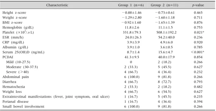
Table 1.Clinical Characteristics between Group 1 and Group 2
Characteristic Group 1 (n=6) Group 2 (n=11) p-value
Height z-score −0.88±1.46 −0.73±0.61 0.465
Weight z-score −1.29±2.80 −1.60±1.18 0.711
BMI z-score −0.92±1.60 −1.65±1.39 0.876
Hemoglobin (g/dL) 11.8±2.6 11.1±1.5 0.753
Platelet (×103/μL) 331.8±79.3 508.1±192.2 0.021*
ESR (mm/h) 24.0±26.3 54.2±40.0 0.236
CRP (mg/dL) 3.9±3.9 4.9±6.0 0.920
Albumin (g/dL) 3.9±1.0 3.6±0.5 0.785
Serum 25(OH)D (ng/mL) 8.7±1.4 15.6±4.7 <0.001*
PCDAI 41.3±9.5 40.0±17.9 0.854
Mild (10-27.5) 0 2 (18.2) 0.266
Moderate (30-37.5) 2 (33.3) 5 (45.5) 0.627
Severe (>40) 4 (66.7) 4 (36.4) 0.232
Abdominal pain 6 (100.0) 9 (81.8) 0.266
Diarrhea 4 (66.7) 8 (72.7) 0.793
Hematochezia 2 (33.3) 2 (18.2) 0.482
Weight loss 4 (66.7) 6 (54.5) 0.627
Extraintestinal manifestations (fever, joint symptom, oral ulcer) 1 (16.7) 5 (45.5) 0.235
Perianal disease 1 (16.7) 4 (36.4) 0.394
Small bowel involvement 6 (100.0) 9 (81.8) 0.266
Values are presented as mean±standard deviation or number (%).
Group 1: 25(OH)D <10 ng/mL, Group 2: 10 ng/mL ≤25(OH)D <30 ng/mL, BMI: body mass index, ESR: erythrocyte sedimentation rate‚ CRP: C-reactive protein, PCDAI: Pediatric Crohn’s Disease Activity Index.
*Statistically significant.
25(OH)D levels of 10-30 ng/mL, moderate vitamin D deficiency/insufficiency [15-17]. Once a patient was diagnosed as vitamin D deficient, they received oral vitamin D supplements.
All data were analyzed using IBM SPSS Statistics software ver. 20.0 (IBM Co., Armonk, NY, USA).
Continuous data of all enrolled patients, such as age and serum 25(OH)D level, are presented as the mean±standard deviation (SD). Categorical data are presented as number (valid percent). Continuous variable in categorized groups, such as anthro- pometric data (Htz, Wtz, BMIz) and laboratory data (Hb, ESR, CRP, and ALB) are expressed as the mean±SD. Changes in anthropometric data (Htz, Wtz, BMIz) for each group were assessed by the Student paired t-test for normally distributed data and one way ANOVA for non-normally distributed data.
Correlations between variables were checked with the Pearson's correlation coefficient. p-values of
<0.05 were considered statistically significant.
This study was approved by the institutional re- view board at Seoul National University Hospital (IRB no. H 1607-078- 776).
RESULTS
A mean serum 25(OH)D level of the 65 CD patients was 15.64±6.9 ng/mL; vitamin D deficiency was found in 81.5% (53/65) and insufficiency in 16.9%
(11/65) of patients. Forty-eight patients were ex- cluded: 34 were not available for follow-up at 6 and 12 months, 13 underwent small bowel resection, and 1 changed induction treatment. Therefore, 17 pa- tients were enrolled in the study. The mean age was 13.7 years (29.4% females). The mean 25(OH)D se- rum level was 13.1±5.1 ng/mL. Vitamin D deficiency and insufficiency were present in 16 patients (94.1%) and 1 patient (5.9%), respectively.
Six patients (35.3%) had severe vitamin D defi-
Table 3.Correlation between Vitamin D Level and Growth Parameter and Other Variables
Vitamin D Hb PLT CRP ESR ALB PCDAI Basal Htz Basal Wtz Basal BMIz Vitamin D −0.143 0.318 0.308 0.395 −0.082 0.085 0.255 0.032 −0.133 Hb −0.143 −0.167 −0.634* −0.383 0.703* −0.553* 0.174 0.092 0.344 PLT 0.318 −0.167 0.096 0.613* −0.107 0.503* −0.166 −0.193 −0.328 CRP 0.308 −0.634* 0.096 0.441 −0.569* 0.650* 0.105 −0.060 −0.307 ESR 0.395 −0.383 0.613* 0.441 0.007 0.579* −0.125 −0.482 −0.544*
ALB −0.082 0.703* −0.107 −0.569* 0.007 −0.301 −0.167 −0.340 −0.007 PCDAI 0.085 −0.553* 0.503* 0.650* 0.579* −0.301 −0.112 −0.196 −0.319 Basal Htz 0.255 0.174 −0.166 0.105 −0.125 −0.167 −0.112 0.659* 0.417 Basal Wtz 0.032 0.092 −0.193 −0.060 −0.482 −0.340 −0.196 0.659* 0.872*
Basal BMIz −0.133 0.344 −0.328 −0.307 −0.544* −0.007 −0.319 0.417 0.872*
Vitamin D: serum 25-hydroxyvitamin D, Hb: hemoglobin, PLT: platelet count, CRP: C-reactive protein, ESR: erythrocyte sedimentation rate‚ ALB: albumin, PCDAI: Paediatric Crohn’s Disease Activity Index, Htz: height z-score, Wtz: weight z-score, BMIz:
body mass index z-score.
*Statistically significant.
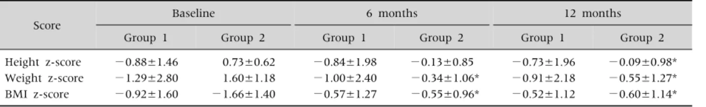
Table 2.Anthropometric Changes in Group 1 and Group 2
Score Baseline 6 months 12 months
Group 1 Group 2 Group 1 Group 2 Group 1 Group 2
Height z-score −0.88±1.46 0.73±0.62 −0.84±1.98 −0.13±0.85 −0.73±1.96 −0.09±0.98*
Weight z-score −1.29±2.80 1.60±1.18 −1.00±2.40 −0.34±1.06* −0.91±2.18 −0.55±1.27*
BMI z-score −0.92±1.60 −1.66±1.40 −0.57±1.27 −0.55±0.96* −0.52±1.12 −0.60±1.14*
Values are presented as mean±standard deviation.
BMI: body mass index.
*All p-values<0.05 compared to baseline.
ciency (Group 1) and 11 patients (64.7%) had mod- erate vitamin D deficiency/insufficiency (Group 2).
The mean serum 25(OH)D level at diagnosis for Group 1 was 8.7±1.4 ng/mL, and the mean level for Group 2 was 15.6±4.7 ng/mL (p<0.001).
There was no significant difference between Group 1 and 2 regarding anthropometric data (Htz, Wtz, BMIz). Compared to Group 1 patients, Group 2 patients had higher PLTs (331.8×10³±79.3/μL vs.
508.1×10³±192.2/μL, p=0.021). Other laboratory data including Hb, ESR, CRP, and ALB were similar between groups. Moreover, PCDAI and frequency of symptom and extraintestinal manifestations be- tween groups did not show a statistically significant difference (Table 1).
Compared to baseline, all anthropometric data (Htz, Wtz, BMIz) in Group 1 showed no significant improvement at 6 and 12 months’ follow-up (Table
2). In the analysis of Group 2, anthropometric data showed significant improvements in Wtz and BMIz sustained throughout the 12 months of follow-up.
Htz showed no significant improvement at 6 months, but there was a significant improvement at 12 months (Table 2).
In an effort to evaluate the confounding relation- ships, we examined the associations between predictors. The Pearson's correlation coefficient found no linear correlation between baseline serum 25(OH)D levels and other variables, including labo- ratory data (Hb, ESR, CRP, and ALB), anthro- pometric data (Htz, Wtz, BMIz), and PCDAI (Table 3).
DISCUSSION
Previous studies reported varying prevalence of hypovitaminosis D in adults with IBD ranging from
16% to 95%, depending on the study. However, rela- tively few studies focused on the vitamin D status of pediatric IBD patients. A study by Pappa et al. [18,19]
reported that the prevalence of vitamin D deficiency was 5.8% to 34.6% in pediatric patients with IBD, whereas a recent study found vitamin D deficiency in 48% of children with IBD, although they did take vi- tamin D supplements [20].
Most current studies evaluate the association of vitamin D status with disease activity index and bio- chemical parameters. However, most of them were done on adults, and few or no studies have evaluated the association of vitamin D status and growth in pe- diatric CD patients. The aim of this study was to ex- amine the association of vitamin D status with growth retardation in Korean pediatric patients with CD. In Korea, vitamin D insufficiency is very com- mon in ordinary person and was found in 47.3% of male and 64.5 % of female. Also, the prevalence of vi- tamin D insufficiency in ordinary person is higher in students and young adults than the elderly [13]. This study identified vitamin D deficiency in 81.5%
(53/65) and insufficiency in 16.9% (11/65) of our pe- diatric patients with CD. That is, 98.5% of our pedia- tric CD patients had hypovitaminosis D. This preva- lence in our study was higher than has been reported previously [18-20].
Vitamin D is widely known as a key regulator of bone health and in children vitamin D deficiency leads to bone deformities and stunted growth [10-12]. More recently, vitamin D has also come to be recognized as an immune regulator and a few studies describe the role of vitamin D in the patho- genesis, clinical outcome, and candidates for ad- junctive treatment of CD [21]. Jørgensen et al. [22]
showed that vitamin D treatment as a maintenance therapy in CD patients reduced the risk of relapse from 13% to 29%. We could assume that high vita- min D levels are associated with a better clinical outcome. A high vitamin D level would have a pos- itive effect on growth and clinical outcome because growth retardation is mainly secondary to disease activity. Recently, an association between low vita- min D level and higher risk for surgery and hospital-
izations for CD [23], and an association between low vitamin D level and loss of response to im- munomodulatory treatments were also reported [24].
In our study, Group 2 (moderate vitamin D defi- ciency/insufficiency) patients showed a significant improvement in Wtz and BMIz after 6 months, which was sustained through to the 12 months’ fol- low-up. Although Htz at 6 months showed no sig- nificant improvement, it reached a significant im- provement at 12 months. However, Group 1’s se- verely vitamin D deficient patients had no significant improvement of Htz, Wtz, and BMIz during fol- low-up (6 and 12 months) despite oral vitamin D supplements. For the patients of Group 1, there was no correlation between vitamin D levels and growth parameters (Htz, Wtz, BMIz) or other laboratory var- iables (Hb, CRP, PLT, ESR‚ ALB) and PCDAI. The findings indicate that baseline vitamin D status has an effect on growth outcome in pediatric CD. We would like to suggest vitamin D as an adjunctive treatment for pediatric CD patients suffering from growth failure.
It is important to note that the limitations of the current study, these include a small sample size and the retrospective nature of the clinical research process. The patient received oral vitamin D supple- ments after study entry, but we did not recheck the serum levels and also failed to verify the vitamin D levels after the 12 months of this research.
In conclusion, baseline vitamin D status is a pre- dictor of growth outcome in pediatric CD patients.
Future prospective studies of a larger cohort would be required to validate these results. Furthermore, a longer follow-up with vitamin D supplementation will be necessary to determine the appropriate sup- plemental dose of vitamin D.
REFERENCES
1. Vortia E, Kay M, Wyllie R. The role of growth hormone and insulin-like growth factor-1 in Crohn's disease: im- plications for therapeutic use of human growth hor- mone in pediatric patients. Curr Opin Pediatr 2011;
23:545-51.
2. Yang SK, Yun S, Kim JH, Park JY, Kim HY, Kim YH, et al. Epidemiology of inflammatory bowel disease in the Songpa-Kangdong district, Seoul, Korea, 1986- 2005: a KASID study. Inflamm Bowel Dis 2008;14:
542-9.
3. Thia KT, Loftus EV Jr, Sandborn WJ, Yang SK. An up- date on the epidemiology of inflammatory bowel dis- ease in Asia. Am J Gastroenterol 2008;103:3167-82.
4. Leong RW, Lau JY, Sung JJ. The epidemiology and phe- notype of Crohn's disease in the Chinese population.
Inflamm Bowel Dis 2004;10:646-51.
5. Alhagamhmad MH, Day AS, Lemberg DA, Leach ST.
An update of the role of nutritional therapy in the man- agement of Crohn's disease. J Gastroenterol 2012;47:
872-82.
6. Lee JJ, Escher JC, Shuman MJ, Forbes PW, Delemarre LC, Harr BW, et al. Final adult height of children with inflammatory bowel disease is predicted by parental height and patient minimum height Z-score. Inflamm Bowel Dis 2010;16:1669-77.
7. Shamir R. Nutritional aspects in inflammatory bowel disease. J Pediatr Gastroenterol Nutr 2009;48 Suppl 2:S86-8.
8. Sawczenko A, Ballinger AB, Savage MO, Sanderson IR.
Clinical features affecting final adult height in patients with pediatric-onset Crohn's disease. Pediatrics 2006;
118:124-9.
9. Theodoratou E, Tzoulaki I, Zgaga L, Ioannidis JP.
Vitamin D and multiple health outcomes: umbrella re- view of systematic reviews and meta-analyses of ob- servational studies and randomised trials. BMJ 2014;
348:g2035.
10. Rosen CJ. Clinical practice. Vitamin D insufficiency. N Engl J Med 2011;364:248-54.
11. Holick MF, Binkley NC, Bischoff-Ferrari HA, Gordon CM, Hanley DA, Heaney RP, et al. Evaluation, treat- ment, and prevention of vitamin D deficiency: an Endocrine Society clinical practice guideline. J Clin Endocrinol Metab 2011;96:1911-30.
12. Holick MF. Vitamin D deficiency. N Engl J Med 2007;357:266-81.
13. Choi HS, Oh HJ, Choi H, Choi WH, Kim JG, Kim KM, et al. Vitamin D insufficiency in Korea--a greater threat to younger generation: the Korea National Health and Nutrition Examination Survey (KNHANES) 2008. J Clin Endocrinol Metab 2011;96:643-51.
14. Levine A, Griffiths A, Markowitz J, Wilson DC, Turner
D, Russell RK, et al. Pediatric modification of the Montreal classification for inflammatory bowel dis- ease: the Paris classification. Inflamm Bowel Dis 2011;17:1314-21.
15. Ulitsky A, Ananthakrishnan AN, Naik A, Skaros S, Zadvornova Y, Binion DG, et al. Vitamin D deficiency in patients with inflammatory bowel disease: associa- tion with disease activity and quality of life. JPEN J Parenter Enteral Nutr 2011;35:308-16.
16. Tajika M, Matsuura A, Nakamura T, Suzuki T, Sawaki A, Kato T, et al. Risk factors for vitamin D deficiency in patients with Crohn's disease. J Gastroenterol 2004;
39:527-33.
17. Andreassen H, Rix M, Brot C, Eskildsen P. Regulators of calcium homeostasis and bone mineral density in pa- tients with Crohn's disease. Scand J Gastroenterol 1998;33:1087-93.
18. Pappa HM, Gordon CM, Saslowsky TM, Zholudev A, Horr B, Shih MC, et al. Vitamin D status in children and young adults with inflammatory bowel disease. Pediatrics 2006;118:1950-61.
19. Pappa HM, Langereis EJ, Grand RJ, Gordon CM.
Prevalence and risk factors for hypovitaminosis D in young patients with inflammatory bowel disease. J Pediatr Gastroenterol Nutr 2011;53:361-4.
20. Laakso S, Valta H, Verkasalo M, Toiviainen-Salo S, Viljakainen H, Mäkitie O. Impaired bone health in in- flammatory bowel disease: a case-control study in 80 pe- diatric patients. Calcif Tissue Int 2012;91:121-30.
21. Mora JR, Iwata M, von Andrian UH. Vitamin effects on the immune system: vitamins A and D take centre stage. Nat Rev Immunol 2008;8:685-98.
22. Jørgensen SP, Agnholt J, Glerup H, Lyhne S, Villadsen GE, Hvas CL, et al. Clinical trial: vitamin D3 treatment in Crohn's disease-a randomized double-blind place- bo-controlled study. Aliment Pharmacol Ther 2010;32:
377-83.
23. Ananthakrishnan AN, Cagan A, Gainer VS, Cai T, Cheng SC, Savova G, et al. Normalization of plasma 25-hydroxy vitamin D is associated with reduced risk of surgery in Crohn's disease. Inflamm Bowel Dis 2013;19:1921-7.
24. Zator ZA, Cantu SM, Konijeti GG, Nguyen DD, Sauk J, Yajnik V, et al. Pretreatment 25-hydroxyvitamin D lev- els and durability of anti-tumor necrosis factor-α ther- apy in inflammatory bowel diseases. JPEN J Parenter Enteral Nutr 2014;38:385-91.