oral aphthous ulcers, and pauciarthritis; those not related to bowel disease activity but simply reflecting the patient’s sus-ceptibility to related autoimmune disorders, including anky-losing spondylitis (AS) and uveitis; and those with an unclear relationship to bowel disease activity, including pyoderma gangrenosum (PG) and primary sclerosing cholangitis (PSC). Extraintestinal complications are initiated by the disease itself and consequently include micronutrient deficiency, osteopo-rosis, peripheral neuropathy, nephrolithiasis, and IBD drug-re-lated side effects.2 EIMs or extraintestinal complications occur in various organs and significantly affect the quality of life of patients with IBD (Table 1). Physicians who treat IBD need to keep track of the progress and treatment of these EIMs or ex-traintestinal complications of the disease through multidisci-plinary approaches. Herein, we will focus on and review the pathogenesis and clinical perspectives of EIMs in IBD.
WHY ARE EIMs IMPORTANT IN IBD?
The prevalence of EIMs is strongly influenced by the age at
di-Pathogenesis and clinical perspectives of extraintestinal
manifestations in inflammatory bowel diseases
Jung Min Kim,1 Jae Hee Cheon1,2,3
1Department of Internal Medicine and Institute of Gastroenterology, Yonsei University College of Medicine, Seoul; 2Avison Biomedical Research Center, Severance Hospital, Seoul; 3Affiliate Faculty, Pohang University of Science and Technology (POSTECH), Pohang, Korea
A considerable number of patients with inflammatory bowel disease (IBD) experience extraintestinal manifestations (EIMs), which can present either before or after IBD diagnosis. Unraveling the pathogenic pathways of EIMs in IBD is challenging because of the lack of reliable criteria for diagnosis and difficulty in distinguishing EIMs from external pathologies caused by drugs or other etiologies. Optimizing treatment can also be difficult. Early diagnosis and management of EIM revolve around multidisciplinary teams, and they should have the resources necessary to make and implement appropriate decisions. In ad-dition, specialists of the affected organs should be trained in IBD treatment. Furthermore, patient awareness regarding the extraintestinal symptoms of IBD is of paramount importance for improving patient understanding of disease and health out-comes. Herein, we review the pathogenesis and clinical perspectives of EIMs in IBD. (Intest Res 2020;18:249-264)
Key Words: Inflammatory bowel diseases; Neuromuscular manifestations; Skin manifestations; Eye manifestations
Received October 11, 2019. Revised November 25, 2019. Accepted March 6, 2020.
Correspondence to Jae Hee Cheon, Department of Internal Medicine and Institute of Gastroenterology, Yonsei University College of Medicine, 50-1 Yonsei-ro, Seodaemun-gu, Seoul 03722, Korea. Tel: +82-2-2228-1990, Fax: +82-2-393-6884, E-mail: [email protected]
INTRODUCTION
Inflammatory bowel disease (IBD), including CD and UC, is a chronic, intestinal inflammatory disease that is often associat-ed with the development of various extraintestinal symptoms. Extraintestinal symptoms can be divided into 2 groups: ex-traintestinal manifestations (EIMs) and exex-traintestinal compli-cations. EIMs are defined in patients with IBD when an inflam-matory pathology is located outside the intestine, the patho-genesis of which depends on the extension/translocation of immune pathways from the intestine, is an independent in-flammatory event sustained by IBD, or shares a common envi-ronmental or genetic predisposition with IBD.1 EIMs can be divided into 3 categories: EIMs directly related to bowel dis-ease activity, including episcleritis, erythema nodosum (EN),
agnosis and sex3,4 and is reported to be 23.0% in CD and 24.4% in UC, according to data from the National Health Insurance System of Korea.3 Up to 50% of patients with IBD experience at least one EIMs, which can develop even before the diagno-sis of IBD.5 However, these results of EIMs differ greatly among study methods and research designs.
Abdominal pain is a common symptom related to IBD, and pain is the principal symptom in IBD.6 Pain is a longstanding problem and can also be seen in EIMs.7 During disease flares, pain is present in 50%–70% of patients with IBD, and the health-related quality of life (HRQOL) of these patients was reported to be significantly lower in patients suffering from pain than in those without pain.6,7
As such, EIMs are a common problem and may significant-ly impact the quality of life of patients with IBD, requiring spe-cific treatments depending on the affected organs. Therefore, successful management of EIMs is essential for improving the quality of life of patients with IBD.
PATHOGENESIS OF EIMs
The pathogenic mechanisms of EIMs have yet to be clearly defined. Defining the pathophysiology of an EIM requires sig-nificant research and consensus due to the lack of consistent
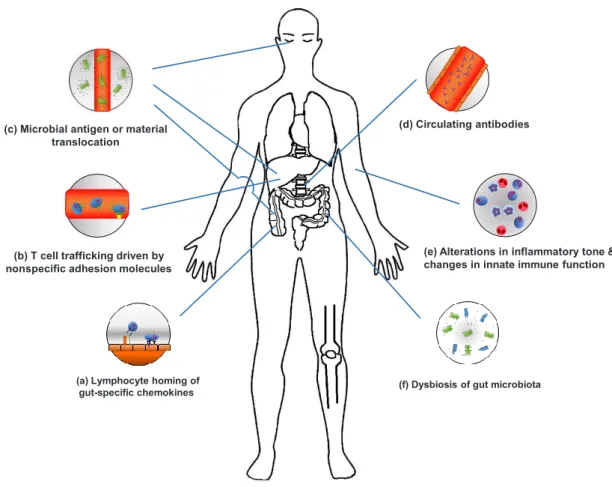
criteria for diagnosing an EIM and difficulty of distinguishing an EIM from extraintestinal complications or a secondary EIM caused by drugs. EIMs are considered to be the result of an antigen-specific immune response from the intestine to an extraintestinal site or an independent inflammatory event that is initiated or persisting from covalent genetic or environmen-tal risk factors in the host, or from IBD (Fig. 1).
1. Dysbiosis of Gut Microbiota and Disrupted Gut Barrier in Intestinal Sites
Short chain fatty acids (SCFAs) enhance intestinal health, cre-ating conditions favorable for resistance to pathogenic bacte-ria and preventing colitis. SCFAs have been found at signifi-cantly lower levels in IBD patients with psoriatic arthritis (PsA).8 IBD patients with PsA had a relative decrease in the abundance of Coprococcus and Ruminococcus species. Ruminococcus is a mucin-degrading bacterium that is especially important for maintaining intestinal homeostasis through SCFA production. The receptor activator of nuclear factor kappa-β ligand (RANKL)/ osteoprotegerin (OPG) system may be a regulator of intestinal microbiomes, and imbalances through the breakage of RANKL in the gut lumen of patients with PsA can promote dissocia-tion of inflammadissocia-tion to a distal site, such as a joint.8
IBD patients with spondyloarthritis (SpA) have an
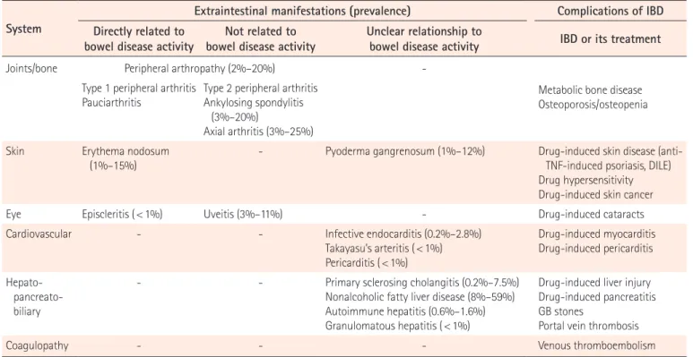
in-Table 1. Extraintestinal Manifestations and Complications Associated with IBD
System
Extraintestinal manifestations (prevalence) Complications of IBD Directly related to
bowel disease activity bowel disease activityNot related to Unclear relationship to bowel disease activity IBD or its treatment
Joints/bone Peripheral arthropathy (2%–20%)
-Type 1 peripheral arthritis
Pauciarthritis Type 2 peripheral arthritisAnkylosing spondylitis (3%–20%)
Axial arthritis (3%–25%)
Metabolic bone disease Osteoporosis/osteopenia
Skin Erythema nodosum
(1%–15%) - Pyoderma gangrenosum (1%–12%) Drug-induced skin disease (anti-TNF-induced psoriasis, DILE)
Drug hypersensitivity Drug-induced skin cancer
Eye Episcleritis ( < 1%) Uveitis (3%–11%) - Drug-induced cataracts
Cardiovascular - - Infective endocarditis (0.2%–2.8%)
Takayasu’s arteritis ( < 1%) Pericarditis ( < 1%) Drug-induced myocarditis Drug-induced pericarditis Hepato- pancreato-biliary
- - Primary sclerosing cholangitis (0.2%–7.5%)
Nonalcoholic fatty liver disease (8%–59%) Autoimmune hepatitis (0.6%–1.6%) Granulomatous hepatitis ( < 1%)
Drug-induced liver injury Drug-induced pancreatitis GB stones
Portal vein thrombosis
Coagulopathy - - - Venous thromboembolism
creased abundance of the genus Dialister, which is positively correlated with disease activity.9 In addition, microbial activa-tion in CD with SpA are associated with interleukin-23 (IL-23) dependent inflammation.10 CD-SpA-derived Escherichia coli induced T helper 17 (Th17) cells polarization and made exac-erbation of colitis or inflammatory arthritis.
Gut dysbiosis in IBD patients with AS induces differentiation and activation of group 3 congenital lymphocytes (ILC3s).11 ILC3s are IL-23-responsive immune cells that play a pro-in-flammatory role in mediating a protective response against extracellular bacterial infections. ILC3s have been identified in the peripheral blood, synovial fluid, and bone marrow tis-sues of patients with AS, most of which express intestinal homing integrin α4β7. Mucosal vascular addressin cell adhe-sion molecule 1 (MAdCAM-1) was highly represented in the
bone marrow and AS patients’ intestinal high endothelial ve-nules. In addition, MAdCAM-1 plays a central role in attracting ILC3s at the inflammation site of active AS and recirculating ILC3s between the gut and inflamed spines.12 In patients with IBD, intestinal effector T cells target gut microbial antigens. Transporting these antigens to the liver through the portal cir-culation allows the recruitment of mucosal effector α4β7 posi-tive T cells through α4β7 MAdCAM-1 interactions.
Patients with PSC showed gut microbial differentiations from those with UC without liver diseases.13 In a Gnotobiotic mice model with PSC/UC, Klebsiella pneumoniae, Proteus mi-rabilis and Enterococcus gallinarum were identified from the mesenteric lymph nodes of PSC/UC Gnotobiotic mice.14 Dis-rupted gut barrier by K. pneumonia is associated with bacteri-al translocation and Th17 priming to the liver. These bacteria
(a) Lymphocyte homing of gut-specific chemokines
(b) T cell trafficking driven by nonspecific adhesion molecules (c) Microbial antigen or material
translocation
(d) Circulating antibodies
(e) Alterations in inflammatory tone & changes in innate immune function
(f) Dysbiosis of gut microbiota
Fig. 1. Mechanisms of extraintestinal manifestations (EIMs). (a) Activation and expansion of effector memory T cells can lead to
induc-tion of mucosal vascular addressin cell adhesion molecule 1 and C-C motif chemokine ligand 25 (CCL25) expression in the liver. (b) Inter-cellular adhesion molecule-1, vascular adhesion protein-1, and vascular cell adhesion molecule 1 are associated with T cell trafficking. (c) Microbial antigens and materials translocate to extraintestinal sites and drive EIMs. (d) Circulating antibodies can provoke an autoim-mune response and peptide(s) expressing common epitopes shared by colon and extra-colonic organs. (e) Neutrophil priming and altera-tion of circulating monocytes and macrophages can induce intestinal inflammaaltera-tion and are associated with innate immune responses. (f) Gut dysbiosis drives non-intestinal inflammation and increases mucosal permeability and lipopolysaccharides.
can lead the progression of hepatobiliary tract disease through the Th17 response in the colon and liver.
Alterations in the composition and metabolic products of the microbiota can affect the production of innate immune cells in patients with IBD.15,16 There were significant differenc-es in the composition of microbiome between HLA-B27+ and HLA-B27– siblings as well as healthy controls, indicating that genetic background can affect the composition and abun-dance of microbiomes.
Faecalibacterium prausnitzii-specific cells (DP8α TREG
clones), found only in the chemokine receptor 6 (CCR6)+ and in the C-X-C motif chemokine receptor 6 (CXCR6)+ fractions of CD4CD8α (DP8α) cells, were significantly decreased in the blood of patients with IBD.17 These gut recirculating markers are expressed in the DP8 cells of the large intestine and remain expressed in F. prausnitzii–reactive DP8α cells in the blood, al-lowing them to move to a specific intestinal position to per-form their functions. These characteristics can be conveyed by the joint expression of CCR6 and CXCR6; the expression of CCR6 and CXCR6 also implies homing to other mucosal loca-tions.
2. Immune Responses in Extraintestinal Sites
Abnormal lymphocyte homing in IBD can explain why EIMs are associated with site specific distributions.18 The α4β7 inte-grin is a lymphocyte receptor for the MAdCAM-1. The interac-tion between α4β7 and MAdCAM-1 plays an important role in regulating lymphocyte homing to mucosal sites.19 Expression of MAdCAM-1 is confined to the intestinal tissue, allowing gut tropic T cells expressing α4β7 integrin to selectively traffic with the intestinal mucosa. Additional tropism for the small bowel comes from C-C motif chemokine ligand 25 (CCL25) which attracts lymphocytes expressing the CCR9 receptor. T cell migration to the small bowel requires integrin α4β719 and CCR9.20 Interestingly, homing to the colon requires α4β7 or α4β1 but not CCR9.21
Both chemokines and adhesion molecules can promote T cell trafficking to extraintestinal sites. The activation and ex-pansion of effector memory T cells can lead to the induction of MAdCAM-1 and CCL25 expression in the liver, thereby pro-moting the recruitment of CCR9+α4β7+ mucosal T cells and development of inflammation.22 Upregulation of inflamma-tion-related adhesion molecules and chemokines also enables the capture of effector T cells, facilitating recruitment to sites other than extraintestinal sites. IL-6 produced during inflam-mation promotes the influx of naïve and memory T cells by
increasing the expression of intercellular adhesion molecule-1 (ICAM-1) in high endothelial venules of the reactive lymph nodes.23 ICAM-1 can also be upregulated with pre-inflamma-tory stimuli in response to TNF-α, IL-1β, and interferon-γ.24
Neutrophils altered by inflammatory cytokines can en-hance their response upon subsequent activation, referred to as “neutrophil priming.”25 Neutrophils in patients with gut in-flammation are primed to secrete high levels of TNF-α and IL-1β after stimulation with lipid polysaccharides.26 However, pa-tients with CD have a defective neutrophil recruitment after the introduction of subcutaneous bacteria, which severely de-lays the subsequent clearance of these bacteria.27 In addition to changes in circulating monocytes and macrophages in pa-tients with IBD, reduced inflammatory cytokine production in response to bacterial stimuli has also been reported.27,28 Neu-trophil-platelet interactions play an important role in the de-gree of tissue neutrophil infiltration through neutrophil integ-rins (αLβ2 and αMβ2) and P-selectin glycoprotein ligand 1; how-ever, neutrophil priming and/or activation in the peripheral site are inhibited by sphingosine.29,30 Ceramide synthase 2 (CerS2) and CerS6 mutually control the aspects of neutrophil recruitment to tissues, and chemotaxis is also regulated by sphingosine-1-phosphate through IL-8 production. In the gut of patients with SpA, IL-23 polarization does not appear to be accompanied by Th17 polarization. However, IL-23 can in-duce gut inflammation and may be associated with the expan-sion and activation of innate immune cells such as ILC3, γδ T cells, and mucosal-associated invariant T cells.11,31,32 Augment-ed expression of IL-23R in γ/δ T cells was also associatAugment-ed with enhanced IL-17 secretion. Stimulation of γ/δ T cells from SpA patients with IL-23 and/or anti-CD3/CD28 strongly induced polarization for IL-17 production.
3. Autoantibodies
On the endothelial side, pre-incubation of the synovial sec-tions with monoclonal antibodies against ICAM-1 and vascu-lar adhesion protein-1 (VAP-1) showed significant inhibition of the binding of small lymphocytes from the IBD gut to the joint vessels.33 Small lamina propria lymphocytes isolated from the intestine of IBD patients adhered to the synovial ves-sels using VAP-1 and their endothelial ligands (CD18 & ICAM-1, and α4β7/α4β1 integrins and vascular cell adhesion mole-cule 1 [VCAM-1]). VAP-1 also plays a pivotal role in the recruit-ment and transmigration of lymphocytes via the hepatic sinu-soidal endothelium, the expression of which is upregulated by inflammation.34 VCAM-1 selectively binds lymphocytes
thr-ough integrin α4β1. It also mediates lymphocyte homing to extraintestinal sites such as the lung, skin, and central nervous systems with chronic inflammation. It is not usually expressed by the intestinal venules but can be upregulated in the blood vessels during inflammation.35 CCR10 with integrin α4β1 and VCAM-1 also mediate the localization of integrin α4β7-nega-tive plasmablasts to non-intestinal mucosal sites (salivary and lacrimal glands and respiratory and urogenital tracts) where CCL28 is also expressed. Among the effector and central memory T cells, CCR10 is limited to integrin α4β1+α4β1– cuta-neous lymphocyte antigen+ skin homing cells (which provide T cell attraction to the CCR10 ligand CCL27 expressed in ke-ratinocytes). Inflammation or mechanical stress in the ex-traintestinal sites can lead to the recruitment of gut-producing effector T cells and further increase the inflammatory pro-cess.34,36
In addition, molecular mimicry, peptide sequences be-tween the gut bacteria and host major histocompatibility complex (MHC) molecules, and reactivation by cross-reactive components of microbiomes or host antigens have been re-ported.37,38
Peptides expressing common epitopes shared by colons and extra-colonic organs, autoimmune responses to the iso-form of tropomyosin (tropomyosin related peptide) expressed in the eyes, skin, joints, biliary epithelia, and intestines can help clarify the pathogenesis of EIMs.39,40 Bacteria can translo-cate across the leaky epithelial barriers and cause adaptive immune responses that are indistinguishable from bacterial epitopes in joints or skin. There is still a lack of clear evidence on the role of circulating antibodies or immune complexes in the pathogenesis of EIMs.
In a mice model, retina-specific T cells causing human blindness due to uveitis received microbial-dependent activa-tion signals, most likely cross-reactive bacterial antigens.41 This result showed that the activation of autoreactive T cells via gut microbes may drive uveitis. In another mice model of autoim-mune uveitis, enhanced leukocytes trafficking between the in-testine and eyes were observed.42 The antigen specificity of T cells responsible for EIMs in humans will require further con-firmation.
4. Genetics of EIMs
Genetic risk loci for both IBD and EIMs overlap extensively. PSC genes and their linked IBD genes are involved in T cell apoptosis (UBASH3A, BCL2L11, FOXO1, and IRF8) and the Janus kinase/signal transducers and activators of transcription
(JAK/STAT) pathways signaling pathways (SOCS1, JAK2, STAT3, and TYK2). In case of AS, T cell apoptosis pathways in-teract with the IBD genes. The AS-related genes, TAPBPL and NPEPPS, function similarly to the AS covalent gene ERAP1 in patients with IBD, which is important for both the develop-ment of AS and IBD. ERAP1 variant K528R associated with CD has involved in aberrant peptide production resulting in suboptimal peptide MHC I complex.43 The CD risk gene, NOD2/CARD15, which encodes a pattern recognition recep-tor, is also associated with sacroiliitis and uveitis.44,45 STAT4 is also responsible for the expansion of Th17 cells activated by IL-23, which promotes chronic inflammation in innate and adaptive immunity and is considerably associated with the development of EIMs involving the joints and eyes in patients with IBD.46 However, it is still unknown whether all relevant loci contribute specifically to the pathology of EIMs or wheth-er thwheth-ere are genes that genwheth-erally cause EIMs by not restricting the inflammatory response to specific body compartments. Therefore, further research is needed.
CLINICAL ASPECTS OF EIMs
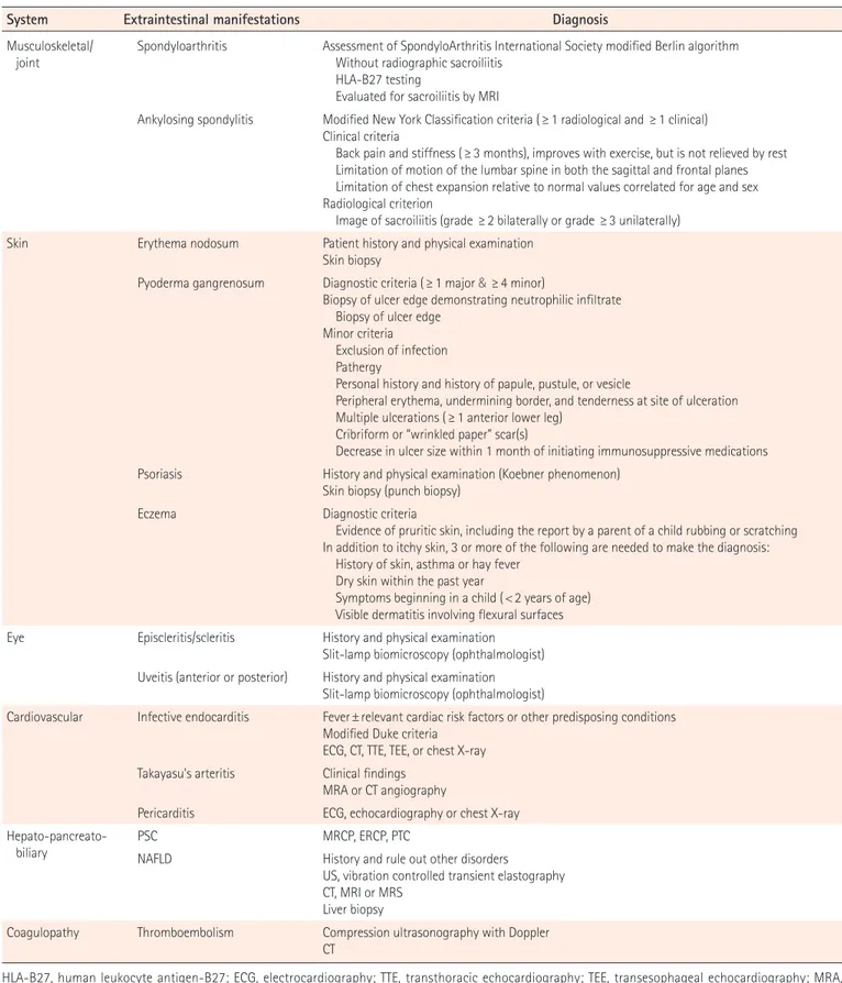
This following section describes the paradigms and tools cur-rently available in clinical studies of the musculoskeletal/joint, skin, eye, vessels, and hepatobiliary EIMs (Table 2).
1. Musculoskeletal/Joint Manifestations
Inflammatory arthrosis of IBD is the most common EIM, with a prevalence of 20%–50% for axial inflammation and 5%–20% for peripheral arthritis.47 Peripheral arthritis in patients with IBD is mainly asymmetric, oligo-articular, and is predomi-nantly found in CD. IBD-related joint symptoms can be subdi-vided into non-inflammatory and inflammatory joint pain. Arthralgia is a major medical problem in patients with IBD. Compared to patients with IBD without arthralgia, non-in-flammatory joint pain was reported in 16% of patients with a significant worsening of their HRQOL determined using the 36-Item Short-Form Health Survey and the Inflammatory Bowel Disease Questionnaire.48 The current gold standard tool for layering SpA is the Ankylosing Spondylitis Disease Ac-tivity Score, which can measure the disease acAc-tivity that may be followed over time.49 In general, the prognosis for peripher-al arthritis is good. The prognosis for axiperipher-al involvement is less favorable and is not related to the prognosis of IBD but to the progression of IBD.
Table 2. Diagnosis of Extraintestinal Manifestations
System Extraintestinal manifestations Diagnosis
Musculoskeletal/
joint Spondyloarthritis Assessment of SpondyloArthritis International Society modified Berlin algorithm Without radiographic sacroiliitis HLA-B27 testing
Evaluated for sacroiliitis by MRI
Ankylosing spondylitis Modified New York Classification criteria (≥ 1 radiological and ≥ 1 clinical) Clinical criteria
Back pain and stiffness (≥ 3 months), improves with exercise, but is not relieved by rest Limitation of motion of the lumbar spine in both the sagittal and frontal planes Limitation of chest expansion relative to normal values correlated for age and sex Radiological criterion
Image of sacroiliitis (grade ≥ 2 bilaterally or grade ≥ 3 unilaterally) Skin Erythema nodosum Patient history and physical examination
Skin biopsy
Pyoderma gangrenosum Diagnostic criteria ( ≥ 1 major & ≥4 minor)
Biopsy of ulcer edge demonstrating neutrophilic infiltrate Biopsy of ulcer edge
Minor criteria Exclusion of infection Pathergy
Personal history and history of papule, pustule, or vesicle
Peripheral erythema, undermining border, and tenderness at site of ulceration Multiple ulcerations (≥ 1 anterior lower leg)
Cribriform or “wrinkled paper” scar(s)
Decrease in ulcer size within 1 month of initiating immunosuppressive medications Psoriasis History and physical examination (Koebner phenomenon)
Skin biopsy (punch biopsy)
Eczema Diagnostic criteria
Evidence of pruritic skin, including the report by a parent of a child rubbing or scratching In addition to itchy skin, 3 or more of the following are needed to make the diagnosis: History of skin, asthma or hay fever
Dry skin within the past year
Symptoms beginning in a child ( < 2 years of age) Visible dermatitis involving flexural surfaces Eye Episcleritis/scleritis History and physical examination
Slit-lamp biomicroscopy (ophthalmologist) Uveitis (anterior or posterior) History and physical examination
Slit-lamp biomicroscopy (ophthalmologist)
Cardiovascular Infective endocarditis Fever ± relevant cardiac risk factors or other predisposing conditions Modified Duke criteria
ECG, CT, TTE, TEE, or chest X-ray Takayasu's arteritis Clinical findings
MRA or CT angiography
Pericarditis ECG, echocardiography or chest X-ray
Hepato-pancreato-biliary PSCNAFLD MRCP, ERCP, PTCHistory and rule out other disorders
US, vibration controlled transient elastography CT, MRI or MRS
Liver biopsy
Coagulopathy Thromboembolism Compression ultrasonography with Doppler CT
HLA-B27, human leukocyte antigen-B27; ECG, electrocardiography; TTE, transthoracic echocardiography; TEE, transesophageal echocardiography; MRA, magnetic resonance angiography; PSC, primary sclerosing cholangitis; MRCP, magnetic resonance cholangio pancreatography; ERCP, endoscopic retrograde cholangiopancreatography; PTC, percutaneous transhepatic cholangiography; NAFLD, nonalcoholic fatty liver disease; US, ultrasound; MRS, magnetic resonance spectroscopy.
2. Skin Manifestations
Diagnoses of skin manifestations are primarily based on clini-cal examinations, and skin biopsies can help in atypiclini-cal cases. EN is associated with IBD activity, arthralgia, or fatigue. The prevalence of EN appears higher in patients with CD than in those with UC.50 EN usually appears on the extensor surface of the lower extremities, mostly in the anterior tibial region,51 and is associated with IBD flares but not always with severity, is self-limited, and usually resolves itself without the formation of ulcers or scarring.52 The appearance of nodules parallels intes-tinal activity, which rarely occurs before the onset of IBD or in the quiescent phase of IBD. IBD treatment leads to the resolu-tion of cutaneous lesions. PG can occur anywhere in the body but occurs most commonly in the shin and adjacent to sto-mas. PG usually has a skin trauma known as “pathergy.” This trauma might even be minimal, such as a venipuncture. Con-troversy exists over whether PG coexists with disease activity. In this case, treatment can be concentrated on the underlying intestinal disease. Patients often show a rapid response, with a stiffening of the lesions within 24 hours of treatment.
Due to the absence of specific tests for diagnosing PG, phy-sicians can determine whether clinical and histopathological images are consistent with PG, excluding other pustules or ul-cerative dermatitis. A further development of assessment tools and standardized classifications are needed.
3. Eye Manifestations
The most common ocular EIMs are dry eye, blepharitis, episcleritis, or anterior uveitis. Some ocular manifestations in IBD can be secondary to treatment and/or effects of the IBD itself. Ophthalmic complications usually originate from in-flammation, but some of these complications may reflect the overall disease activity.53 Episcleritis can be unilateral or bilat-eral and is more common in women. It usually does not need specific treatments other than those for the original disease. Scleritis is chronic and is characterized by edema and cellular infiltration of the scleral tissues. Uveitis is not common but can have potentially more serious consequences. It occurs acutely or subacutely and is usually very painful. Anterior uveitis is also commonly referred to as iritis, which indicates pain and photophobia and can be associated with blurred vi-sion or floaters. The slit-lamp test confirms the diagnosis of an ocular EIM. In case of vision impairment, the patient must be immediately referred to the ophthalmologist to avoid vision loss.
4. Vascular Manifestations
Cardiovascular manifestations in IBD mostly occur as an im-mune-related pathogenesis including pericarditis, myocardi-tis, venous and arterial thromboembolisms, arrhythmias and conduction disorders, infective endocarditis, and Takayasu’s arteritis. Pericarditis is the most frequent cardiovascular EIM in patients with IBD but has not been associated with IBD ac-tivity.54 The clinical picture of myocarditis or pericarditis relat-ed to IBD is nonspecific. These patients may have symptoms of an acute coronary syndrome, heart failure, or arrhythmias and may also have cardiogenic shock or sudden death.
If this clinical picture occurs within the first 28 days after starting treatment with 5-aminosalicylic acid or its derivatives, drug toxicity is suspected.55 Because 12-lead electrocardio-grams and biomarkers of cardiac injury are also nonspecific, transthoracic echocardiography, coronary angiography, or cardiovascular magnetic resonance imaging may offer more precise information for all patients with clinical presentations suggesting myopericarditis.56 Furthermore, endomyocardial biopsies can be considered a gold standard for the diagnosis of myocarditis.57
In meta-analysis studies, the risk of arterial thromboembolic events or cardiovascular mortality in patients with IBD does not increase but is associated with an increase in the risk of cardiovascular morbidities from cerebrovascular accidents and ischemic heart disease, especially in women.58,59 Lower rates of peripheral vascular disease in IBD do not appear to in-crease the risk of peripheral arterial thromboembolic diseases. However, hyperhomocysteinemia, a risk factor for arterial and venous thrombosis, is more common in patients with IBD than in the general population.60 In addition, an increased risk of arterial thrombosis due to systemic inflammation and hy-percoagulability involves premature atherosclerosis.61
5. Hepatobiliary Manifestations
Approximately 20% to 30% of patients with IBD show abnor-mal hepatic biochemistries.62 PSC is highly correlated with IBD, and about 50%–80% of patients with PSC have an ac-companying IBD.63 Patients with IBD and PSC often have pre-dominantly right-sided, high-frequency “backwash” ileitis, and rectal sparing.64 Patients with UC, when diagnosed with PSC, are negatively associated with smoking and older ages.65 Symptoms are intermittent and can be confusing due to the underlying IBD symptoms. In patients with IBD, PSC is usual-ly asymptomatic, which requires an awareness of high clinical association. Although typically imaged using magnetic
reso-nance cholangiography and confirmed using endoscopic ret-rograde cholangiopancreatography (ERCP), the Crohn’s and Colitis Organisation consensus group suggests that ERCP be performed only for intervention in stricture dilatation and/or if brush cytology specimen sampling is indicated.66
Autoimmune hepatitis (AIH)/PSC overlapping syndrome is seen in UC patients than CD.67 It is suspected that the charac-teristics of AIH and PSC in the same patient are present, and a definitive diagnosis of AIH is required according to the inter-national AIH group criteria.68
The prevalence of AIH in pediatric IBD patients is 1.6%.69 AIH can be divided into 2 types. AIH type 1 is positive for anti-nuclear antibodies and/or smooth muscle antibodies, where-as AIH type 2 is liver/kidney microsomal type 1 and/or anti-liver cytosol type 1 is positive. Even if antinuclear antibodies or smooth muscle antibodies is 1:20 or liver/kidney microso-mal type 1 is 1:10 in children, it is associated with AIH in chil-dren.70
Granulomatous hepatitis is a rare EIM of CD. The estimated prevalence is less than 1% of IBD. It may manifest fever, alka-line phosphatase, hepatosplenomegaly. Granulomatous hepa-titis may be induced by mesalazine and sulfasalazine therapy.71 In nonalcoholic fatty liver disease, IBD-specific risk factors include intraperitoneal abscesses, fistula disease, colitis sever-ity, malnutrition, protein loss, and drug use.
Thiopurine and gallstones are the most common causes of
acute pancreatitis in patients with IBD, and acute pancreatitis caused by thiopurine is generally not complicated and is self-limiting.72 Most chronic pancreatitis cases in patients with IBD are idiopathic, and PSC is a less common cause. Exocrine pancreatic insufficiency appears to be the most common pan-creatic symptom in IBD, but its clinical significance remains unknown.
TREATMENT FOR EIMs INCLUDING BIOLOGICS Optimizing treatments is often challenging. There is lack of high-quality evidence to support the most commonly used treatment methods for EIMs and other treatment strategies that can complement them. Treatment of EIMs should be based on the severity of symptoms and their association with IBD activity. The primary goal is symptom control and preser-vation of mobility and function. In some cases, symptomatic treatment with physiotherapy may be sufficient (Table 3).
1. Joint Manifestations
Treatment of IBD-related arthropathy is based on SpA studies, predominantly AS. Patients with axial SpA should be co-man-aged by rheumatologists because of the potentially disruptive disease progress. Severity of type 1 arthritis usually runs paral-lel to IBD activity, and type 2 arthritis usually requires long-term treatment. Therefore, effective treatment of underlying
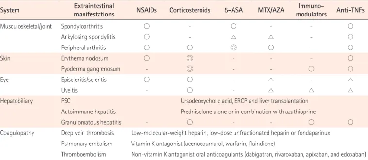
Table 3. Treatments for Extraintestinal Manifestations
System Extraintestinal manifestations NSAIDs Corticosteroids 5-ASA MTX/AZA modulatorsImmuno- Anti-TNFs
Musculoskeletal/joint Spondyloarthritis ○ - ○ - - ○
Ankylosing spondylitis ○ - △ △ - ○
Peripheral arthritis ○ ○ ◎ ○ - ○
Skin Erythema nodosum ○ ◎ - - - ○
Pyoderma gangrenosum - ◎ - - ○ ○
Eye Episcleritis/scleritis ○ ○ - △ - △
Uveitis - ○ - △ △ △
Hepatobiliary PSC Ursodeoxycholic acid, ERCP and liver transplantation
Autoimmune hepatitis Prednisolone alone or in combination with azathioprine
Granulomatous hepatitis - ○ - - ○ ○
Coagulopathy Deep vein thrombosis Low-molecular-weight heparin, low-dose unfractionated heparin or fondaparinux Pulmonary embolism Vitamin K antagonist (acenocoumarol, warfarin, fluindione)
Thromboembolism Non-vitamin K antagonist oral anticoagulants (dabigatran, rivaroxaban, apixaban, and edoxaban) 5-ASA, 5-aminosalicylic acid; MTX, methotrexate; AZA, azathioprine; PSC, primary sclerosing cholangitis; ERCP, endoscopic retrograde cholangiopancreatography; ◎, recommended; ○, available; △, controversial.
gut inflammation is important for the treatment of peripheral arthritis.
Intensive physiotherapy may be helpful in AS, and NSAIDs should be used for a short time period due to their potential to activate underlying IBD.73 Cyclooxygenase-2 inhibitors such as etoricoxib and celecoxib may be safer and have a lower risk of disease flare than conventional NSAIDs.74 Sulfasalazine, methotrexate (MTX), and azathioprine are generally ineffec-tive or only slightly effecineffec-tive in the treatment of AS. However, sulfasalazine is effective in treating patients with peripheral joint disease, especially those with a short disease duration.75 Sulfasalazine is an optional treatment in patients with SpA and peripheral disease and may be effective for large joint ar-thropathy.76 MTX and azathioprine are considered marginally effective in the treatment of peripheral arthropathy. For pa-tients with intolerance, unresponsiveness, or poor response to NSAIDs, anti-TNF therapy is the preferred treatment because of the limited efficacy of MTX and thiopurine.77
2. Skin Manifestations
Treatment of EN consists of treating the underlying IBD, and systemic steroids may be helpful. For patients with resistant or frequent relapses, immunomodulators and anti-TNF therapy can be considered alone or in combination.78
PG treatment strategies are the same between IBD and non-IBD patients. Systemic corticosteroids are considered as the first-line treatment, and oral or intravenous tacrolimus with oral cyclosporines may be considered for refractory cases.79,80 Anti-TNF therapy represents a new and effective treatment option for medically refractory PG cases.
3. Eye Manifestations
Due to the potential loss of vision, ophthalmologists must evaluate each suspected case to confirm the diagnosis and help guide the treatment. Dry eyes may be treated with topical lubricants. Episcleritis may be self-limiting and usually re-sponds to underlying IBD treatment along with topical NSAIDs and glucocorticoids.81 Uveitis requires urgent consul-tations with ophthalmologists and needs prompt treatment because of the risk of vision loss. Treatment usually consists of steroids. Other therapies (azathioprine, MTX, or anti-TNF therapy) have been discussed only for resistant cases.82
4. Vascular Manifestations
Because thromboembolism occurs due to multifactorial causes and acquired risk factors, precautions (hydration,
cor-rection of vitamin deficiencies, and wearing gradual compres-sion stockings) can help prevent venous thromboembolisms (VTEs). Prophylactic anticoagulation therapy is recommend-ed for high-risk patients (hospitalizrecommend-ed due to an active phase or surgery) with VTEs.83 There is still no evidence for the pro-phylaxis of active disease in outpatients with IBD. However, because many VTEs occur in outpatients with IBD, guidelines are required for VTE prophylaxis when patients with IBD have acutely or chronically active diseases. Acute deep vein throm-boses and pulmonary embolisms are treated using anticoagu-lants according to international guidelines. Low-molecular-weight heparin is commonly used, and low-dose unfraction-ated heparin or fondaparinux can be used as an alternative option. Long-term treatment includes oral anticoagulants such as vitamin K antagonists (acenocoumarol, warfarin, flu-indione) or non-vitamin K antagonist oral anticoagulants, such as dabigatran, rivaroxaban, apixaban, or edoxaban.
5. Hepatobiliary Manifestations
Ursodeoxycholic acid improves abnormal liver function tests and is widely used in patients with PSC, but prognosis improve-ments are controversial. Other therapeutic agents (steroid, cy-closporine, tacrolimus, MTX, or anti-TNF therapy) still lack ev-idence for improvements in clinical endpoints.84 ERCPs play a role in the management of biliary strictures and should be performed after antibiotic prophylaxis in patients with PSC. It can reduce the risk of cholangitis, bacteremia, and septicemia after ERCP.85 Patients with PSC and end-stage liver disease should be evaluated to determine whether liver transplanta-tions should be performed according to the standard guide-lines. PSC has a significantly greater risk for the development of colorectal dysplasia and carcinoma in patients with UC.86 Therefore, surveillance colonoscopies with target biopsies are recommended at the diagnosis and at every 1–2 years there-after.87
Immunosuppressive therapy with prednisolone alone or in combination with azathioprine is the main therapeutic regi-men of AIH and has been to show the prolongation of life ex-pectancy.67 In granulomatous hepatitis, corticosteroids and immunosuppressive drugs are used for treatment.
6. Biologics and Probiotics for EIMs
Biologics have been tested to prove their efficacy in the treat-ment of EIMs as well as IBD. Clinical benefits of infliximab or adalimumab may not be limited to gut treatment effects but may extend to improve EIMs.88 Infliximab and adalimumab
therapy also have potential beneficial effects on metabolic bone diseases, hematologic diseases, or vascular EIMs.88 How-ever, anti-TNF treatments can cause paradoxical cutaneous inflammations, which are considered a class drug effect, and are generally reversible upon drug transition or discontinua-tion. This paradoxical cutaneous inflammation manifests itself or appears in most cases as psoriasis, which accounts for why anti-TNF-α drug use is increasingly recognized as a cause of drug-induced skin lesions.89 Paradoxical psoriasis skin lesions are characterized by selective overexpression of type I inter-feron, skin accumulation of plasmacytoid dendritic cells (pDCs), and reduced T cell counts.90 Anti-TNF-α treatment prolongs the production of type 1 interferons by pDC by in-hibiting pDC maturation. Type I interferon overexpression re-sults in a skin phenotype of paradoxical psoriasis, indepen-dent of T cells, unlike classical psoriasis. TNF-α inhibition may reduce the accumulation of Th1 and Th17 cells at the site of inflammation but may induce compensatory expansion at other locations, resulting in skin lesions.91
The role of vedolizumab in EIM treatment has not been de-fined yet. Vedolizumab inhibits the binding of α4β7+ integrins to the intestinal mucosal cell adhesion molecule (MAd-CAM-1) but not to VCAM-1.92 The binding mechanism seems to be selective for cutaneous intestinal lymphocyte migration. In contrast, lymphocyte migration by VCAM-1 is unaffected. Nevertheless, the overlapping binding behavior of α4β7 with MAdCAM-1 and VCAM-1 may be due to partly different affin-ities.92,93 According to these results, vedolizumab has no direct therapeutic effect on cutaneous EIMs and can play a role in EIM management by treating disease activity in EIMs that parallels IBD.94 In a systematic review, vedolizumab was sug-gested to reduce the incidence of new EIMs but also be effec-tive in the treatment of preexisting EIMs (particularly PSC, rheumatic and cutaneous manifestations).95
Data on other biologicals are limited. Ustekinumab is a fully human IgG1 kappa monoclonal antibody that blocks the IL-12/23 p40 subunit. IL-12 and IL-23 are associated with adap-tive immune responses, and blocking IL-12/IL-23 helps im-prove chronic inflammatory conditions. Ustekinumab has been reported for the management of cutaneous and joint EIMs (psoriasis, PsA).96,97 In addition, it is effective for the treat-ment of anti-TNF antibody-related paradoxical cutaneous in-flammation.98 However, paradoxical arthralgic or cutaneous manifestations have also been reported.99,100 In addition, it did not reduce the occurrence of new EIMs.101 Therefore, usteki-numab requires additional safety and efficacy evidence from
randomized controlled trials and open-label, real-world co-horts. Ustekinumab will be an important treatment option in EIMs and IBD because it is superior to vedolizumab in terms of overall efficacy, rapid onset, and efficacy in small intestine disease.
Tofacitinib is a selective JAK inhibitor and the first oral med-ication approved for use in treating moderate-to-severe UC. Its efficacy of treating cutaneous EIMs has been reported. STAT3 was significantly upregulated in PG/EN compared to psoriasis and revealed potential benefits from JAK inhibition in these patients.94,102 In a double-blind, head-to-head, non-in-feriority, randomized controlled trial in patients with rheuma-toid arthritis who had insufficient response to MTX treatment, combination therapy with tofacitinib and MTX showed simi-lar efficacy to that with adalimumab and MTX, and tofacitinib monotherapy was shown to be inferior to either combina-tion.103 Because tofacitinib is expected to be free of anti-drug antibodies due to its low molecular weight, it results in a rapid therapeutic effect in patients with acute severe UC. Further studies on the efficacy and safety of the combination therapy with tofacitinib and other drugs are needed. In addition, there is a need to compare the efficacy and side-effects of upadaci-tinib and tofaciupadaci-tinib, which are selective JAK1 inhibitors.
Data on probiotics for EIMs are limited. Several studies sug-gested that the use of probiotics has a beneficial effect for cu-taneous or arthralgic EIMs but requires further confirmation from randomized controlled trials or systematic reviews eval-uating the effects of probiotics on EIMs.104,105
KEY RECOMMENDATIONS FOR IMPROVING THE DISEASE COURSE AND PRINCIPLES TO BE APPLIED TO TREATMENT
Physicians should always consider EIMs in the management of patients with IBD. Early diagnosis and treatment can pre-vent permanent tissue destruction and the development of complications in patients with IBD with developing EIMs. Di-agnostic delay is considered a risk factor for the development of EIM complications and abdominal surgery.106 Uveitis can result in visual impairment if the diagnosis is delayed. Due to the diagnostic delay accompanying these inflammatory con-ditions, the need for proper approaches and tools that can quickly provide accurate and viable information is further em-phasized. Sex, age, ethnicity, geography, socioeconomic sta-tus, duration, and extension of disease, disease activity, and a history of medications are associated with the diagnoses of
EIMs. Genetic factors and dysbiosis of gut microbiomes also play an important role in identifying EIMs. Physicians should be careful because specific features of the clinical presenta-tions may signal the possibility of future EIMs. Screening and surveillance of EIMs are difficult for physicians in only one specialty to include in their treatment protocols. Therefore, it is necessary to follow-up with multidisciplinary care. Educat-ing patients on EIMs and their complications will improve the effectiveness of medications and follow-ups. However, re-search has shown that certain EIMs, such as VTEs, PSCs, and nephrolithiasis, may be less emphasized in patient educa-tion.107 Patients’ perception of thromboembolisms or cardio-vascular EIMs can have a significant impact on the associated morbidity and mortality. VTE has a higher incidence in pa-tients with IBD than in the general population, and IBD dis-ease activity is associated with an incrdis-eased risk of VTE.108 However, only 12% of patients were aware that the risk of VTE in the outpatient environment was high and less than half of the patients were convinced that they recognized the signs and symptoms of deep vein thromboses and pulmonary em-bolisms,107 indicating a need to reconsider the awareness of EIMs in patients with IBD.
The role of mobile applicable for the self-management of IBD by patients will continue to increase by providing a more efficient, effective, and good quality healthcare system at low-er costs.109 Daily monitoring of UC through TrueColours UC improved UC management while also reducing the burden placed on individual and healthcare services.110 These applica-tions can not only remotely monitor patients via electronic patient-reported outcome measures (PROs) or device-gener-ated data (such as activity trackers) but also improves patient engagement.111 The goal of recent IBD therapies is moving to-ward a therapeutic-targeted strategy using PROs, biomarkers of inflammation, and mucosal healing to optimize therapeu-tics and should also be used for the treatment of EIMs. CONCLUSIONS
For EIMs of IBD, new mechanisms are being uncovered in a number of fields, including immunology, microbiology, and genetics, and can reveal a wide range of therapeutic targets. It is fundamentally important to ensure proper diagnosis and management of EIMs, which can often result in a much more debilitating and lower quality of life than actual IBD. Better pa-tient images require new tools to monitor inflammation from various angles, and managements for complex and diverse
EIMs should be discussed, treated, and tracked through multi-disciplinary care. In addition to establishing the required diag-nosis and treatment process, patients should be encouraged to participate more actively in the various stages of medical care, measurements, implementations, and evaluations of the quality of their care.
FINANCIAL SUPPORT
The authors received no financial support for the research, au-thorship, and/or publication of this article.
CONFLICT OF INTEREST
Cheon JH has been the Editor of Intestinal Research since 2013. However, he was not involved in the peer reviewer se-lection, evaluation, or decision of this article. Except for that, no potential conflict of interest relevant to this article was re-ported.
AUTHOR CONTRIBUTION
Conceptualization, methodology, writing - original draft prep-aration: Kim JM. Writing - review and editing: Cheon JH. Su-pervision project administration: Cheon JH. Approval of final manuscript: all author.
ORCID
Kim JM https://orcid.org/0000-0003-3910-9380
Cheon JH https://orcid.org/0000-0002-2282-8904
REFERENCES
1. Hedin CRH, Vavricka SR, Stagg AJ, et al. The pathogenesis of extraintestinal manifestations: implications for IBD research, diagnosis, and therapy. J Crohns Colitis 2019;13:541-554. 2. Vavricka SR, Schoepfer A, Scharl M, Lakatos PL, Navarini A,
Rogler G. Extraintestinal manifestations of inflammatory bowel disease. Inflamm Bowel Dis 2015;21:1982-1992. 3. Yang BR, Choi NK, Kim MS, et al. Prevalence of
extraintesti-nal manifestations in Korean inflammatory bowel disease patients. PLoS One 2018;13:e0200363.
4. Severs M, Spekhorst LM, Mangen MJ, et al. Sex-related differ-ences in patients with inflammatory bowel disease: results of 2 prospective cohort studies. Inflamm Bowel Dis 2018;24:
1298-1306.
5. Vavricka SR, Rogler G, Gantenbein C, et al. Chronological or-der of appearance of extraintestinal manifestations relative to the time of IBD diagnosis in the swiss inflammatory bowel disease cohort. Inflamm Bowel Dis 2015;21:1794-1800. 6. Wagtmans MJ, Verspaget HW, Lamers CB, van Hogezand
RA. Crohn’s disease in the elderly: a comparison with young adults. J Clin Gastroenterol 1998;27:129-133.
7. Zeitz J, Ak M, Müller-Mottet S, et al. Pain in IBD patients: very frequent and frequently insufficiently taken into account. PLoS One 2016;11:e0156666.
8. Scher JU, Ubeda C, Artacho A, et al. Decreased bacterial di-versity characterizes the altered gut microbiota in patients with psoriatic arthritis, resembling dysbiosis in inflammatory bowel disease. Arthritis Rheumatol 2015;67:128-139. 9. Tito RY, Cypers H, Joossens M, et al. Brief report: dialister as a
microbial marker of disease activity in spondyloarthritis. Ar-thritis Rheumatol 2017;69:114-121.
10. Viladomiu M, Kivolowitz C, Abdulhamid A, et al. IgA-coated E. coli enriched in Crohn’s disease spondyloarthritis promote TH17-dependent inflammation. Sci Transl Med 2017;9: eaaf9655.
11. Ciccia F, Guggino G, Rizzo A, et al. Type 3 innate lymphoid cells producing IL-17 and IL-22 are expanded in the gut, in the peripheral blood, synovial fluid and bone marrow of pa-tients with ankylosing spondylitis. Ann Rheum Dis 2015;74: 1739-1747.
12. Cuthbert RJ, Fragkakis EM, Dunsmuir R, et al. Brief report: group 3 innate lymphoid cells in human enthesis. Arthritis Rheumatol 2017;69:1816-1822.
13. Kummen M, Holm K, Anmarkrud JA, et al. The gut microbial profile in patients with primary sclerosing cholangitis is tinct from patients with ulcerative colitis without biliary dis-ease and healthy controls. Gut 2017;66:611-619.
14. Nakamoto N, Sasaki N, Aoki R, et al. Gut pathobionts under-lie intestinal barrier dysfunction and liver T helper 17 cell im-mune response in primary sclerosing cholangitis. Nat Micro-biol 2019;4:492-503.
15. Dodd D, Spitzer MH, Van Treuren W, et al. A gut bacterial pathway metabolizes aromatic amino acids into nine circu-lating metabolites. Nature 2017;551:648-652.
16. King SJ, McCole DF. Epithelial-microbial diplomacy: escalat-ing border tensions drive inflammation in inflammatory bowel disease. Intest Res 2019;17:177-191.
17. Godefroy E, Alameddine J, Montassier E, et al. Expression of CCR6 and CXCR6 by gut-derived CD4+/CD8α+ T-regulatory
cells, which are decreased in blood samples from patients with inflammatory bowel diseases. Gastroenterology 2018; 155:1205-1217.
18. Adams DH, Eksteen B. Aberrant homing of mucosal T cells and extra-intestinal manifestations of inflammatory bowel disease. Nat Rev Immunol 2006;6:244-251.
19. Berlin C, Berg EL, Briskin MJ, et al. Alpha 4 beta 7 integrin mediates lymphocyte binding to the mucosal vascular ad-dressin MAdCAM-1. Cell 1993;74:185-195.
20. Svensson M, Marsal J, Ericsson A, et al. CCL25 mediates the localization of recently activated CD8alphabeta(+) lympho-cytes to the small-intestinal mucosa. J Clin Invest 2002;110: 1113-1121.
21. Mora JR, von Andrian UH. T-cell homing specificity and plas-ticity: new concepts and future challenges. Trends Immunol 2006;27:235-243.
22. Chapman RW. Aetiology and natural history of primary scle-rosing cholangitis: a decade of progress? Gut 1991;32:1433-1435.
23. Chen Q, Fisher DT, Clancy KA, et al. Fever-range thermal stress promotes lymphocyte trafficking across high endothe-lial venules via an interleukin 6 trans-signaling mechanism. Nat Immunol 2006;7:1299-1308.
24. Henninger DD, Panés J, Eppihimer M, et al. Cytokine-in-duced VCAM-1 and ICAM-1 expression in different organs of the mouse. J Immunol 1997;158:1825-1832.
25. Condliffe AM, Kitchen E, Chilvers ER. Neutrophil priming: pathophysiological consequences and underlying mecha-nisms. Clin Sci (Lond) 1998;94:461-471.
26. Nikolaus S, Bauditz J, Gionchetti P, Witt C, Lochs H, Schreiber S. Increased secretion of pro-inflammatory cytokines by cir-culating polymorphonuclear neutrophils and regulation by interleukin 10 during intestinal inflammation. Gut 1998;42: 470-476.
27. Smith AM, Rahman FZ, Hayee B, et al. Disordered macro-phage cytokine secretion underlies impaired acute inflam-mation and bacterial clearance in Crohn’s disease. J Exp Med 2009;206:1883-1897.
28. Sanders TJ, McCarthy NE, Giles EM, et al. Increased produc-tion of retinoic acid by intestinal macrophages contributes to their inflammatory phenotype in patients with Crohn’s dis-ease. Gastroenterology 2014;146:1278-1288.
29. Espaillat MP, Kew RR, Obeid LM. Sphingolipids in neutrophil function and inflammatory responses: mechanisms and im-plications for intestinal immunity and inflammation in ulcer-ative colitis. Adv Biol Regul 2017;63:140-155.
30. Soehnlein O, Steffens S, Hidalgo A, Weber C. Neutrophils as protagonists and targets in chronic inflammation. Nat Rev Immunol 2017;17:248-261.
31. Ciccia F, Accardo-Palumbo A, Alessandro R, et al. Interleu-kin-22 and interleuInterleu-kin-22-producing NKp44+ natural killer cells in subclinical gut inflammation in ankylosing spondyli-tis. Arthritis Rheum 2012;64:1869-1878.
32. Kenna TJ, Davidson SI, Duan R, et al. Enrichment of circulat-ing interleukin-17-secretcirculat-ing interleukin-23 receptor-positive γ/δ T cells in patients with active ankylosing spondylitis. Ar-thritis Rheum 2012;64:1420-1429.
33. Salmi M, Jalkanen S. Human leukocyte subpopulations from inflamed gut bind to joint vasculature using distinct sets of adhesion molecules. J Immunol 2001;166:4650-4657. 34. Lalor PF, Edwards S, McNab G, Salmi M, Jalkanen S, Adams
DH. Vascular adhesion protein-1 mediates adhesion and transmigration of lymphocytes on human hepatic endotheli-al cells. J Immunol 2002;169:983-992.
35. Soriano A, Salas A, Salas A, et al. VCAM-1, but not ICAM-1 or MAdCAM-1, immunoblockade ameliorates DSS-induced colitis in mice. Lab Invest 2000;80:1541-1551.
36. Jacques P, McGonagle D. The role of mechanical stress in the pathogenesis of spondyloarthritis and how to combat it. Best Pract Res Clin Rheumatol 2014;28:703-710.
37. Scofield RH, Kurien B, Gross T, Warren WL, Harley JB. HLA-B27 binding of peptide from its own sequence and similar peptides from bacteria: implications for spondyloarthropa-thies. Lancet 1995;345:1542-1544.
38. Ramos M, Alvarez I, Sesma L, Logean A, Rognan D, López de Castro JA. Molecular mimicry of an HLA-B27-derived ligand of arthritis-linked subtypes with chlamydial proteins. J Biol Chem 2002;277:37573-37581.
39. Oshitani N, Watanabe K, Nakamura S, Higuchi K, Arakawa T. Extraintestinal complications in patients with ulcerative coli-tis. Nihon Rinsho 2005;63:874-878.
40. Bhagat S, Das KM. A shared and unique peptide in the hu-man colon, eye, and joint detected by a monoclonal anti-body. Gastroenterology 1994;107:103-108.
41. Horai R, Zárate-Bladés CR, Dillenburg-Pilla P, et al. Microbio-ta-dependent activation of an autoreactive T cell receptor provokes autoimmunity in an immunologically privileged site. Immunity 2015;43:343-353.
42. Nakamura YK, Janowitz C, Metea C, et al. Short chain fatty acids ameliorate immune-mediated uveitis partially by alter-ing migration of lymphocytes from the intestine. Sci Rep 2017;7:11745.
43. Reeves E, James E. The role of polymorphic ERAP1 in autoin-flammatory disease. Biosci Rep 2018;38:BSR20171503. 44. Martin TM, Smith JR, Rosenbaum JT. Anterior uveitis:
cur-rent concepts of pathogenesis and interactions with the spondyloarthropathies. Curr Opin Rheumatol 2002;14:337-341.
45. Peeters H, Vander Cruyssen B, Laukens D, et al. Radiological sacroiliitis, a hallmark of spondylitis, is linked with CARD15 gene polymorphisms in patients with Crohn’s disease. Ann Rheum Dis 2004;63:1131-1134.
46. Moon CM, Cheon JH, Kim SW, et al. Association of signal transducer and activator of transcription 4 genetic variants with extra-intestinal manifestations in inflammatory bowel disease. Life Sci 2010;86:661-667.
47. De Vos M, De Keyser F, Mielants H, Cuvelier C, Veys E. Re-view article: bone and joint diseases in inflammatory bowel disease. Aliment Pharmacol Ther 1998;12:397-404.
48. Palm O, Moum B, Ongre A, Gran JT. Prevalence of ankylosing spondylitis and other spondyloarthropathies among patients with inflammatory bowel disease: a population study (the IBSEN study). J Rheumatol 2002;29:511-515.
49. van der Heijde D, Lie E, Kvien TK, et al. ASDAS, a highly dis-criminatory ASAS-endorsed disease activity score in patients with ankylosing spondylitis. Ann Rheum Dis 2009;68:1811-1818.
50. Nguyen GC, Torres EA, Regueiro M, et al. Inflammatory bowel disease characteristics among African Americans, Hispanics, and non-Hispanic Whites: characterization of a large North American cohort. Am J Gastroenterol 2006;101: 1012-1023.
51. Greenstein AJ, Janowitz HD, Sachar DB. The extra-intestinal complications of Crohn’s disease and ulcerative colitis: a study of 700 patients. Medicine (Baltimore) 1976;55:401-412. 52. Trost LB, McDonnell JK. Important cutaneous manifesta-tions of inflammatory bowel disease. Postgrad Med J 2005; 81:580-585.
53. Hopkins DJ, Horan E, Burton IL, Clamp SE, de Dombal FT, Goligher JC. Ocular disorders in a series of 332 patients with Crohn’s disease. Br J Ophthalmol 1974;58:732-737.
54. Kupferschmidt H, Langenegger T, Krähenbühl S. Pericarditis in chronic inflammatory bowel disease: underlying disease or side effects of therapy? Clinical problem solving. Schweiz Med Wochenschr 1996;126:2184-2190.
55. Brown G. 5-Aminosalicylic acid-associated myocarditis and pericarditis: a narrative review. Can J Hosp Pharm 2016;69: 466-472.
56. Kindermann I, Barth C, Mahfoud F, et al. Update on myocar-ditis. J Am Coll Cardiol 2012;59:779-792.
57. Cooper LT, Baughman KL, Feldman AM, et al. The role of en-domyocardial biopsy in the management of cardiovascular disease: a scientific statement from the American Heart Asso-ciation, the American College of Cardiology, and the Europe-an Society of Cardiology. Endorsed by the Heart Failure Soci-ety of America and the Heart Failure Association of the Euro-pean Society of Cardiology. J Am Coll Cardiol 2007;50:1914-1931.
58. Fumery M, Xiaocang C, Dauchet L, Gower-Rousseau C, Pey-rin-Biroulet L, Colombel JF. Thromboembolic events and cardiovascular mortality in inflammatory bowel diseases: a meta-analysis of observational studies. J Crohns Colitis 2014;8:469-479.
59. Singh S, Singh H, Loftus EV Jr, Pardi DS. Risk of cerebrovascu-lar accidents and ischemic heart disease in patients with in-flammatory bowel disease: a systematic review and meta-analysis. Clin Gastroenterol Hepatol 2014;12:382-393. 60. Oussalah A, Guéant JL, Peyrin-Biroulet L. Meta-analysis:
hy-perhomocysteinaemia in inflammatory bowel diseases. Ali-ment Pharmacol Ther 2011;34:1173-1184.
61. Tan VP, Chung A, Yan BP, Gibson PR. Venous and arterial dis-ease in inflammatory bowel disdis-ease. J Gastroenterol Hepatol 2013;28:1095-1113.
62. Mendes FD, Levy C, Enders FB, Loftus EV Jr, Angulo P, Lindor KD. Abnormal hepatic biochemistries in patients with in-flammatory bowel disease. Am J Gastroenterol 2007;102: 344-350.
63. Fausa O, Schrumpf E, Elgjo K. Relationship of inflammatory bowel disease and primary sclerosing cholangitis. Semin Liver Dis 1991;11:31-39.
64. Loftus EV Jr, Harewood GC, Loftus CG, et al. PSC-IBD: a unique form of inflammatory bowel disease associated with primary sclerosing cholangitis. Gut 2005;54:91-96.
65. Park YE, Cheon JH, Park JJ, et al. Risk factors and clinical courses of concomitant primary sclerosing cholangitis and ulcerative colitis: a Korean multicenter study. Int J Colorectal Dis 2018;33:1497-1500.
66. Harbord M, Annese V, Vavricka SR, et al. The first European evidence-based consensus on extra-intestinal manifestations in inflammatory bowel disease. J Crohns Colitis 2016;10:239-254.
67. Woodward J, Neuberger J. Autoimmune overlap syndromes. Hepatology 2001;33:994-1002.
68. Gregorio GV, Portmann B, Karani J, et al. Autoimmune
hepa-titis/sclerosing cholangitis overlap syndrome in childhood: a 16-year prospective study. Hepatology 2001;33:544-553. 69. Jose FA, Garnett EA, Vittinghoff E, et al. Development of
ex-traintestinal manifestations in pediatric patients with inflam-matory bowel disease. Inflamm Bowel Dis 2009;15:63-68. 70. Manns MP, Czaja AJ, Gorham JD, et al. Diagnosis and
man-agement of autoimmune hepatitis. Hepatology 2010;51:2193-2213.
71. Braun M, Fraser GM, Kunin M, Salamon F, Tur-Kaspa R. Me-salamine-induced granulomatous hepatitis. Am J Gastroen-terol 1999;94:1973-1974.
72. Ramos LR, Sachar DB, DiMaio CJ, Colombel JF, Torres J. In-flammatory bowel disease and pancreatitis: a review. J Crohns Colitis 2016;10:95-104.
73. Klein A, Eliakim R. Non steroidal anti-inflammatory drugs and inflammatory bowel disease. Pharmaceuticals (Basel) 2010;3:1084-1092.
74. El Miedany Y, Youssef S, Ahmed I, El Gaafary M. The gastro-intestinal safety and effect on disease activity of etoricoxib, a selective cox-2 inhibitor in inflammatory bowel diseases. Am J Gastroenterol 2006;101:311-317.
75. Chen J, Liu C. Sulfasalazine for ankylosing spondylitis. Co-chrane Database Syst Rev 2005;(2):CD004800.
76. Dougados M, vam der Linden S, Leirisalo-Repo M, et al. Sul-fasalazine in the treatment of spondyloarthropathy: a ran-domized, multicenter, double-blind, placebo-controlled study. Arthritis Rheum 1995;38:618-627.
77. Baraliakos X, Braun J. Biologic therapies for spondyloarthri-tis: what is new? Curr Rheumatol Rep 2012;14:422-427. 78. Kaufman I, Caspi D, Yeshurun D, Dotan I, Yaron M, Elkayam
O. The effect of infliximab on extraintestinal manifestations of Crohn’s disease. Rheumatol Int 2005;25:406-410.
79. Baumgart DC, Wiedenmann B, Dignass AU. Rescue therapy with tacrolimus is effective in patients with severe and refrac-tory inflammarefrac-tory bowel disease. Aliment Pharmacol Ther 2003;17:1273-1281.
80. Brooklyn T, Dunnill G, Probert C. Diagnosis and treatment of pyoderma gangrenosum. BMJ 2006;333:181-184.
81. Mintz R, Feller ER, Bahr RL, Shah SA. Ocular manifestations of inflammatory bowel disease. Inflamm Bowel Dis 2004;10: 135-139.
82. Fries W, Giofré MR, Catanoso M, Lo Gullo R. Treatment of acute uveitis associated with Crohn’s disease and sacroileitis with infliximab. Am J Gastroenterol 2002;97:499-500. 83. Nguyen GC, Bernstein CN, Bitton A, et al. Consensus
thromboembolism in inflammatory bowel disease: Canadi-an Association of Gastroenterology. Gastroenterology 2014; 146:835-848.
84. Chandok N, Hirschfield GM. Management of primary scle-rosing cholangitis: conventions and controversies. Can J Gas-troenterol 2012;26:261-268.
85. Brand M, Bizos D, O’Farrell P Jr. Antibiotic prophylaxis for patients undergoing elective endoscopic retrograde cholan-giopancreatography. Cochrane Database Syst Rev 2010;(10): CD007345.
86. Soetikno RM, Lin OS, Heidenreich PA, Young HS, Blackstone MO. Increased risk of colorectal neoplasia in patients with primary sclerosing cholangitis and ulcerative colitis: a meta-analysis. Gastrointest Endosc 2002;56:48-54.
87. Van Assche G, Dignass A, Bokemeyer B, et al. Second Euro-pean evidence-based consensus on the diagnosis and man-agement of ulcerative colitis part 3: special situations. J Crohns Colitis 2013;7:1-33.
88. Peyrin-Biroulet L, Van Assche G, Gómez-Ulloa D, et al. Sys-tematic review of tumor necrosis factor antagonists in ex-traintestinal manifestations in inflammatory bowel disease. Clin Gastroenterol Hepatol 2017;15:25-36.
89. de Gannes GC, Ghoreishi M, Pope J, et al. Psoriasis and pus-tular dermatitis triggered by TNF-{alpha} inhibitors in pa-tients with rheumatologic conditions. Arch Dermatol 2007; 143:223-231.
90. Conrad C, Di Domizio J, Mylonas A, et al. TNF blockade in-duces a dysregulated type I interferon response without au-toimmunity in paradoxical psoriasis. Nat Commun 2018;9:25. 91. Greuter T, Navarini A, Vavricka SR. Skin manifestations of in-flammatory bowel disease. Clin Rev Allergy Immunol 2017; 53:413-427.
92. Soler D, Chapman T, Yang LL, Wyant T, Egan R, Fedyk ER. The binding specificity and selective antagonism of vedoli-zumab, an anti-alpha4beta7 integrin therapeutic antibody in development for inflammatory bowel diseases. J Pharmacol Exp Ther 2009;330:864-875.
93. Sody E, Körber A. Psoriasis induced by vedolizumab. In-flamm Bowel Dis 2017;23:E9-E11.
94. Vavricka SR, Galván JA, Dawson H, et al. Expression patterns of TNFα, MAdCAM1, and STAT3 in intestinal and skin mani-festations of inflammatory bowel disease. J Crohns Colitis 2018;12:347-354.
95. Chateau T, Bonovas S, Le Berre C, Mathieu N, Danese S, Pey-rin-Biroulet L. Vedolizumab treatment in extra-intestinal manifestations in inflammatory bowel disease: a systematic
review. J Crohns Colitis 2019;13:1569-1577.
96. Kavanaugh A, Puig L, Gottlieb AB, et al. Efficacy and safety of ustekinumab in psoriatic arthritis patients with peripheral arthritis and physician-reported spondylitis: post-hoc analy-ses from two phase III, multicentre, double-blind, placebo-controlled studies (PSUMMIT-1/PSUMMIT-2). Ann Rheum Dis 2016;75:1984-1988.
97. Greb JE, Gottlieb AB, Goldminz AM. High-dose ustekinumab for the treatment of severe, recalcitrant pyoderma gangreno-sum. Dermatol Ther 2016;29:482-483.
98. Tillack C, Ehmann LM, Friedrich M, et al. Anti-TNF antibody-induced psoriasiform skin lesions in patients with inflamma-tory bowel disease are characterised by interferon-γ-expressing Th1 cells and IL-17A/IL-22-interferon-γ-expressing Th17 cells and respond to anti-IL-12/IL-23 antibody treatment. Gut 2014; 63:567-577.
99. Čarija A, Ivić I, Marasović-Krstulović D, Puizina-Ivić N.
Para-doxical psoriatic arthritis in a patient with psoriasis treated with ustekinumab. Rheumatology (Oxford) 2015;54:2114-2116.
100. Gregoriou S, Kazakos C, Christofidou E, Kontochristopoulos G, Vakis G, Rigopoulos D. Pustular psoriasis development af-ter initial ustekinumab administration in chronic plaque psoriasis. Eur J Dermatol 2011;21:104-105.
101. Biemans VBC, van der Meulen-de Jong AE, van der Woude CJ, et al. Ustekinumab for Crohn’s disease: results of the ICC registry, a nationwide prospective observational cohort study. J Crohns Colitis 2020;14:33-45.
102. Peyrin-Biroulet L, Danese S, Louis E, et al. DOP50 Effect of upadacitinib on extra-intestinal manifestations in patients with moderate to severe Crohn’s disease: data from the CELEST study. J Crohns Colitis 2019;13 Suppl 1:S057. 103. Fleischmann R, Mysler E, Hall S, et al. Efficacy and safety of
tofacitinib monotherapy, tofacitinib with methotrexate, and adalimumab with methotrexate in patients with rheumatoid arthritis (ORAL Strategy): a phase 3b/4, double-blind, head-to-head, randomised controlled trial. Lancet 2017;390:457-468.
104. Satta R, Pes GM, Rocchi C, Pes MC, Dore MP. Is probiotic use beneficial for skin lesions in patients with inflammatory bowel disease? J Dermatolog Treat 2019;30:612-616.
105. Sanges M, Valente G, Rea M, et al. Probiotics in spondyloar-thropathy associated with ulcerative colitis: a pilot study. Eur Rev Med Pharmacol Sci 2009;13:233-234.
106. Manser CN, Borovicka J, Seibold F, et al. Risk factors for com-plications in patients with ulcerative colitis. United European
Gastroenterol J 2016;4:281-287.
107. Huang V, Mishra R, Thanabalan R, Nguyen GC. Patient awareness of extraintestinal manifestations of inflammatory bowel disease. J Crohns Colitis 2013;7:e318-e324.
108. Murthy SK, Nguyen GC. Venous thromboembolism in in-flammatory bowel disease: an epidemiological review. Am J Gastroenterol 2011;106:713-718.
109. Kelso M, Feagins LA. Can smartphones help deliver smarter
care for patients with inflammatory bowel disease? Inflamm Bowel Dis 2018;24:1453-1459.
110. Walsh A, Matini L, Hinds C, et al. Real-time data monitoring for ulcerative colitis: patient perception and qualitative anal-ysis. Intest Res 2019;17:365-374.
111. Atreja A, Otobo E, Ramireddy K, Deorocki A. Remote patient monitoring in IBD: current state and future directions. Curr Gastroenterol Rep 2018;20:6.