An angiomyolipoma (AML) is a benign tumor occur- ring predominantly in the kidney. Extrarenal AMLs are rare, and those occurring in the retroperitoneal per- inephric space are extremely rare, with only 13 previ- ously reported cases (1-3). An AML in the perinephric space is a fat-containing tumor and is difficult to differ- entiate from other fatty lesions including lipomatosis, lipoma, and liposarcoma. We report a case of AML of the perinephric space found incidentally during the in- vestigation of flank pain and abdominal distension.
Diagnosis was confirmed by radical nephrectomy.
Case Report
A 13-year-old boy presented with left flank pain and abdominal distension, first noticed five months earlier.
Over a 48-month period, since the diagnosis of chronic renal failure following hemolytic uremic syndrome, he had undergone hemodialysis once of twice a week.
There were no neurologic symptoms, but physical ex- amination revealed moderate left costovertebral angle tenderness at palpation and abdominal distension, mainly in the left flank area.
Abdominal ultrasonography revealed an ill-defined hyperechoic mass in the left renal pelvis and per- inephric space (Fig. 1A), while contrast-enhanced CT demonstrated a huge hypoattenuated mass and thick- ened renal capsule, with multiple enhancing linear strands, in the perinephric space surrounding the left kidney and extending into the left renal sinus (Fig. 1B).
Aneurysmal dilatation of intratumoral vessels was noted (Fig. 1C), as were multiple tiny calcifications accompa- nying a perinephric fatty mass in the left perinephric space. No mass was detected in the renal parenchyma, though well-marginated hypoattenuated lesions were present in the left hepatic lobe (Fig. 1D). Gun biopsy re- vealed only fatty change.
The patient underwent radical left nephrectomy.
Grossly, the perirenal fatty mass, measuring 20×12×
11 cm, encompassed the whole of the left kidney, form- ing a large solid mass. Its cut surface revealed homoge- neously yellowish adipose tissue with thin fibrous strands, a focal area of whitish gray myxoid tissue, and a fibrous region (Fig. 1E). The upper ureter was surround-
J Korean Radiol Soc 2002;47:647-650
─ 647 ─
Angiomyolipoma of the Perinephric Space: Case Report1
Ho Seob Shin, M.D., Seong Kuk Yoon, M.D., Jin Hwa Lee, M.D., Chan Sung Kim, M.D., Jong Young Oh, M.D., Tae Beom Shin, M.D., Ki-Nam Lee, M.D.,
Kyung Jin Nam, M.D., Dae Cheol Kim, M.D.2
Angiomyolipomas commonly originate from renal parenchyma but extremely rarely from perinephric space. We report a case of angiomyolipoma of the perinephric space confirmed by radical nephrectomy. A 13-year-old boy presented with left flank pain and abdominal distension, first experienced five months earlier. Ultrasonography and CT indicated that in the space surrounding the left kidney, a huge fat-containing mass with linear strands was present.
Index words : Angiomyolipoma
Retroperitoneal space, CT
Retroperitoneal space, neoplasms
1Department of Diagnostic Radiology, Dong-A University College of Medicine
2Department of Pathology, Dong-A University College of Medicine Received October 4, 2002 ; Accepted November 5, 2002
Address reprint requests to : Seong Kuk Yoon, M.D., Department of Diagnostic Radiology, Dong-A University College of Medicine, 1,3-ga, Dongdaesin-dong, Seo-gu, Busan 604-714, Korea.
Tel. 82-51-240-5368 Fax. 82-51-253-4931 E-mail: [email protected]
Ho Seob Shin, et al: Angiomyolipoma of the Perinephric Space
─ 648 ─
A B
C D
E F
L R
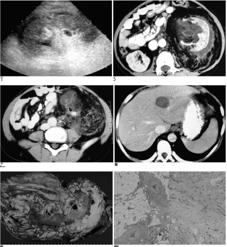
Fig. 1. Perinephric AML in a 13-year-old boy.
A. Ultrasonogram shows an ill-defined hyperechoic lesion in the left renal sinus and perinephric space.
B. Contrast-enhanced CT scan demonstrates a large fat-containing mass and thickened renal capsule with multiple linear strands in the left perinephric space and renal pelvis.
C. Contrast-enhanced CT scan (slightly lower level than B) shows a focal aneurysmal dilatation of intratumoral vessel (arrow) in the left perinephric space.
D. Contrast-enhanced CT scan shows a well-marginated mass of hypoattenuation in the left hepatic lobe. A gun biopsy specimen revealed only a fatty change.
E. The cut surface of the gross specimen shows homogeneously yellowish adipose tissues with thick fibrous capsule and thin fi- brous strands.
F. Pathologic examination reveals mature fat tissues, variable-sized vessels (left), and immature smooth muscle bundles (right) (original magnification, ×100; hematoxylin-eosin staining).
ed by the perirenal fatty mass which, microscopically, was composed of mature fatty tissues, variable-sized blood vessels, and smooth muscle bundles (Fig. 1F).
Immunohistochemical studies using smooth muscle actin, desmin, and oil red-O stain indicated smooth mus- cle components and fatty tissues.
Discussion
An AML is an uncommon benign tumor named for its three constituents: blood vessels, fat, and smooth mus- cle (4). The color of a cut section of the mass varies from yellow to gray, and areas of pure fat, necrosis, and hem- orrhage may be apparent at gross inspection.
Microscopic examination reveals that the tumor consists of mature fat cells, sheets of smooth muscle, and areas of tortuous thick-walled blood vessels. Although one or two of these tissues may predominate, careful examina- tion will generally lead to identification of all three types (5). The histopathologic appearance of renal AMLs is characterized by the fat component, making them - with the exception of rare cases without visible fat - easily identifiable at CT (6).
An AML most often originates from renal parenchy- ma: extrarenal AMLs are rare, though the liver is the most common extrarenal site; other rare sites reported include the spleen, lymph nodes, uterus, vagina, penis, nasal cavity, hard palate, abdominal wall, fallopian tube, spermatic cord, and colon (7). In our case, CT re- vealed multiple, focal fatty hypoattenuated lesions in the left hepatic lobe, but gun biopsy revealed only fatty change. Since, however, the gun biopsy may reveal only one portion of the hepatic mass, the possibility of an he- patic angiomyolipoma cannot be excluded.
AMLs extremely rarely originate from perinephric space rather than the more common renal parenchyma, and only 13 such cases have been reported in the litera- ture (1-3). Laws et al. (8) summarized the features of the first five cases reported: the patients ranged in age from 23 to 64 years, and all but one of the tumors were in the perinephric space within Gerota’s fascia, were solitary, varied from 3 to 11 cm, and showed no renal involvement or tuberous sclerosis.
If an AML involves or originates in the renal sinus, it is difficult to differentiate from other fatty tumors of the perinephric space such as lipomatosis, lipoma, and li-
posarcoma (4). Because of uncertainty as to whether the tumor was in fact benign, previous cases of perinephric AML have usually involved surgical exploration and nephrectomy, and there is still a belief that because of its malignant nature, perinephric liposarcoma should be treated with radical surgery as soon as possible. Murphy et al. (9) suggested that unlike an AML, a retroperitoneal liposarcoma was usually located outside of Gerota’s fas- cia. Wang et al. (3), however, suggested that a retroperi- toneal liposarcoma was typically bulky and extended in- to the perinephric space, and that careful analysis of the CT characteristics of linear vascularity, aneurysmal di- latation of intratumoral vessels, the bridging vessel sign, hematoma, the beak sign, and discrete intrarenal fatty tumors could be helpful in diagnosing perinephric AML.
In our case, CT also demonstrated intratumoral linear vascularity, aneurysmal dilatation of intratumoral ves- sels, and calcifications.
In summary, AML originating in the perinephric space is an exceptionally rare disease entity but AML originating in the perinephric space and renal sinus should be borne in mind as a differential diagnosis when imaging studies show a fat-containing mass in the perinephric and renal sinus.
References
1. Angulo JC, Lopez JI, Carnicero JA, Flores N. Extrarenal retroperi- toneal angiomyolipoma. Urol Int 1994;52:58-60
2. Peh WC, Lim BH, Tam PC. Case report: Perinephric angiomy- olipomas in tuberous sclerosis. Br J Radiol 1994;67:1026-1029 3. Wang LJ, Wong YC, Chen CJ, See LC. Computerized tomography
characteristics that differentiate angiomyolipomas from liposarco- mas in the perinephric space. J Urol 2002;167:490-493
4. Michael JM, Parvati R, Marc PB, et al. Angiomyolipoma of the re- nal sinus: diagnosis by percutaneous biopsy. Urology 2000;55:286 5. Pollack HM, McClennan BL. Clinical Urography, 2nd ed. In Byrn
W, Bernard FK, Benign neoplams of the Renal parenchyma.
Philadelphia: Saunders, 2000;1425-1429
6. Helenon O, Merran S, Paraf F, et al. Unusual fat-containing tu- mors of the kidney: a diagnostic dilimma. RadioGraphics 1997;17:
129
7. Hanna RM, Dahniya MH, Al-Marzouk N, Grexa E. Extrarenal an- giomyolipomas of the perinephric space in tuberose sclerosis.
Australas Radiol 1997;41:339-341
8. Laws SY. Fok M, Shek WH, Ma LT, Wong J. Retroperitoneal ex- trarenal angiomyolipoma. Aust N Z J Surg 1994;64:449-451 9. Murphy DP, Glazier DB, Chenven ES, et al. Extrarenal retroperi-
toneal angiomyolipoma: nonoperative management. J Urol 2000;163:234-235
J Korean Radiol Soc 2002;47:647-650
─ 649 ─
Ho Seob Shin, et al: Angiomyolipoma of the Perinephric Space
─ 650 ─
대한방사선의학회지 2002;47:647-650
신주위공간의 혈관근지방종: 증례 보고1
1동아대학교 의과대학 진단방사선과학교실
2동아대학교 의과대학 병리학교실
신호섭・윤성국・이진화・김찬성・오종영・신태범・이기남・남경진・김대철2
신실질과 달리 신주위공간에서 발생하는 혈관근지방종은 매우 드물다. 이에 저자들은 근치적 신절제술로 확진된 신 주위공간에서 발생한 혈관근지방종 1예를 보고한다. 13세 남자 환아가 5개월간의 좌측 옆구리 통증과 복부팽만을 호 소하였다. 초음파검사와 전산화단층촬영 소견상 좌신을 둘러싸는 신주위공간에서 선상 선음영을 동반한 거대 지방 함 유종괴를 보였다.