• 검색 결과가 없습니다.

Prehypertension and Left Ventricular Diastolic Dysfunction in Middle-Aged Koreans

N/A
N/A
Protected

Academic year: 2022

Share "Prehypertension and Left Ventricular Diastolic Dysfunction in Middle-Aged Koreans"

Copied!
8
0
0

로드 중.... (전체 텍스트 보기)

전체 글

(1)

Korean Circulation Journal

Introduction

Hypertension (HT) is a major global public health burden,

1)

and it is an important risk factor for cardiovascular disease (CVD) and

cerebrovascular accident.

2)

Prehypertensive subjects are at risk of progression to HT, and prehypertension is associated with excessive morbidity and mortality due to CVD.

3)

Echocardiography is an important non-invasive diagnostic tool for investigation of left ventricular function. Left ventricular diastolic dysfunction due to various myocardial diseases occurs frequently in hypertension, and it is a common cause of heart failure in patients with hypertension.
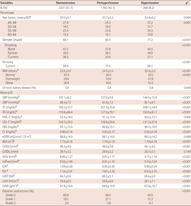
4)

However, few studies have shown an association between diastolic dysfunction grade and blood pressure classification, especially mild hypertension or prehypertension (preHT). We sought to investigate the association between diastolic dysfunction and preHT in apparently healthy middle-aged adults.

Subjects and Methods

Study population and protocol

The present study included 4261 Koreans (male:female ratio 7:3),

Print ISSN 1738-5520 • On-line ISSN 1738-5555

Prehypertension and Left Ventricular Diastolic Dysfunction in Middle-Aged Koreans

Shin Yi Jang, PhD 1 , Sujin Kim, BSN 1 , Chang Kwan Lee, PhD 2 , Eun Jeong Cho, MD 3 , Soo Jin Cho, MD 4 , and Sang-Chol Lee, MD 1

1

Division of Cardiology, Department of Medicine, Heart Vascular Stroke Institute, Samsung Medical Center, Sungkyunkwan University School of Medicine,

2

Department of Nursing, KC University, Seoul,

3

Division of Cardiology, Department of Internal Medicine, Cardiology Clinic, National Cancer Center, Goyang,

4

Health Promotion Center, Samsung Medical Center, Sungkyunkwan University School of Medicine, Seoul, Korea

Background and Objectives: Left ventricular diastolic dysfunction is known to be a marker of myocardial damage, in particular myocardial fibrosis resulting from hypertension (HT). However, few studies have shown an association between the grade of diastolic dysfunction and blood pressure classification. We investigated the association between diastolic dysfunction and prehypertension (preHT) in apparently healthy adults who underwent routine health examinations.

Subjects and Methods: The study sample included 4261 Koreans, 45 to 64 years of age with no previous history of HT, diabetes mellitus, malignancy, proven coronary artery disease, or valvular heart disease based on echocardiography, who underwent routine health examinations including echocardiography. The subjects were classified into three groups based on resting blood pressure:

prehypertensive, hypertensive, and normotensive.

Results: The prevalence of preHT in our study was 42.1%. After adjusting for age, gender, smoking status, alcohol consumption, fasting blood sugar, serum lipid profile, and body mass index, left ventricular diastolic dysfunction grades 1 and 2 were significantly more frequent in subjects with preHT (odds ratio [OR] 1.66 [95% confidence interval {CI} 1.40-1.96] and 1.37 [95% CI 0.95-1.97], respectively).

When analyzed according to gender, the increased OR was especially notable in males.

Conclusion: Left ventricular diastolic dysfunction appears to be significantly associated with preHT in Korean middle-aged males. (Korean Circ J 2016;46(4):536-541)

KEY WORDS: Prehypertension; Ventricular dysfunction, left; Echocardiography; Middle-aged.

Received: July 15, 2015

Revision Received: October 13, 2015 Accepted: November 26, 2015

Correspondence: Sang-Chol Lee, MD, Division of Cardiology, Department of Medicine, Imaging Center, Heart Vascular Stroke Institute, Samsung Medical Center, Sungkyunkwan University School of Medicine, 81 Irwon-ro, Gangnam-gu, Seoul 06351, Korea

Tel: 82-2-3410-3419, Fax: 82-2-2148-7973 Email: [email protected]

• The authors have no financial conflicts of interest.

This is an Open Access article distributed under the terms of the Creative Commons Attribution Non-Commercial License (http://creativecommons.

org/licenses/ by-nc/3.0) which permits unrestricted non-commercial use,

distribution, and reproduction in any medium, provided the original work

is properly cited.

(2)

45 to 64 years of age with no previous history of HT, diabetes mellitus, malignancy, proven coronary artery disease, or valvular heart disease, who underwent routine health examinations including echocardiography at the Health Promotion Center of Samsung Medical Center from January 2009 to June 2010. This study was approved by the Samsung Medical Center Institutional Review Board, and the requirement for obtaining informed consent was waived for this retrospective study.

Systolic blood pressure (SBP) and diastolic blood pressure (DBP) were measured after the subject had rested for ten minutes in the sitting position. Blood samples were collected from the antecubital vein after overnight nil per os (NPO). Total cholesterol (TC), triglyceride (TG), high density lipoprotein-cholesterol (HDL-C), low density lipoprotein-cholesterol (LDL-C), fasting blood sugar (FBS), and serum creatinine (Cr) levels were measured. The estimated glomerular filtration rate (Egfr) was calculated using the Chronic Kidney Disease Epidemiology Collaboration(CKD EPI) equation.

Information on the presence of HT, type 2 diabetes mellitus, and history of smoking and alcohol consumption was obtained from personal questionnaires.

Echocardiographic evaluation

Subjects underwent comprehensive transthoracic echocardiography (2-dimensional, M-mode, and tissue Doppler echocardiography) using commercially available equipment (Vivid 7, GE Medical Systems, Milwaukee, WI, USA). Well-trained echocardiographers interpreted all echocardiograms and obtained the following measurements: left ventricle internal dimension measured at end diastole (LVIDd), left ventricle internal dimension measured at end systole (LVIDs), interventricular septal wall thickness at end diastole (IVSd), left ventricle posterior wall thickness at end diastole (LVPWd), and left ventricular mass index (LVMI), left ventricular mass divided by body surface area (BSA), height (cm)

0.725

×weight

(kg)

0.425

×0.007184 measured with M-mode; early diastolic mitral

inflow velocity/late diastolic mitral inflow velocity (E/A), early diastolic mitral inflow velocity/peak early diastolic mitral septal annular velocity (E/e’) measured by flow and tissue Doppler analysis;

left ventricular ejection fraction (LVEF) measured on 2-dimensional (2-D) echocardiography using biplane Simpson’s method; and left atrial volume index (LAVI) measured on 2-D echocardiography calculated by the area-length method and indexed to BSA. Left ventricular diastolic dysfunction was categorized as normal, grade 1, grade 2, grade 3, or grade 4 according to the guidelines of the American Society of Echocardiography. All echocardiographic data were measured and obtained in accordance with the American Society of Echocardiography guidelines and the European Association of Echocardiography guidelines.

5)6)

Unless otherwise

specified in this paper, diastolic dysfunction referenced herein refers to left ventricular diastolic dysfunction.

Blood pressure classification

PreHT was defined as SBP of 120-139 mmHg and/or DBP 80-89 mmHg according to the Seventh Report of the Joint National Committee (JNC7)

7)

criteria and without the use of any anti- hypertensive drug and/or non-pharmacological interventions. HT was defined as SBP≥140 mmHg and/or DBP≥90 mmHg and/or with active antihypertensive drug therapy. If subjects had SBP≥140 mmHg and/or DBP≥90 mmHg, the blood pressure was retaken, and the mean blood pressure value was used. Normotension was defined as SBP<120 mmHg and DBP<80 mmHg without the use of antihypertensive medication. The prevalence of preHT in our study was 42.1%. This value was slightly lower than that in previous reports among Koreans, in which Jang et al. showed that the prevalence of preHT was over 50% in general Korean middle-aged adults.

8)

Obesity

Body mass index (BMI) was calculated as weight in kilograms divided by the square of height in meters. Obesity was defined as BMI≥25.

9)

Health behavioral factors

Subjects were identified as current smokers if they had smoked within one year of the survey date and as former smokers if they had not smoked for at least one year before the survey date.

Alcohol drinking was defined as current drinking within one year of the survey date as reported in the health interview questionnaire.

Statistical analyses

The general characteristics analyses among HT, preHT and normotension groups were performed using one-way analysis of variance (ANOVA) and Bonferroni method in multiple comparisons testing for the continuous variables and χ

2

tests for the categorical variables. Multinomial logistic regression analysis was conducted to show the associations among HT, preHT and normotension and diastolic dysfunction adjusted for variables including age, gender, smoking, BMI, TG, HDL-C, and FBS.

This study was approved by the Samsung Medical Center Institutional Review Board, and the requirement for obtaining informed consent was waived for this retrospective study (IRB File Number 2009-09-088).

Results

Clinical characteristics of the subjects are summarized in Table 1.

(3)

Table 1. General characteristics, health behaviors, and obesity grade of participants compared by blood pressure classification (N=4261)

Variables Normotensive Prehypertensive Hypertensive p*

N (%) 2201 (51.7) 1792 (42.1) 268 (6.2)

Percentage

Age (years; mean±SD) 53.2±5.1 53.7±5.2 53.4±5.2 0.004

45-49 50-54 55-59 60-64

27.9 34.5 22.4 15.2

24.3 34.0 23.6 18.1

27.2 31.7 24.3 16.8

0.061

Gender (male) 68.1 80.4 77.2 <0.001

Smoking None Former Current

47.2 26.5 26.3

37.9 36.2 25.9

40.0 36.9 23.1

<0.001

Drinking

Current 59.4 71.4 68.1 <0.001

BMI obesity Normal Overweight Obese

23.5±2.5 43.5 29.6 26.9

24.5±2.5 26.5 33.0 40.5

25.2±2.5 20.2 27.6 52.2

<0.001

<0.001

Chronic kidney disease (%) 0.6 0.8 0.8 0.645

Mean±SD

SBP (mmHg) 107.1±8.3 127.0±9.9 149.5±12.9 <0.001

DBP (mmHg) 68.4±7.5 81.8±7.2 92.1±9.7 <0.001

TC (mg/dL) 198.3±33.7 201.9±33.6 208.7±34.9 <0.001

TG (mg/dL) 119.9±66.4 137.0±79.1 150.5±81.7 <0.001

HDL-C (mg/dL) 52.5±14.0 51.3±13.4 50.5±12.1 0.008

LDL-C (mg/dL) § 124.3±29.2 126.8±29.6 131.3±31.9 <0.001

FBS (mg/dL) 93.1±13.4 96.8±15.1 99.7±19.5 <0.001

Cr (mg/dL) 0.90±0.16 0.93±0.17 0.92±0.16 <0.001

eGFR (mL/min/1.73 m

2

) 88.6±14.0 88.1±14.0 88.3±14.2 0.499

BSA (m

2

) § 1.73±0.16 1.79±0.16 1.79±0.16 <0.001

LVIDd (mm) § 48.3±4.0 49.0±3.8 49.1±4.0 <0.001

LVIDs (mm) 28.7±3.2 28.9±3.2 28.7±3.3 0.140

IVSd (mm) § 8.66±1.27 8.97±1.17 9.11±1.16 <0.001

LVPwd (mm) 8.55±1.06 8.91±1.10 9.10±1.04 <0.001

E/A 1.04±0.26 0.95±0.26 0.90±0.24 <0.001

E/e’ 7.34±2.07 7.87±2.35 8.53±3.75 <0.001

LVEF (%) § 64.7±5.0 65.2±5.1 65.6±5.0 <0.001

LAVI (mL/m

2

)

||

26.5±6.5 26.6±6.6 28.1±7.1 <0.001

LVMI (g/m

2

) 81.8±16.4 84.9±15.9 87.8±16.7 <0.001

Diastolic dysfunction (%) Grade 0

Grade 1 Grade 2

80.9 16.2 2.9

69.0 27.1 3.9

62.0 31.3 6.7

<0.001

2

-test or one way analysis of variance (ANOVA) for prehypertensive (preHT) vs. hypertensive (HT) vs. normotensive group; p was calculated by one way ANOVA test and Bonferroni multiple comparisons tests for continuous variables (p<0.05), preHT vs. normotensive, preHT vs. normotensive, HT vs. normo- tensive, and preHT vs. HT, § preHT vs. normotensive, HT vs. normotensive,

||

HT vs. normotensive and preHT vs. HT. SD: standard deviation, BMI: body mass in- dex, SBP: systolic blood pressure, DBP: diastolic blood pressure, TC: total cholesterol, TG: triglyceride, HDL-C: high density lipoprotein-cholesterol, LDL-C:

low density lipoprotein-cholesterol, FBS: fasting blood sugar, Cr: creatinine, eGFR: estimated glomerular filtration rate, BSA: body surface area, LVIDd: left

ventricular internal dimension diastolic, LVIDs: left ventricular internal dimension systolic, IVSd: interventricular septal wall thickness at end diastole, LVP-

wd: LV posterior wall thickness at end diastole, E: early diastolic mitral inflow velocity, A: late diastolic mitral inflow velocity, E’: early diastolic mitral annu-

lar velocity, LVEF: left ventricular ejection fraction by Simpson’s biplane method, LAVI: left atrial volume index, LVMI: left ventricular mass index calculated

by the American Society of Echocardiography guidelines

(4)

Approximately 40% of our subjects were prehypertensive. The proportion of diastolic dysfunction grade 1 or 2 was significantly higher in preHT (31.0% overall) and HT (38.0% overall) groups compared to the normotensive group (19.1% overall) (p<0.01). Male gender was more prevalent in the preHT group compared to normotensive or HT groups (p<0.001). The proportion of current smokers, current alcohol drinkers, and obese subjects was significantly different among the 3 groups (p<0.001). The mean values of SBP, DBP, TC, TG, LDL-C, FBS, LVIDd, IVSd, LVPwd, E/e’, LVEF, LAVI, and LVMI were higher in patients with HT than in subjects with normotension or preHT (p<0.001). The mean values of HDL-C and Cr were lower in the HT group than in the normotensive or preHT group (p=0.008, p<0.001, respectively).

Data showed that the statistically significant adjusted odds ratio (OR) for diastolic dysfunction with preHT was as follows: male gender, 1.59 (95% confidence interval [CI] 1.30-1.95); current smoking, 0.80 (95% CI 0.66-0.98); BMI, 1.14 (95% CI 1.11-1.18); and the OR was 1.66 (95% CI 1.40-1.96) for grade 1 diastolic dysfunction and 1.37 (95% CI 0.95-1.97) for grade 2 diastolic dysfunction, respectively. Particularly, in males, the adjusted OR for diastolic dysfunction with preHT was 1.74 (95% CI 1.44-2.10) for grade 1 diastolic dysfunction and 1.31 (95% CI 0.84-2.04) for grade 2 diastolic dysfunction. In females, the adjusted OR for diastolic dysfunction with preHT was 1.32 (95% CI 0.91-1.90) for grade 1 diastolic dysfunction and 1.47 (95% CI 0.76- 2.76) for grade 2 diastolic dysfunction, respectively (Table 2).

Discussion

Our overall findings showed that diastolic dysfunction appears to be significantly associated with preHT in apparently healthy middle-aged Korean population. Results of the present study correspond well with those in a previous study, which showed that preHT was associated with LV diastolic function.

10)

The results of the present study are also consistent with another report on diastolic dysfunction and blood pressure classification in healthy Turkish adults, which showed that diastolic function was slightly impaired in prehypertensive subjects.

11)

In other reports, blood pressure control is known to improve diastolic dysfunction in hypertensive subjects.

12-14)

The association between diastolic dysfunction and HT has been reported previously.

15-17)

Our data showed a relatively higher grade of diastolic dysfunction in healthy middle- aged prehypertensive subjects. As diastolic dysfunction is an important pathophysiological intermediate between HT and heart failure, preventive strategies are necessary to reduce morbidity in prehypertensive subjects who show a risk for diastolic dysfunction.

Examination of heart function using echocardiography has been recommended for assessing target organ damage in hypertensive patients, but our research suggests that echocardiographic examination should be extended to include prehypertensive subjects, and a more aggressive lifestyle change and/or treatment for middle-aged prehypertensive subjects may be recommended according to their heart function.

To date, few prior studies regarding the association between Table 2. Odds ratios (and 95% confidence interval) by blood pressure classification*

Variables All Male Female

Prehypertension Hypertension Prehypertension Hypertension Prehypertension Hypertension Age 1.01 (0.99, 1.02) 0.98 (0.96-1.01) 0.99 (0.98, 1.01) 0.96 (0.93, 0.99) 1.04 (1.01, 1.07) 1.05 (0.99, 1.11)

Gender (male vs. female) 1.59 (1.30, 1.95) 1.11 (0.76, 1.60) - - - -

Smoking None Former Current

1.0

1.09 (0.90, 1.31) 0.80 (0.66, 0.98)

1.0

1.11 (0.76, 1.60) 0.66 (0.44, 1.01)

1.0

1.14 (0.94, 1.38) 0.82 (0.67, 1.01)

1.0

1.17 (0.80, 1.72) 0.67 (0.44, 1.03)

1.0

0.47 (0.15, 1.47) 0.48 (0.15, 1.58)

1.0 - 0.66 (0.08, 5.57) BMI 1.14 (1.11, 1.18) 1.25 (1.18, 1.32) 1.13 (1.01, 1.17) 1.13 (1.10, 1.17) 1.15 (1.08, 1.21) 1.24 (1.12, 1.38) TG 1.01 (1.001, 1.02) 1.01 (1.002, 1.02) 1.01 (1.001, 1.02) 1.01 (1.001, 1.02) 1.01 (1.001, 1.02) 1.01 (0.99, 1.02) HDL-C 1.01 (1.008, 1.02) 1.01 (1.005, 1.02) 1.01 (1.009, 1.02) 1.02 (1.01, 1.03) 1.01 (1.001, 1.02) 1.01 (0.97, 1.02) FBS 1.01 (1.005, 1.02) 1.02 (1.01, 1.03) 1.01 (1.004, 1.01) 1.01 (1.004, 1.02) 1.01 (1.002, 1.03) 1.02 (0.99, 1.04) Diastolic dysfunction

Grade 0 Grade 1 Grade 2

1.0

1.66 (1.40, 1.96) 1.37 (0.95, 1.97)

1.0

2.10 (1.54, 2.87) 2.51 (1.41, 4.44)

1.0

1.74 (1.44, 2.10) 1.31 (0.84, 2.04)

1.0

2.24 (1.58, 3.18) 2.96 (1.52, 5.76)

1.0

1.32 (0.91, 1.90) 1.47 (0.76, 2.76)

1.0

1.54 (0.79, 3.02) 1.47 (0.46, 4.69)

*Estimated by multiple logistic regression analysis of the variables indicated in the above table. BMI: body mass index, TG: triglyceride, HDL-C: high density

lipoprotein-cholesterol, FBS: fasting blood sugar

(5)

diastolic dysfunction and preHT according to gender have been presented. We showed an association between diastolic dysfunction and preHT, which was especially prominent in males.

This is consistent with previous reports

18)

showing an overall higher incidence of diastolic dysfunction in healthy adult men compared to women. According to previous study

19)

on diastolic dysfunction in patients with HT which showed that some of the echocardiographic parameters for diastolic function demonstrated a trend towards more impaired diastolic function in male hypertensives, this finding can be explained by the relatively larger size of the mitral annulus of the heart resulting in decreased ventricular filling velocities and deceleration time. Another explanation for this finding, at least in middle-aged people, is the protective effect of female hormones, as indices of left ventricular filling showed a significant decline with age in premenopausal and postmenopausal women in a previous study.

20)

However, as the design of this study was retrospective and our subjects were people who voluntarily visited our health promotion center for health examinations, a well-designed, age- and gender-matched prospective echocardiographic study may be needed for classification.

The prevalence of LV diastolic dysfunction in the normotensive group in our study was approximately 20%, and an additional analysis showed a higher prevalence of LV diastolic dysfunction in the older age group (Supplementary Tables 1 and 2 in the online- only Data Supplement). As already known, LV diastolic dysfunction can develop as the age increases in the absence of heart disease including HT.

21)22)

In addition, higher arterial stiffness has also been reported to be associated with worse LV diastolic function.

23)24)

Meanwhile, there was a significant difference in BMI among the groups, and this may have been one of the mechanisms that may be associated with diastolic dysfunction in prehypertensive subjects.

Fenk et al.

25)

showed that in severe obesity, weight reduction was associated with improved LV diastolic function, and the Northern Manhattan Study, a population-based prospective cohort study in the US, showed that increased BMI was associated with worse LV diastolic function.

26)

Although prehypertensive subjects are at risk of progression to HT,

27)

pharmacological therapy in preHT subjects without comorbidities remains debatable with respect to whether drug treatment should be initiated. However, as our data suggest, these prehypertensive subjects are at higher risk of developing early and significant left ventricular diastolic dysfunction, and they may need to modify their health behavior such as reducing sodium intake, losing weight, and participating in regular physical activity.

Limitations of the present study

This study has several limitations. Firstly, this was a retrospective

study performed on subjects who voluntarily visited our center for health examinations. Thus, we could not observe any causal relationships between diastolic dysfunction and preHT. Secondly, because echocardiography was performed in middle-aged subjects who wanted a detailed evaluation of CVD, our study may reflect some level of selection bias. However, selection bias in this study could be low because we excluded patients with previous history of HT, diabetes mellitus, or malignancy based on medical history and those with proven coronary artery disease or valvular heart disease. Finally, the proportion of males was higher than that of females in our study. To compensate for this limitation, logistic regression was performed after adjusting for gender. Although the study was conducted at a single center with asymptomatic subjects who did not have CVD, we believe that this study in a substantially large group of subjects accurately represents Korean middle-aged adults who underwent echocardiography.

Conclusions

This study showed that diastolic dysfunction is significantly associated with preHT in middle-aged Korean males.

Acknowledgments

This study was supported by Samsung Medical Center grant [#SMO113-15-01].

SYJ made study design, analyzed statistical data, discussed interpretation, wrote this paper, and took full responsibility for this paper. SJK discussed interpretation. CKL discussed interpretation.

EJC discussed interpretation. SJC discussed interpretation. SCL made study design, discussed interpretation and took full responsibility for this paper. All of the authors are responsible for the intellectual content of the manuscript.

Supplementary Materials

The online-only Data Supplement is available with this article at http://dx.doi.org/10.4070/kcj.2016.46.4.536.

References

1. Lawes CM, Vander Hoorn S, Rodgers A; International Society of Hypertension. Global burden of blood-pressure-related disease, 2001. Lancet 2008;371:1513-8.

2. Julius S, Nesbitt SD, Egan BM, et al. Feasibility of treating

(6)

prehypertension with an angiotensin-receptor blocker. N Engl J Med 2006;354:1685-97.

3. Mainous AG 3rd, Everett CJ, Liszka H, King DE, Egan BM.

Prehypertension and mortality in a nationally representative cohort.

Am J Cardiol 2004;94:1496-500.

4. Schulz R, Güther B, Mutert S, Kuhn J. Obesity in Bavarian adolescents: prevalence in trend, sociodemographic structural features and subjective health. Gesundheitswesen 2010;72:88-98.

5. Lang RM, Bierig M, Devereux RB, et al. Recommendations for chamber quantification: a report from the American Society of Echocardiography’s Guidelines and Standards Committee and the Chamber Quantification Writing Group, developed in conjunction with the European Association of Echocardiography, a branch of the European Society of Cardiology. J Am Soc Echocardiogr 2005;18:1440-63.

6. Nagueh SF, Appleton CP, Gillebert TC, et al. Recommendations for the evaluation of left ventricular diastolic function by echocardiography.

Eur J Echocardiogr 2009;10:165-93.

7. Chobanian AV, Bakris GL, Black HR, et al. The seventh report of the Joint National Committee on prevention, detection, evaluation, and treatment of high blood pressure: the JNC 7 report. JAMA 2003;289:2560-72.

8. Jang SY, Ju EY, Choi S, et al. Prehypertension and obesity in middle- aged Korean men and women: the third Korea national health and nutrition examination survey (KNHANES III) study. J Public Health (Oxf) 2012;34:562-9.

9. Weisell RC. Body mass index as an indicator of obesity. Asia Pac J Clin Nutr 2002;11 Suppl 8:S681-4.

10. Ahn HS, Kim SJ, Kim MK, et al. The difference of left ventricular hypertrophy and the diastolic function between prehypertensive and normotensive. Korean Circ J 2006;36:437-42.

11. Erdogan D, Caliskan M, Yildirim I, et al. Effects of normal blood pressure, prehypertension and hypertension on left ventricular diastolic function and aortic elastic properties. Blood Press 2007;16:114-21.

12. Almuntaser I, Mahmud A, Brown A, et al. Blood pressure control determines improvement in diastolic dysfunction in early hypertension.

Am J Hypertens 2009;22:1227-31.

13. Celentano A, Crivaro M, Perticone F, et al. Anti-hypertensive effect of manidipine: 24 hours monitoring evaluation and Doppler- echocardiographic remarks. Blood Press Suppl 1996;5:29-35.

14. Brilla CG, Funck RC, Rupp H. Lisinopril-mediated regression of myocardial fibrosis in patients with hypertensive heart disease.

Circulation 2000;102:1388-93.

15. Erdogan D, Caliskan M, Gullu H, et al. Aortic elastic properties and left ventricular diastolic function in white-coat hypertensive individuals. Blood Press Monit 2006;11:191-8.

16. Bhatia RS, Tu JV, Lee DS, et al. Outcome of heart failure with preserved ejection fraction in a population-based study. N Engl J Med 2006;355:260-9.

17. Almuntaser I, Brown A, Murphy R, et al. Comparison of echocardiographic measures of left ventricular diastolic function in early hypertension.

Am J Cardiol 2007;100:1771-5.

18. Li X, Cao RX, Lv RR, Cao WH. Study on the trend of obesity prevalence among primary and middle school students in Beijing, from 1985 to 2005. Zhonghua Liu Xing Bing Xue Za Zhi 2008;29:469- 72.

19. Regenauer A. Does the obesity “epidemic” stop the life expectancy trend? Versicherungsmedizin 2008;60:32-3.

20. Eisenmann JC. Insight into the causes of the recent secular trend in pediatric obesity: common sense does not always prevail for complex, multi-factorial phenotypes. Prev Med 2006;42:329-35.

21. Zabalgoitia M, Ur Rahman SN, Haley WE, et al. Role of left ventricular hypertrophy in diastolic dysfunction in aged hypertensive patients. J Hypertens 1997;15:1175-9.

22. Miller TR, Grossman SJ, Schectman KB, Biello DR, Ludbrook PA, Ehsani AA. Left ventricular diastolic filling and its association with age. Am J Cardiol 1986;58:531-5.

23. Russo C, Jin Z, Palmieri V, et al. Arterial stiffness and wave reflection:

sex differences and relationship with left ventricular diastolic function. Hypertension 2012;60:362-8.

24. Jang SY, Ju EY, Huh EH, Kim JH, Kim DK. Determinants of brachial- ankle pulse wave velocity and carotid-femoral pulse wave velocity in healthy Koreans. J Korean Med Sci 2014;29:798-804.

25. Fenk S, Fischer M, Strack C, et al. Successful weight reduction improves left ventricular diastolic function and physical performance in severe obesity. Int Heart J 2015;56:196-202.

26. Russo C, Jin Z, Homma S, et al. Effect of obesity and overweight on left ventricular diastolic function: a community-based study in an elderly cohort. J Am Coll Cardiol 2011;57:1368-74.

27. Vasan RS, Larson MG, Leip EP, Kannel WB, Levy D. Assessment of

frequency of progression to hypertension in non-hypertensive

participants in the Framingham Heart Study: a cohort study. Lancet

2001;358:1682-6.

(7)

Supplementary Table 1. General characteristics, health behaviors and, obese grade of participants compared by blood pressure classification (N=4261)

Variables Grade 0 Grade 1 Grade 2 p*

N (%) 3185 (74.8) 926 (21.7) 150 (3.5)

Percentage

Age (years, mean±SD) 52.5±4.9 56.1±4.9 55.6±4.7 <0.001

45-49 50-54 55-59 60-64

31.5 36.4 19.9 12.2

11.4 25.7 32.6 32.3

9.3 36.0 29.3 25.4

<0.001

Gender (male) 73.1 77.4 67.3 0.005

Smoking Non Former Current

42.6 29.8 27.6

42.5 36.2 21.3

48.0 20.6 31.3

<0.001

Drinking

Current 64.9 65.2 63.5 0.926

BMI obesity Normal Overweight Obese

23.8±2.6 38.4 29.9 31.7

24.5±2.3 24.1 34.6 41.3

24.8±2.7 25.3 31.3 43.4

<0.001

<0.001

BP classification Normotensive preHT HT

56.0 38.8 5.2

38.4 52.5 9.1

42.0 46.0 12.0

<0.001

Chronic kidney disease (%) 0.47 1.08 2.00 0.012

Mean±SD

SBP (mmHg) 117.0±15.2 122.7±16.1 124.8±17.6 <0.001

DBP (mmHg) 74.5±10.7 79.0±10.3 76.3±11.0 <0.001

TC (mg/dL) § 199.5±33.7 203.9±33.8 199.6±35.2 0.002

TG (mg/dL) § 125.4±70.7 140.9±81.1 131.4±23.5 <0.001

HDL-C (mg/dL) § 52.4±13.8 50.0±12.7 51.8±13.3 0.008

LDL-C (mg/dL) § 124.8±29.5 129.5±29.5 123.6±30.4 <0.001

FBS (mg/dL) 93.9±13.3 98.4±18.0 96.9±15.3 <0.001

Cr (mg/dL) 0.91±0.16 0.93±0.16 0.89±0.30 <0.001

eGFR (mL/min/1.73m

2

) 88.7±13.8 86.4±14.1 90.9±16.7 0.499

BSA (m

2

) 1.76±0.16 1.77±0.15 1.75±0.17 0.028

LVIDd (mm) 48.6±3.9 48.6±4.0 49.1±4.1 0.267

LVIDs (mm) 28.7±3.2 28.8±3.2 28.5±3.3 0.603

IVSd (mm)

||

8.74±1.16 8.99±1.18 9.52±2.32 <0.001

LVPwd (mm) 8.66±1.09 8.92±1.07 9.06±1.22 <0.001

E/A 1.08±0.22 0.68±0.13 1.09±0.25 <0.001

E/e’

7.50±1.9 7.40±2.0 12.0±5.6 <0.001

LVEF (%)

64.9±5.0 64.9±5.0 66.1±5.1 0.011

LAVI (mL/m2)

||

26.5±6.1 25.4±6.4 36.8±8.0 <0.001

LVMI (g/m2)

||

82.7±16.0 84.7±16.3 91.9±19.1 <0.001

2

-test or one way analysis of variance (ANOVA) for prehypertensive (preHT) vs hypertensive (HT) vs. normotensive group; p-value was calculated by one way ANOVA test and Bonferroni multiple comparisons tests for continuous variables (p-value<0.05), preHT vs. normotensive and HT vs. normotensive,

‡ preHT vs. normotensive and preHT vs. HT, § preHT vs. normotensive,

||

preHT vs. normotensive, HT vs. normotensive, and preHT vs. HT

HT vs. normotensive and preHT vs. HT. SD: standard deviation, BMI: body mass index, SBP: systolic blood pressure, DBP: diastolic blood pressure, TC: total cholesterol, TG: triglyc- eride, HDL-C: high density lipoprotein-cholesterol, LDL-C: low density lipoprotein-cholesterol, FBS: fasting blood sugar, Cr: creatinine, eGFR: estimated glo- merular filtration rate, BSA: body surface area, LVIDd: left ventricular internal dimension diastolic, LVIDs: left ventricular internal dimension systolic, IVSd:

interventricular septal wall thickness at end diastole, LVPwd: LV posterior wall thickness at end diastole, E: early diastolic mitral inflow velocity, A: late dia-

stolic mitral inflow velocity, E’: early diastolic mitral annular velocity, LVEF: left ventricular ejection fraction by Simpson’s biplane method, LAVI: left atrial

volume index, LVMI: left ventricular mass index calculated by the American Society of Echocardiography guidelines

(8)

Supplementary Table 2. Table distribution of diastolic dysfunction by age group (p<0.001) Left ventricle

diastolic dysfunction

Age group (years)

45-49 (%) 50-54 (%) 55-59 (%) 60-64 (%)

All (n=4261) n=1124

26.4 n=1453

34.1 n=981

23.0 n=703

16.5

Normal 89.3 79.9 64.7 54.8

Grade 1 9.4 16.4 30.8 39.8

Grade 2 1.3 3.7 4.5 5.4

Normotension (n=2201) n=616

28.0 n=759

34.5 n=493

22.4 n=333

15.1

Normal 92.5 85.5 73.6 60.1

Grade 1 6.4 11.7 23.4 33.9

Grade 2 1.1 2.8 3.0 6.0

수치

Table 1. General characteristics, health behaviors, and obesity grade of participants compared by blood pressure classification (N=4261)

참조

관련 문서

In this study, we compared the concentration of total cholesterol (CHOL), high density lipoprotein (HDL) cholesterol, low density lipoprotein (LDL) cholesterol and

The low density lipoprotein cholesterol (LDL), high density lipoprotein cholesterol (HDL), total cholesterol (TC), triglyceride (TG), systolic blood pressure (SBP),

Fasting serum levels of glucose, insulin, triglyceride, total cholesterol (TC), low density lipoprotein cholesterol (LDL-C), high density lipoprotein cholesterol (HDL-C), leptin

Comparison of serum total cholesterol (TC), triglyceride (TG), high-density lipoprotein cholesterol (HDL-C), and low-density lipoprotein cholesterol (LDL-C) according

In conclusiosn, significant relationships of reflux esophagitis with obesity, low high-density lipoprotein (HDL) cholesterol, high triglyceride, high BP, and elevated fasting

BMI, body mass inde; DBP, diastolic blood pressure; DM, diabetes mellitus; FBS, fasting blood glucose; HDL-C, high-density lipoprotein cholesterol; HOMA-IR,

BMI, body mass index; DBP, diastolic blood pressure; FPG, fasting plasma glucose; HDL, high density lipoprotein cholesterol; LDL, low density lipoprotein

BMI, body mass index; BP, blood pressure; LDL-C, low density lipoprotein cholesterol; HDL-C, high density lipoprotein cholesterol; HbA1c, gly- cated hemoglobin; HOMA-IR,