INTRODUCTION
Gastroesophageal reflux disease (GERD) is a major clinical problem in Western countries. Several recent endoscope-based studies have suggested that the overall prevalence of reflux esophagitis (RE) in Western countries was around 10-20% (1). In contrast, GERD has traditionally been considered to be less common in Asia (2). However, more recent studies suggest that the prevalence of GERD in Asia is increasing. In Japan, the overall prevalence of RE among the adult pop-ulation is roughly 16% (3). In Taiwan, the prevalence of RE in patients evaluated for upper gastrointestinal tract symp-toms is 15% (4). These findings are similar to those report-ed in the West.
RE has been considered fairly rare among ethnic Koreans. However, recent studies have shown that the incidence is in-creasing in the Korean population. The prevalence of RE in subjects undergoing a routine check-up was reported to 2.36% in 1993 (2), and 3.4% in 1997 (5). In 1999, the prevalence of RE was found to be 5.3% in subjects with gastrointestinal symptoms (6). The incidence is expected to increase not only due to developments in endoscopic examination and increas-ing awareness of the condition, but also because of changes in
preference to a more Westernized diet, as well as changes in lifestyle. However, despite awareness of the increasing preva-lence of RE, few studies have been performed to investigate the risk factors of RE in Korea.
This study was performed to estimate the prevalence of RE in healthy Korean subjects and to elucidate the differences in clinical characteristics between subjects with RE and those without.
MATERIALS AND METHODS
Subject population
We retrospectively examined the medical records of healthy subjects who were examined by endoscopy in a routine check-up program of a single university hospital from October 2004 to September 2005. A total of 6,082 subjects were enrolled in the study. Because this was a retrospective study, reflux sy-mptoms such as heartburn, dyspepsia, epigastric pain, and belching were not investigated. Evaluation about smoking and alcohol drinking was performed. The presence of
Heli-cobacter pylori (H. pylori) infection was diagnosed by rapid ure-197
Hyun Joo Song, Ki-Nam Shim, Su Jin Yoon, Seong-Eun Kim, Hee Jung Oh, Kum Hei Ryu, Chang Yoon Ha, Hye Jung Yeom, Ji Hyun Song, Sung-Ae Jung, and Kwon Yoo
Department of Internal Medicine, Ewha Medical Research Institute, School of Medicine, Ewha Womans University, Seoul, Korea
Address for correspondence Ki-Nam Shim, M.D.
Department of Internal Medicine, Ewha Womans University School of Medicine, 911-1 Mok-dong, Yangcheon-gu, Seoul 158-710, Korea Tel : +82.2-2655-2076, Fax : +82.2-2650-2632 E-mail : [email protected]
DOI: 10.3346/jkms.2009.24.2.197
The Prevalence and Clinical Characteristics of Reflux Esophagitis in
Koreans and Its Possible Relation to Metabolic Syndrome
The prevalence of reflux esophagitis is increasing in Korea. To estimate the preva-lence and clinical characteristics of reflux esophagitis in healthy subjects, we retro-spectively examined the medical records of healthy subjects undergoing a routine check-up from October 2004 to September 2005. A total of 6,082 (3,590 men, mean age 44±10 yr) subjects were enrolled in this study. The prevalence of reflux esophagi-tis in healthy subjects was 10.5%. According to the univariate analysis, male sex (odds ratio [OR] 3.49, 95% confidence interval [CI] 2.84-4.30), smoking history (OR 1.91, 95% CI 1.60-2.28), body mass index (BMI) >30 kg/m2(OR 2.13, 95% CI 1.37-3.33), total cholesterol >250 mg/dL (OR 1.50, 95% CI 1.05-2.14), low-density lipopro-tein (LDL) cholesterol ≥160 mg/dL (OR 1.52, 95% CI 1.08-2.14), triglyceride ≥150 mg/dL (OR 1.92, 95% CI 1.61-2.30), high blood pressure (BP) (OR 1.46, 95% CI 1.20-1.76), and fasting glucose ≥110 mg/dL (OR 1.45, 95% CI 1.13-1.86) were significantly associated with reflux esophagitis (all p<0.05). However, age, alcohol drinking and Helicobacter pylori infection were not associated with reflux esophagi-tis. In conclusiosn, significant relationships of reflux esophagitis with obesity, low high-density lipoprotein (HDL) cholesterol, high triglyceride, high BP, and elevated fasting glucose suggested that reflux esophagitis might represent the disease spec-trum of the metabolic syndrome.
Key Words : Esophagitis, Peptic; Endoscopy; Metabolic Syndrome
Received : 30 September 2007 Accepted : 26 June 2008
ase test (Asan Helicobacter test, Asan pharm.co., LTD., Seoul, Korea) or histologic findings from the biopsy specimens. The esophagogastroduodenoscopy (EGD) was a component of a complete medical examination that includes routine studies of blood, urine, stool, and an ultrasound of the abdomen. The body mass index (BMI) was calculated using the following formula: BMI=weight (kg)/height2(m2). Data for blood
pres-sure (BP), total cholesterol, low-density lipoprotein (LDL) cholesterol, high-density lipoprotein (HDL) cholesterol, tri-glyceride, and fasting glucose were also collected. High BP was defined as ≥130/85 mmHg or documented use of anti-hypertensive therapy according to National Cholesterol Edu-cation Program (NCEP) criteria (7).
Metabolic syndrome was defined based on the World Health Organization (WHO) criteria and NCEP criteria. Under the WHO criteria, the diagnosis of metabolic syndrome can be made in subjects with type 2 DM, impaired glucose tolerance, or insulin resistance, and also requires at least two of the fol-lowing four components: 1) hypertension, either treated with medication or ≥160/90 mmHg untreated; 2) dyslipidemia with elevated plasma triglyceride (≥150 mg/dL) and/or low HDL (<35 mg/dL in men, <39 mg/dL in women); 3) obe-sity with BMI ≥30 kg/m2or central adiposity (waist-hip ratio
>0.90 in men or >0.85 in women); and 4) microalbuminuria (urinary average excretion rate ≥20 μg/min or albumin-cre-atinine ratio ≥20 mg/g). The NCEP criteria for metabolic syndrome require at least three of the following: waist circum-ference >40 inch in men or >35 inch in women, plasma tri-glyceride≥150 mg/dL, HDL cholesterol <40 mg/dL in men or <50 mg/dL in women, blood pressure ≥130/85 mmHg, and fasting plasma glucose ≥110 mg/dL (7).
Endoscopic findings
The severities of RE were defined by the Los Angeles clas-sification (1). The criteria for the diagnosis of esophagitis were: grade A, one or more mucosal breaks confined to the mucosal folds, each no longer than 5 mm; grade B, at least one mucosal break more than 5 mm long confined to the mucosal folds; grade C, at least one mucosal break continuing between the
tops of two or more mucosal folds but not circumferential; grade D, circumferential mucosal break. Minimal change esophagitis was excluded because of low interobserver agree-ment (8).
Statistical analysis
The differences of mean value in age, BMI, total cholesterol, LDL cholesterol, HDL cholesterol, triglyceride, and fasting glucose were evaluated using Student’s t test. Categorical vari-ables such as sex, smoking, alcohol drinking and H. pylori in-fection were evaluated using Pearson chi-square test. The risk of reflux esophagitis was calculated by logistic regression anal-ysis with regards to several variables, including age, sex, smok-ing, alcohol drinking and H. pylori infection, BMI, high BP, total cholesterol, LDL cholesterol, HDL cholesterol, triglyc-eride, and fasting glucose. A p value below 0.05 was consid-ered to be statistically significant. The software package used for analysis was SPSS version 13.0 (SPSS Inc., Chicago, IL, U.S.A.).
RESULTS
Prevalence and age distribution
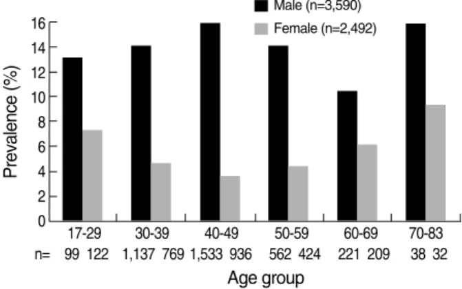
Among a total of 6,082 subjects (3,590 male and 2,492 female, mean age 44±10 yr, 17-83 yr) (Fig. 1), 639 subjects were found to have RE, and the overall prevalence was 10.5%. The prevalence of RE in male was 14.6% (523 of 3,590), the prevalence of RE in female was only 4.7% (116 of 2,376) (p< 0.01). When the age group was stratified by 17-29, 30-39, 40-49, 50-59, 60-69, and 70-83 yr, the prevalence of RE ra-nged from 10.4 % to 15.8% in each age group among male. In contrast, the prevalence of RE ranged 3.7% 9.4% in each age group among female. The prevalence of RE was similar in each age group between both sexes (Fig. 2).
Fig. 1. Age distribution of the subjects.
Number of persons 1,600 1,400 1,200 1,000 800 600 400 200 0 17-29 30-39 40-49 50-59 60-69 70-83 Age group Male (n=3,590) Female (n=2,492)
Fig. 2. Prevalence of reflux esophagitis in each age group. The pr-evalence of reflux esophagitis was similar in each age group betwe-en both sexes. Prevalence (%) 16 14 12 10 8 6 4 2 0 17-29 30-39 40-49 50-59 60-69 70-83 n= 99 122 1,137 769 1,533 936 562 424 221 209 38 32 Age group Male (n=3,590) Female (n=2,492)
Endoscopic findings of subjects with RE
Most of the subjects showed mild grade RE with 519 (81 %) of the subjects presenting with grade A, followed by 112 (17.5%) with grade B and 8 (1.3%) with grade C. No sub-jects presented with grade D (Table 1).
Clinical features of subjects with and without RE
The difference in mean age between subjects with and wi-thout RE was not statistically significant (43.9±9.6 yr vs. 43.2±9.7 yr, p=0.5). Male sex was predominant in subjects with RE (81.8%) than those without (56.4%) (p<0.01). Smok-ing history was significantly higher in subjects with RE (34.6 %) than those without (21.6%) (p<0.01). However, alcohol drinking (91.7% vs. 89.9%), and H. pylori infection (2.7% vs. 4.1%) were not significantly different between the two groups.
LA classification Male Female Total
n (%) n (%) n (%) A 410 (64.2) 109 (17.1) 519 (81.2) B 106 (16.6) 6 (0.9) 112 (17.5) C 7 (1.1) 1 (0.2) 8 (1.3) D 0 (0.0) 0 (0.0) 0 (0.0) Total 523 (81.8) 116 (8.2) 639 (100.0)
LA, los argeles.
Table 1. LA classification of subjects with reflux esophagitis ac-cording to sex
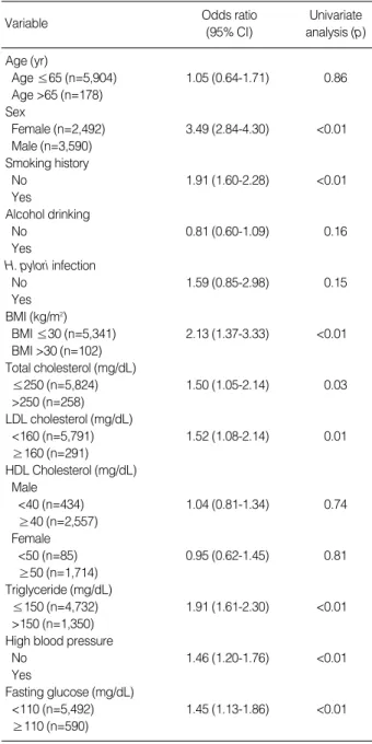
Variable Odds ratio Univariate
(95% CI) analysis (p) Age (yr) Age ≤65 (n=5,904) 1.05 (0.64-1.71) 0.86 Age >65 (n=178) Sex Female (n=2,492) 3.49 (2.84-4.30) <0.01 Male (n=3,590) Smoking history No 1.91 (1.60-2.28) <0.01 Yes Alcohol drinking No 0.81 (0.60-1.09) 0.16 Yes H. pylori infection No 1.59 (0.85-2.98) 0.15 Yes BMI (kg/m2) BMI ≤30 (n=5,341) 2.13 (1.37-3.33) <0.01 BMI >30 (n=102) Total cholesterol (mg/dL) ≤250 (n=5,824) 1.50 (1.05-2.14) 0.03 >250 (n=258) LDL cholesterol (mg/dL) <160 (n=5,791) 1.52 (1.08-2.14) 0.01 ≥160 (n=291) HDL Cholesterol (mg/dL) Male <40 (n=434) 1.04 (0.81-1.34) 0.74 ≥40 (n=2,557) Female <50 (n=85) 0.95 (0.62-1.45) 0.81 ≥50 (n=1,714) Triglyceride (mg/dL) ≤150 (n=4,732) 1.91 (1.61-2.30) <0.01 >150 (n=1,350) High blood pressure
No 1.46 (1.20-1.76) <0.01
Yes
Fasting glucose (mg/dL)
<110 (n=5,492) 1.45 (1.13-1.86) <0.01
≥110 (n=590)
BMI, body mass index; CI, confidence interval; high blood pressure, ≥130/85 mmHg or documented use of antihypertensive therapy. LDL, low-density lipoprotein; HDL, high-density lipoprotein.
Table 3. Logistic regression analysis of covariables for reflux eso-phagitis
Subjects Subjects
Variable with reflux without reflux p value
esophagitis esophagitis (n=639) (n=5,443) Age (yr) 43.9±9.6 43.2±9.9 0.5 Sex Male (%) 523 (81.8) 3,067 (56.4) <0.01 Female (%) 116 (18.2) 2,376 (43.6) Smoking history Yes (%) 221 (34.6) 1,180 (21.7) <0.01 No (%) 418 (65.4) 4,263 (78.3) Alcohol drinking Yes (%) 586 (91.7) 4,895 (89.9) 0.16 No (%) 53 (8.3) 548 (10.1)
Helicobacter pylori infection*
Positive (%) 11 (2.7) 106 (4.1) 0.15 Negative (%) 404 (97.3) 2,451 (95.9) BMI (kg/m2) 24.7±2.8 23.6±2.9 <0.01 Total cholesterol (mg/dL) 191.2±33.6 188.3±32.6 0.04 LDL cholesterol (mg/dL) 112.2±28.1 109.7±27.5 0.03 HDL cholesterol (mg/dL) 50.7±12.1 53.5±12.7 <0.01 Triglyceride (mg/dL) 134.6±80.1 113.5±83.2 <0.01 High blood pressure�
Yes (%) 163 (25.6%) 1,035 (19.1%) <0.01
No (%) 473 (74.4%) 4,375 (80.9%)
Fasting glucose (mg/dL) 98.1±16.7 96.3±18.8 0.02 *Data on Helicobacter pylori infection were unavailable for 224 (35.1%) subjects with reflux esophagitis and for 2,886 (53.0%) subjects without reflux esophagitis; �
Data on high blood pressure were unavailable for 3 (0.5%) subjects with reflux esophagitis and for 33 (0.6%) subjects with-out reflux esophagitis.
BMI, body mass index; high blood pressure, ≥130/85 mmHg or doc-umented use of antihypertensive therapy.
LDL, low-density lipoprotein; HDL, high-density lipoprotein.
Table 2. A comparison of clinical characteristics between sub-jects with reflux esophagitis and those without
And BMI was also found to be significantly higher in sub-jects with RE than in those without (24.7±2.8 kg/m2vs.
23.6±2.9 kg/m2, p<0.01), as were total cholesterol (191.2
±33.6 mg/dL vs. 188.3±32.6 mg/dL, p=0.04), LDL choles-terol (112.2±28.1 mg/dL vs. 109.7±27.5 mg/dL, p=0.03), triglyceride (134.6±80.1 mg/dL vs. 113.5±83.2 mg/dL,
p<0.01), high BP (25.6% vs. 19.1%, p<0.01), and fasting
glucose (98.1±16.7 mg/dL vs. 96.3±18.8 mg/dL, p=0.02). HDL cholesterol (50.7±12.1 mg/dL vs. 53.5±12.7 mg/dL,
p<0.01) was significantly lower in subjects with RE than in
those without (Table 2). Risk factors of RE
According to univariate analysis, male sex (odds ratio [OR]
3.49, 95% confidence interval [CI]2.84-4.30), smoking his-tory (OR 1.91, 95% CI 1.60-2.28), BMI >30 kg/m2(OR
2.13, 95% CI 1.37-3.33), total cholesterol >250 mg/dL (OR 1.50, 95% CI 1.05-2.14), LDL cholesterol ≥160 mg/dL (OR 1.52, 95% CI 1.08-2.14), triglyceride ≥150 mg/dL (OR 1.92, 95% CI 1.61-2.30), high BP (OR 1.46, 95% CI 1.20-1.76), and fasting glucose ≥110 mg/dL (OR 1.45, 95% CI 1.13-1.86) were significant risk factors of RE (all
p<0.05). However, old age (age >65 yr, OR 1.05, 95% CI
0.64-0.71), alcohol drinking (OR 0.81, 95% CI 0.60-1.09) and H. pylori infection (OR 1.59, 95% CI 0.85-2.98) were not shown to be significant risk factors of RE. Low HDL cholesterol (male <40 mg/dL, female <50 mg/dL) was not a significant risk factor in males (OR 1.04, 95% CI 0.81-1.34) and females (OR 0.95, 95% CI 0.62-1.45) (Table 3).
DISCUSSION
GERD is one of the most prevalent digestive diseases expe-rienced in Western countries. In the United States, the preva-lence is about 1,900,000 cases/yr and the annual cost of diagnosis and treatment is 93,000,000,000 dollars (9). The prevalence of GERD-related complications, including RE, Barrett’s esophagus, and esophageal adenocarcinoma has been steadily increasing in the United States and Western Europe. As the reasons for the increase in GERD and its complica-tions, changes in diet, smoking, and alcohol intake as well as the declining prevalence of H. pylori infection have been pro-posed (10). In our results, smoking was an important risk fac-tor, however, alcohol drinking and H. pylori infection had no relationships with GERD. Studies have also hypothesized that the increasing trend of obesity in Western countries has par-alleled the increase in esophageal adenocarcinoma and may be an important factor in this change (11).
In Korea, it is generally accepted that the nationwide preva-lence of RE is lower than that in Western countries. However, recent studies have shown that the numbers of patients with RE have shown an increasing trend (2, 5, 6). In this study,
endoscopic examination indicated that the overall prevalence of RE was 10.5% close to that of Western countries (10-20%) (1). The possible causes for the increasing prevalence in RE observed in the Korean population are of great interest. Patient selection, increased referrals for endoscopy, and marked impro-vement in diagnostic techniques may partially explain this trend. Furthermore, obesity shows an increasing trend as a cause of RE in Korea. A recently reported study indicated that the prevalence of overweight (25≤BMI≤30 kg/m2)
and obesity (BMI >30 kg/m2) was 23.4% and 1.7% in males
and 24.9% and 3.2% in females in 1998 (11). However, in 2001, the prevalence of overweight or obesity in Korean adults was 30.6% (32.4% in males and 29.4% in females) (12). In our study, the prevalence of overweight and obesity were 36.9 %, 2.1% in males and 21.3%, 2.1% in females, respectively, and BMI was found to be significantly higher in subjects with RE than that in those without (24.7±2.8 kg/m2vs. 23.6±
2.9 kg/m2, p<0.01). BMI >30 kg/m2was found to be a
signifi-cant risk factor of RE by univariate analysis (OR 2.13, 95% CI 1.37-3.33).
Obesity may affect esophageal reflux through mechanical factors and humoral factors. Such mechanical factors include increased intra-abdominal pressures, impaired gastric emp-tying, decreased lower esophageal sphincter pressure, and in-creased frequency of transient lower esophageal sphincter relax-ation, thus leading to increased esophageal acid exposure (10). The humoral mechanisms are not well known. It has been hypothesized that estrogen also influences reflux in obese pa-tients. Two studies conducted by the same group of investi-gators from Sweden observed that the association between obe-sity and GERD might be mediated by estrogen (13, 14). The first study reported a statistically significant association be-tween obesity and esophagitis in women, which was poten-tiated by the use of estrogen in postmenopausal women (13). The second study found that obese women were at increased risk of experiencing symptoms of GERD compared with obese men, and the risk was highest in premenopausal women and postmenopausal women receiving estrogen therapy (14).
Few reports have assessed the association between dyslipi-demia and GERD. Total cholesterol was found to be higher in subjects with RE with borderline significance than in the general population, but HDL cholesterol levels did not dif-fer (15). However, in this study, we found that dyslipidemia can be a possible causative factor of RE. Using univariate logis-tic regression analysis, it was found that total cholesterol >250 mg/dL, LDL cholesterol ≥160 mg/dL, and triglyceride ≥150 mg/dL were associated with the occurrence of RE. HDL choles-terol was significantly lower in subjects with RE than in those without the condition, but low HDL cholesterol (male <40 mg/dL, female <50 mg/dL) was not found to be a significant risk factor. This could potentially be explained by relatively low proportion of the subject’s population with low HDL ch-olesterol.
with age (16, 17). In this study, the difference in mean age between subjects with and without esophagitis was not sta-tistically significant. This is may be due to the enrollment of a relatively young subject population (age ≤65: n=5,904 vs. age >65: n=178). When we divided the age by subgroup, there were no significant differences within the both sexes. There is a male preponderance in RE, which ranges from 2:1 to 3:1 in comparison to females (18). The preponderance of males with RE in the present study was more evident (M:F= 4.5:1). According to univariate analysis, male sex was signif-icant risk factor of RE (OR 3.49, 95% CI 2.84-4.30).
Hypertension has been reported as frequently present in patient with RE (15). The present study showed that high BP was risk factor of RE as well (OR 1.46, 95% 1.20-1.76), but we did not investigate drug therapy in the subjects who underwent endoscopic examination. Calcium antagonists are used to treat hypertension, but these drugs also decrease the lower esophageal sphincter pressure and inhibit muscle con-traction in the esophagus itself (19). In Korea, calcium antag-onists are widely used to treat hypertension, so it is possible that antihypertensive therapy had an influence on our findings.
An association between diabetes mellitus (DM) and GERD has been reported previously (20, 21). There are several pos-sible mechanisms underlying DM-related GERD. In asymp-tomatic diabetic patients, there is a higher prevalence of abnor-mal gastroesophageal reflux than among the general popu-lation (22). The incidence of symptomatic GERD tended to be higher in diabetic patients with neuropathy than in those without neuropathy (20). However, the relationship between glucose intolerance and GERD has not yet been reported. In the present study, fasting glucose was significantly higher in subjects with RE than in those without (98.1±16.7 mg/dL vs. 96.3±18.8 mg/dL, p=0.02), and fasting glucose ≥110 mg/dL was found to be a significant risk factor of RE (OR 1.45, 95% CI 1.13-1.86). Hyperglycemia may influence eso-phageal motor and sensory function (23). In healthy volun-teers, hyperglycemia decreases lower esophageal sphincter pres-sure and the velocity of esophageal peristalsis (24).
Metabolic syndrome has important clinical and public he-alth implications. In the United States, the unadjusted and age-adjusted prevalence of metabolic syndrome has been esti-mated to be 21.8% and 23.7%, respectively (25). In Japan, the prevalence of metabolic syndrome is 30.6% for men and 3.6% for women (26). In Korea, the prevalence of the metabol-ic syndrome was reported to be 13.5% for men and 15.0% for women (27). In the present study, the diagnostic compo-nents of metabolic syndrome (obesity, high triglyceride, low HDL cholesterol, high BP and elevated fasting glucose) based on NCEP criteria (7) were found to be significantly associat-ed with RE. In metabolic syndrome, insulin resistance is an important pathogenic factor. Insulin resistance induces sever-al metabolic changes, including hyperglycemia and dyslipi-demia (28). Alternative mechanisms might include an asso-ciation between metabolically active compounds and RE (29).
These and other mechanisms by which obesity may affect risk for RE, such as growth factors, humoral factors like leptin, and hormonal factors like estrogen, were not examined in this study (30). A recent report showed components of the meta-bolic syndrome such as obesity, hyperglycemia, and hyper-tension are associated with the occurrence of RE in Japan (31). However, there was no report about the treatment of metabol-ic syndrome and improvement of RE until now.
Potential limitations still remain in our study. In this study, people who attended annual medical check-ups were enrolled for the assessment of RE. Therefore, the results of this study were similar, but not identical to those of population-based studies. We could not evaluate the reflux symptom such as heartburn, regurgitation, dyspepsia, epigastric pain, belch-ing, and non-cardiac chest pain because of the retrospective study. In addition, we did not assess physical data such as waist-to-hip ratio, so we could not elucidate the prevalence of metabolic syndrome in our study population. This also prevented us from making inferences regarding the causali-ty between metabolic syndrome and RE. However, this is first study to show the possibility that reflux esophagitis may be part of the disease spectrum of metabolic syndrome in a large sample of the Korean population.
In conclusion, the prevalence of RE in healthy subjects who had a routine check-up was 10.5% in Korea, and nearly ap-proaches that reported in Western countries. Significant cor-relations between RE and obesity, high triglyceride, low HDL cholesterol, high BP, and elevated fasting glucose levels sug-gested that RE might be part of the disease spectrum of me-tabolic syndrome. Further studies are needed to evaluate the relationship between RE and metabolic syndrome.
REFERENCES
1. Inamori M, Togawa J, Nagase H, Abe Y, Umezawa T, Nakajima A, Saito T, Ueno N, Tanaka K, Sekihara H, Kaifu H, Tsuboi H, Kaya-ma H, Tominaga S, Nagura H. Clinical characteristics of Japanese reflux esophagitis patients as determined by Los Angeles classifica-tion. J Gastroenterol Hepatol 2003; 18: 172-6.
2. Goh KL, Chang CS, Fock KM, Ke M, Park HJ, Lam SK. Gastro-oesophageal reflux disease in Asia. J Gastroenterol Hepatol 2000; 15: 230-8.
3. Furukawa N, Iwakiri R, Koyama T, Okamoto K, Yoshida T, Kashi-wagi Y, Ohyama T, Noda T, Sakata H, Fujimoto K. Proportion of reflux esophagitis in 6010 Japanese adults: prospective evaluation by endoscopy. J Gastroenterol 1999; 34: 441-4.
4. Yeh C, Hsu CT, Ho AS, Sampliner RE, Fass R. Erosive esophagitis and Barrett’s esophagus in Taiwan: a higher frequency than expect-ed. Dig Dis Sci 1997; 42: 702-6.
5. Lee SJ, Song CW, Jeen YT, Chun HJ, Lee HS, Um SH, Lee SW, Choi JH, Kim CD, Ryu HS, Hyun JH. Prevalence of endoscopic reflux eso-phagitis among Koreans. J Gastroenterol Hepatol 2001; 16: 373-6. 6. Yeom JS, Park HJ, Cho JS, Lee SI, Park IS. Reflux esophagitis and
its relationship to hiatal hernia. J Korean Med Sci 1999; 14: 253-6.
7. Miranda PJ, DeFronzo RA, Califf RM, Guyton JR. Metabolic
syn-drome: definition, pathophysiology, and mechanisms. Am Heart J 2005; 149: 33-45.
8. Hongo M. Minimal changes in reflux esophagitis: red ones and white
ones. J Gastroenterol 2006; 41: 95-9.
9. Sandler RS, Everhart JE, Donowitz M, Adams E, Cronin K, Good-man C, Gemmen E, Shah S, Avdic A, Rubin R. The burden of
select-ed digestive diseases in the Unitselect-ed States. Gastroenterology 2002; 122: 1500-11.
10. Hampel H, Abraham NS, El-Serag HB. Meta-analysis: obesity and
the risk for gastroesophageal reflux disease and its complications. Ann Intern Med 2005; 143: 199-211.
11. Kim Y, Suh YK, Choi H. BMI and metabolic disorders in South
Kore-an adults: 1998 Korea National Health Kore-and Nutrition Survey. Obes Res 2004; 12: 445-53.
12. Kim DM, Ahn CW, Nam SY. Prevalence of obesity in Korea. Obes
Rev 2005; 6: 117-21.
13. Nilsson M, Lundegardh G, Carling L, Ye W, Lagergren J. Body mass
and reflux oesophagitis: an oestrogen-dependent association? Scand J Gastroenterol 2002; 37: 626-30.
14. Nilsson M, Johnsen R, Ye W, Hveem K, Lagergren J. Obesity and
estrogen as risk factors for gastroesophageal reflux symptoms. JAMA 2003; 290: 66-72.
15. Gudlaugsdottir S, Verschuren W, Dees J, Stijnen T, Wilson J.
Hyper-tension is frequently present in patients with reflux esophagitis or Bar-rett’s esophagus but not in those with non-ulcer dyspepsia. Eur J Intern Med 2002; 13: 369-75.
16. Pilotto A, Franceschi M, Leandro G, Scarcelli C, D’Ambrosio LP, Seripa D, Perri F, Niro V, Paris F, Andriulli A, Di Mario F. Clinical
features of reflux esophagitis in older people: a study of 840 consec-utive patients. J Am Geriatr Soc 2006; 54: 1537-42.
17. Lind T, Havelund T, Carlsson R, Anker-Hansen O, Glise H, Hern-qvist H, Junghard O, Lauritsen K, Lundell L, Pedersen SA, Stubberod A. Heartburn without oesophagitis: efficacy of omeprazole therapy
and features determining therapeutic response. Scand J Gastroenterol 1997; 32: 974-9.
18. Behar J, Biancani P, Sheahan DG. Evaluation of esophageal tests in
the diagnosis of reflux esophagitis. Gastroenterology 1976; 71: 9-15.
19. Hongo M, Traube M, McAllister RG Jr, McCallum RW. Effects of
nifedipine on esophageal motor function in humans: correlation with plasma nifedipine concentration. Gastroenterology 1984; 86: 8-12.
20. Nishida T, Tsuji S, Tsujii M, Arimitsu S, Sato T, Haruna Y, Miyamo-to T, Kanda T, Kawano S, Hori M. Gastroesophageal reflux disease
related to diabetes: analysis of 241 cases with type 2 diabetes melli-tus. J Gastroenterol Hepatol 2004; 19: 258-65.
21. Kinekawa F, Kubo F, Matsuda K, Inoue H, Kuriyama S.
Gastroe-sophageal reflux disease in diabetic patients. Nippon Rinsho 2004; 62: 1546-52.
22. Lluch I, Ascaso JF, Mora F, Minguez M, Pena A, Hernandez A, Be-nages A. Gastroesophageal reflux in diabetes mellitus. Am J
Gastroen-terol 1999; 94: 919-24.
23. Rayner CK, Samsom M, Jones KL, Horowitz M. Relationships of
upper gastrointestinal motor and sensory function with glycemic con-trol. Diabetes Care 2001; 24: 371-81.
24. De Boer SY, Masclee AA, Lam WF, Lamers CB. Effect of acute
hy-perglycemia on esophageal motility and lower esophageal sphincter pressure in humans. Gastroenterology 1992; 103: 775-80.
25. Ford ES, Giles WH, Dietz WH. Prevalence of the metabolic syndrome
among US adults: findings from the third National Health and Nutri-tion ExaminaNutri-tion Survey. JAMA 2002; 287: 356-9.
26. Miyatake N, Wada J, Kawasaki Y, Nishii K, Makino H, Numata T.
Relationship between metabolic syndrome and cigarette smoking in the Japanese population. Intern Med 2006; 45: 1039-43.
27. Park HS, Lee SY, Kim SM, Han JH, Kim DJ. Prevalence of the
me-tabolic syndrome among Korean adults according to the criteria of the International Diabetes Federation. Diabetes Care 2006; 29: 933-4.
28. Wassink AM, Olijhoek JK, Visseren FL. The metabolic syndrome:
metabolic changes with vascular consequences. Eur J Clin Invest 2007; 37: 8-17.
29. Corley DA, Kubo A. Body mass index and gastroesophageal reflux
disease: a systematic review and meta-analysis. Am J Gastroenterol 2006; 101: 2619-28.
30. El-Serag HB, Graham DY, Satia JA, Rabeneck L. Obesity is an
inde-pendent risk factor for GERD symptoms and reflux esophagitis. Am J Gastroenterol 2005; 100: 1243-50.
31. Moki F, Kusano M, Mizuide M, Shimoyama Y, Kawamura O, Tak-agi H, Imai T, Mori M. Association between reflux esophTak-agitis and
features of the metabolic syndrome in Japan. Aliment Pharmacol Ther 2007; 26: 1069-75.