https://doi.org/10.20307/nps.2016.22.4.263
263
Rhynchophylline, One of Major Constituents of Uncariae Ramulus et Uncus Enhances Pentobarbital-induced Sleep Behaviors and Rapid Eye Movement Sleep
in Rodents
Jae Hyeon Yoo, Tae-Woo Ha, Jin Tae Hong, and Ki-Wan Oh*
1
College of Pharmacy, Chungbuk National University, Cheongju, 28644 Republic of Korea
Abstract − Rhynchophylline (RP) is a major tetracyclic oxindole alkaloid of Uncariae Ramulus et Uncus which has been used to treat hypertension, seizures, pain and anxiety in the oriental countries. A recent report revealed that RP attenuated ischemia-induced neuronal damage and kainite-induced convulsions in animals. This study was performed to investigate whether RP enhances pentobarbital-induced sleep behaviors and modulates sleep architecture in mice. Locomotor activity was significantly inhibited by RP at 0.25 and 0.5 mg/kg, similar to 2 mg/
kg diazepam (a benzodiazepine agonist) in mice. RP shortened sleep latency and increased total sleep time in a dose-dependent manner when administrated with pentobarbital (42 mg/kg, i.p.). RP also increased the number of sleeping mice and total sleep time by concomitant administration with the sub-hypnotic dosage of pentobarbital (28 mg/kg, i.p.). On the other hand, RP (0.25 mg/kg, p.o.) itself significantly inhibited sleep-wake cycles, prolonged total sleep time, and rapid eye movement in rats. In addition, RP also increased chloride influx in the primary cultured hypothalamic neuronal cells. In addition, we found that glutamic acid decarboxylase (GAD
65/67) was activated by RP. In conclusion, RP augments pentobarbital-induced sleeping behaviors, and can be a candidate for treating insomnia.
Keywords − Uncariae Ramulus et Uncus, Rhynchophylline, Pentobarbital, GABA
Areceptors subunits, Electroencephalogram, REM sleep
Introduction
Insomnia can be defined as the inability to initiate or maintain sleep. It is also one of the most common health problems in modern society. Furthermore, chronic insomnia disturbs daily sleep and causes diverse problems in daily living such as chronic fatigue, decreased work efficiency, lack of alertness, and poor cognitive abilities.
Over the last decade, scientists have shown increased interest in herbal medicines, which contain phytochemicals that promote health. Many herbs such as St, John’s wort, kava kava, valerian, and passion flower have been introduced in European countries.
1Herbs as sleep aids are becoming more popular as alternative medicines.
γ-Aminobutyric acid (GABA), the main inhibitory neurotransmitter of the central nervous system (CNS), is the most prevalent target for treating insomnia. It is well established that activation of GABA
A-ergic neurons plays
an important role in sleep. Glutamic acid decarboxylase (GAD
65/67), an enzyme responsible for the synthesis of GABA also plays a crucial role in sleep. On the other hand, GABA is released to the synapse, the extracellular space between the neurons. The GABA
Areceptors complex consists of a Cl
−ionophore principally coupled to GABA, barbiturate, benzodiazepine, steroid, and picrotoxin binding sites.
2,3Basic subunits are composed to α (1~6), β (1~3) and γ (1~3).
4These binding sites trigger the chloride channel’s opening with resulting membrane hyperpolari- zation
5. GABA
A-ergic drugs have induced sedative-hypnotic effects in animals and humans.
6Rhynchophylline (RP, Fig. 1) is a major tetracyclic oxindole alkaloid from the Uncaira species, which has been long used medicinally. Recently, it has been reported that RP attenuates ischemia-induced neuronal damage in the hippocampus by the noncompetitive antagonistic effect of N-methly-D-aspartate (NMDA). Moreover, RP reduced kainic acid-induced epileptic seizure. The Uncaira species has been traditionally used to treat neurological disorders such as seizures, pain, and anxiety in the oriental countries
7. We are interested in whether RP enhances
*Author for correspondence
Ki-Wan Oh, College of Pharmacy, Chungbuk National University, Cheongju 28644 Republic of Korea
Tel: +82-43-261-2827; E-mail: [email protected]
pentobarbital-induced sleeping behaviors and modulates sleep architecture because it has shown neuroprotective and inhibitory pharmacological effects on the CNS
8-10. Furthermore, the possible mechanisms of RP as a candidate for insomnia treatment are suggested from these experiments.
Experimental
Reagents and chemicals − Rhynchophylline (Fig. 1) was purchased from Tokyo Chemical Industry Co., Ltd.
(Tokyo, Japan). Diazepam, pentobarbital, and muscimol were acquired, respectively, from the following companies:
Samjin Pharm. (Seoul, Korea), Hanlim Pharm. Co., Ltd.
(Seoul, Korea), and Tocris (Cookson, UK or Ellisville, MO, USA). Fetal bovine serum and DMEM were obtained from GIBCO (Grand Island, NY, USA). The Clsensitive fluorescence probe N-(ethoxycarbonyl-methyl)- 6-methoxyquinolinium bromide (MQAE) was purchased from Sigma-Aldrich (St. Louis, MO, USA). The specific rabbit polyclonal antibodies against GABA
Areceptors subunits, GAD
65/67and the corresponding conjugated anti- rabbit immunoglobulin G-horseradish peroxidase were obtained from Abcam Inc. (Cambridge, UK).
Animals − The animals used for experiments were 4- week-old ICR male mice and 8-week-old male Sprague Dawley (SD) rats weighing 20 - 25 g and 300 - 320 g, respectively (purchased from Samtako, Osan, Korea). All rodents were housed in acrylic cages (45 × 60 × 23 cm) and were kept at least 1 week for acclimation time. The room condition was maintained at 22 ± 2
oC, relative humidity (50 - 52%), and a 12-h light/dark cycle with ad libitum feeding. The behavioral experiments were perfor- med between 10:00 and 17:00 and were carried out in accordance with the Principle of Laboratory Animal Care (NIH publication No. 85-23, revised 1985). This experiment was performed in accordance with the Animal Care and Use Guidelines of Chungbuk National University, Korea.
Locomotor activity measurement − Spontaneous loco- motor activity was measured by a tilting-type ambulometer (AMB-10, O’Hara, Tokyo, Japan) for 1 h
11. The mice in each group had 10 min of adaptation time in the activity cages (20 cm in diameter and 18 cm in height). Diazepam (2 mg/kg, p.o.) and RP (0.125 and 0.25 mg/kg, p.o.) dissolved in distilled water were administered 30 min and 60 min prior to the experiment, respectively.
Pentobarbital-induced sleeping behaviors measure- ment − All mice were fasted for a day, and all experi- ments were carried out between 1:00 and 5:00 p.m.
Pentobarbital was diluted in 0.9% physiological saline.
Muscimol (0.2 mg/kg) and RP (0.125 and 0.25 mg/kg) were orally administered before 15 min and 60 min, respectively, and then pentobarbital (42 mg/kg) was injected intraperitoneally. After the pentobarbital, the mice were moved to another cage. Sleep latency was recorded as time elapsed after the pentobarbital injection. Sleep was recorded as the time between the elapse and the righting of animals. The mice that failed sleep within 15 min were excluded from the experiment
12, 13.
EEG telemetry transmitter implantation and data collection − After pentobarbital (50 mg/kg, i.p.) was administered, the rats were placed on a pad in the stereotaxic apparatus under aseptic conditions. Transmitters (Data Sciences International TA11CTA-F40, MN, USA) were implanted under the skin after the scalp incision. In detail, the skull periosteum was removed, and then two holes forward drilled to insert electric lines (A: 2.0 [Bregma], L: 1.5; P: 7.0 [Bregma], L: 1.5 contra-lateral)
14. The transmitter lines were subcutaneously connected to the skull, and dental cement was used to fix the electric lines to the skull. The incisions were sewn up with a silk 4 – 0 suture. An antibiotic was given to all rats after surgery (5 million unit potassium penicillin-G Injection, Keunwha, Korea). After the transmitters were implanted, the rats were given a week of recovery time. RP (0.25 mg/kg, p.o) was administered to rats. All signals were transmitted by AD converter (Eagle PC30, USA), and stored in the computer, and the computer could also graphically display the results. Fast Fourier transform (FFT) analysis generated power density values from 0 to 20.0 Hz with a resolution of 0.5 Hz. Mean FFT was also in the range of 0 and 20.0 Hz for every 10 sec. EEG data in all rats were recorded from 11:00 a.m. to 5:00 p.m.
15.
Data analysis − Sleep cycles were graphically recorded
and saved in Sleep-Sign 2.1software (KISSEI Comtec Co
Ltd, Matsumoto, Japan). Data were classified into wake-
fulness, non-rapid eye movement (NREM), and rapid eye
movement (REM) for every 10 sec.
16Wakefulness and
Fig. 1. Chemical structure of rhynchophylline.
NREM states were found in high-frequency and slow waves, respectively. δ-Wave (0.75~4.0 Hz) and θ-wave (5.0~9.0 Hz, peak at 7.5 Hz) were increased in the low EEG waves during REM sleep. Wakefulness, NREM, REM, and total sleep time (NREM+REM) were recorded for each rat for 6 h. The EEG power was set up at 0.5 – 20.0 Hz in 0.5 Hz bins. Sleep architecture was evaluated in three waves in the range of 8.0 – 13.0 Hz
17. Data were calculated as relative values in Microsoft Excel.
Cell culture − Primary cultures of the SD rats’ hypo- thalamus cells were tested for 7-8 days
18. The cells were seeded at 1.0 × 10
5cells in 96-well microplates coated with poly-L-lysine (50 µg/mL; Sigma, St. Louis, MO, USA). The DMEM used for cell cultures contained 10%
fetal bovine serum, glutamine (2.0 mM), gentamicin (100 μg/mL), antibiotic-antimycotic solution (10 μg/mL; Sigma), and potassium chloride (25 mM). Cells were incubated at 37
oC in a humidified atmosphere of 5% CO
2/95% air.
After 16 h, the 96-well plates were added into cytosine arabinofuranoside (final concentration 10 µM; Sigma) to inhibit non-neuronal cell growth.
Measurement of intracellular Cl
−influx − MQAE (a sensitive fluorescent substance for Cl
−) was used to measure Cl
−influx in the rats’ cerebellum cells following the method of West and Molloy
19. After overnight MQAE treatment, the cells were washed three times in a buffer (pH 7.4) that contained 2.4 mM HPO
42−, 0.6 mM H
2PO
4−, 10 mM HEPES, 10 mM D-glucose, and 1.0 mM MgSO
4. The fluorescence data were measured according to excitation wavelength 320 nm and emission wavelength 460 nm by Elisa Reader (SpectraMax M2e Multi-Mode, PA, USA)
20. The data were calculated as F/F
0based on the Cl
−data ratios. F is the fluorescence of each sample, and F
0is the fluorescence without Cl
−ions.
Western blotting − Protein samples were extracted from the rat’s hypothalamus cell cultures. RP (final concentration 0.25 mg/ml) was dissolved in 0.01% DMSO.
The control sample was treated in the same solvent as that used in the RP treatment. After diazepam or RP administration, the cells were extracted and treated with a cold lysis buffer [25 mM Tris–HCl (pH 7.4), 150 mM NaCl, 1.0 mM CaCl
2, 1% Triton X-100, 1.0 mM PMSF, 10 µl/mL aprotinin, 1.0 mM NaF, and 2.0 mM sodium orthovanadate]. Supernatant extracts were recovered after centrifugation at 13,000 × g at 4
oC for 20 minutes. Protein concentration was measured using Bradford protein analysis and stored at −20
oC. The same amounts of protein were placed in 10% SDS-polyacrylamide gel, and then the electrophoresis was loaded. The protein was transferred to PVDF membranes (Hybond-P, GE Healthcare,
Amersham, UK) using semi-dry transfer. The blots were blocked for 1 h at room temperature with 5.0% (w/v) BSA [applied to all primary antibodies except for glyceraldehyde 3-phosphate dehydrogenase (GAPDH)], and 5.0% (w/v) skim milk (only applied to GAPDH) in tris-buffered saline solution (TBS) containing 0.1% Tween- 20. Both specific rabbit polyclonal antibodies against GABA
Areceptor subunits and rabbit anti-GAD
65/67polyclonal antibody at the appropriate dilution in TBST and 5.0% BSA (1:2,500 for all the primary antibodies used) were incubated overnight at 4°C. After washing with TBST, the blots were treated 1:3,000 dilution of a secondary antibody at room temperature for 4 hours (goat anti-rabbit, IgG). A the secondary antibody was detected using ECL light-emitting substrate (Roche Diagnostics, Mannheim, Germany)
21.
Statistical analysis − All statistical analysis was performed with SigmaStat software (SPSS Inc., Chicago, USA). Experimental results are shown as mean ± SEM, and significance was measured with analysis of variance (ANOVA). When there were significant differences, values were compared with Student's t-test. However, in sub- hypnotic pentobarbital-induced sleep, the falling asleep/
total was compared using Chi-square test. P was considered significant at less than 0.05.
Resrult and Discussion
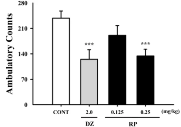
Effect of RP on locomotor activity in mice − Locomotor activity was significantly inhibited by RP at 0.25 mg/kg.
Moreover, diazepamat 2.0 mg/kg significantly decreased locomotor activity in the tested animals (Fig. 2). From
Fig. 2. Effects of RP on locomotor activity test. Ambulation activity was measured for 1 h, 30 min after oral administration of diazepam and 1 h after administration of RP. Each column shows the mean ± SEM. The significance of the compound’s effects was assessed using ANOVA. Where there was significant variability, the individual values were compared using Student's t-test.
***
P < 0.005 compared with the control.
these preliminary experiments, we suggest that RP might be sedative.
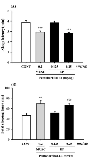
Effect of RP on pentobarbital-induced sleeping behaviors in mice − RP (0.25 mg/kg) reduced sleep latency time and prolonged sleeping time induced by pentobarbital (42 mg/kg, i.p.). Pretreatment with mucsimol (0.2 mg/kg, i.p.) as a positive control 30 min before the pentobarbital (42 mg/kg, i.p.) also increased sleeping time and decreased sleep latency (Fig. 3). We suggest that RP could reduce sleep latency and increase total sleep.
Effect of RP on sleep onset by sub-hypnotic dosage of pentobarbital in mice − RP (0.25 mg/kg) reduced the sleep onset time and prolonged the sleep duration induced by a sub-hypnotic pentobarbital dose (28 mg/kg, i.p.).
Similarly, muscimol (a GABA receptor agonist) signifi- cantly is affected pentobarbital-induced sleep (Table 1).
We suggest that RP would interact with GABA
Areceptors.
Fig. 3. Effects of RP on sleep onset and duration in pentobarbital- treated mice. Mice were deprived of sleep for 24 h prior to the experiment. Pentobarbital (42 mg/kg, i.p.) was administered to mice following administration of muscimol or RP, and sleep latency (A) and sleep time (B) were measured. Each column shows the mean ± SEM. The significance of the compounds’
effects was assessed using ANOVA. Where there was significant variability, the individual values were compared using Student's t- test.
**P < 0.01,
***P < 0.005 compared with the control.
Table 1. Effects of RP on sleep onset of mice treated by sub- hypnotic dose of pentobarbital (28 mg/kg, i.p.)
Group Dose (mg/kg) No. falling asleep/total Sleep time (min)
Control 0 712 35.1 ± 2.5
Muscimol 0.2 12/12* 59.3 ± 5.8**
RP 0.125 9/12 41.5 ± 3.6
0.25 11/12 51.2 ± 5.4*
Each value reflects the mean ± S.E.M. Where there was signifi- cant variability, the individual values were compared using Chi- square and Student's t-test. *P < 0.05, **P < 0.01 compared with the control.
Fig. 4. Effects of RP on numbers of sleep-wake cycles. Where there was significant variability, the individual values were compared using Student's t-test.
***P < 0.005 compared with the control.
Fig. 5. Effects of RP on rat sleep architecture. The data show the
mean ± SEM of time spent, which separated the wakefulness and
sleep (NREM and REM) states. The significance of the com-
pounds’ effects was assessed using ANOVA. Where there was
significant variability, the individual values were compared using
Student's t-test.
*P < 0.05,
**P < 0.01 compared with that of the
naïve control.
Effect of RP on sleep-wake cycles − RP (0.25 mg/kg) significantly reduced sleep-wake cycles (Fig. 4); that is, RP reduced wakefulness.
Effect of RP on sleep architectures − After EEG analysis, we found that RP (0.25 mg/kg) significantly prolonged total sleep time, especially REM (slow-wave) sleep (Fig. 5). RP also decreased wakefulness.
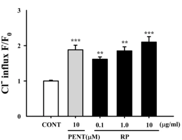
Effect of RP on intracellular Cl
−influx in primary cultured hypothalamus cells − RP (1.0 μg/ml) significantly
increased intracellular Cl
−influx, resulting in the hyper- polarization of the neuronal cell membrane. In addition, Pentobarbital (10 μM) also significantly increased intra- cellular Cl
−influx in primary cultured hypothalamus cells (Fig. 6).
Effect of RP on expression of GAD
65/67− GAD
65/67expression was induced by RP (0.25 mg/kg) in the rats’
primary hypothalamic neuron cells (Fig. 7). We suggest that RP activates GAD
65/67.
Effect of RP on expression of GABA
Areceptors subunits − From these experiments, the GABA
Areceptor subtypes activation was measured by western blotting. All subtypes of GABA
Areceptors except γ3 subtype were overexpressed with the RP (0.25 mg/kg). Pentobarbital as a positive control also showed similar patterns (Fig. 8).
Discussion
Uncariae ramulus et Uncus (UR) is a traditional Chinese herb that has been used to treat epileptic seizures.
RP, one of the major components of UR, displays neuroprotective and anti-convulsive actions
8, 10. This review expanded upon a previous paper that reviewed the Fig. 6. Effects of RP on Cl
−influx in primary cultured cerebellar
granule cells. After the hypothalamic neuronal cells were cultured for 8 days, the cells were incubated with MQAE overnight, and then RP (0.01, 0.1, and 1 µg/ml) and pentobarbital (10 µM) were added 1 h prior to measurement. Each column shows the mean ± SEM. The significance of the compounds’
effects was assessed using ANOVA. Where there was significant variability, the individual values were compared using Student's t-test.
**P < 0.01,
***P < 0.005 compared with that of the control.
Fig. 7. Effects of RP on the expression of GAD; the GAD65/67 expression was induced by RP (0.25 mg/kg) in the hypothalamic neuronal cells of the mice. GAPDH levels were needed in order to normalize the protein expression. Each column shows the mean ± SEM. The significance of the compounds’ effects was assessed using ANOVA. Where there was significant variability, the individual values were compared using Student's t-test.
***
P < 0.005 compared with the control.
Fig. 8. Effects of RP on expression of GABA
Areceptor subunits.
Immunoblots are shown of lysed hypothalamic neuronal cells
that were treated for 1 h following RP. GAPDH levels were
needed in order to normalize the protein expression. Each column
shows the mean ± SEM. The significance of the effects of the
compounds was assessed using ANOVA. Where there was signi-
ficant variability, the individual values were compared using
Student's t-test.
*P < 0.05,
**P < 0.01,
***P < 0.005, compared with
that of the control.
pharmacological actions of the Uncaria alkaloids, rhynchophylline, and isorhynchophylline
22. The current review encompasses many studies on rhynchophylline and provides information on its use in treating cardiovas- cular and central neurological disorders while highlighting ion channel and central neurotransmitters as potential therapeutic targets
23. Based on previous studies, we focused on the hypnotic effect of RP as the ultimate goal of the experiment. The preliminary experiment results demonstrate that RP inhibited locomotor activity, showing sedative effects in mice. We investigated the effects of different dosages of RP and muscimol in rodents with pentobarbital treatment and found that RP enhanced pentobarbital-induced sleep, similar to muscimol. It is suggested that potentiation of RP’s hypnotic effect can interact with GABA
A-ergic systems.
The sleep architectures of rat after oral RP adminis- tration were also analyzed. The spontaneous electrical activity of rat brain can be recorded by SSG over a short period of time, and sleep/wake cycles can be measured using EEG frequency analysis. We found that RP reduced sleep/wakefulness cycles, which is important in treating insomnia. Sleep can be divided into two major stages, REM and NREM. REM sleep is a distinctive sleep stage that alternates with episodes of NREM sleep
24-26. During the early years of sleep research, REM sleep was charac- terized by fast-wave sleep along with muscle atonia, brain activation, and eye movement. NREM sleep was discovered to play a role in restoring physiological functions
27. We especially focused on determining whether RP increased REM, NREM, and total sleep time and altered sleep architectures. Our experimental data show that RP caused significant reduction in the number of sleep-wake cycles.
Furthermore, RP increased total sleep and REM sleep.
Activating GABA
A–ergic transmission is important for treating insomnia. First, GABA is synthesized from glutamate exclusively in GABA
A-ergic neurons by GAD, which consists of two isoforms with molecular weights of 65-kDa and 67-kDa.
28,29Protein expression levels of GAD
65/67were measured in primary cultured hypothalamic neuronal cells; RP increased protein expression levels in these cells. It is suggested that RP activates GAD
65/67.