• 검색 결과가 없습니다.

Ethanol Extracts of Angelica decursiva Induces Apoptosis in Human Oral Cancer Cells

N/A
N/A
Protected

Academic year: 2021

Share "Ethanol Extracts of Angelica decursiva Induces Apoptosis in Human Oral Cancer Cells"

Copied!
6
0
0

로드 중.... (전체 텍스트 보기)

전체 글

(1)

Copyright ⓒ 2010, The Korean Academy of Oral Biology

215

Journal of Oral Biology

Ethanol Extracts of Angelica decursiva Induces Apoptosis in Human Oral Cancer Cells

Myoung-Hwa Lee

3†

, Myung Mi Kim

1†

, Joong-Ki Kook

1

, Do Kyung Kim

2

, Hye Ryun Kim

2

, Heung-Joong Kim

3

, and Chun Sung Kim

1

*

1

Department of Oral Biochemistry and Oral Biology Research Institute, School of Dentistry, Chosun University, 375 Seosuk-Dong, Dong-Gu, Gwangju 501-759, Korea

2

Department of Oral Physiology and Oral Biology Research Institute, School of Dentistry, Chosun University, 375 Seosuk-Dong, Dong-Gu, Gwangju 501-759, Korea

3

Department of Oral Anatomy and Oral Biology Research Institute, School of Dentistry, Chosun University, 375 Seosuk-Dong, Dong-Gu, Gwangju 501-759, Korea

(received December 7, 2010 ; revised December 10, 2010 ; accepted December 16, 2010)

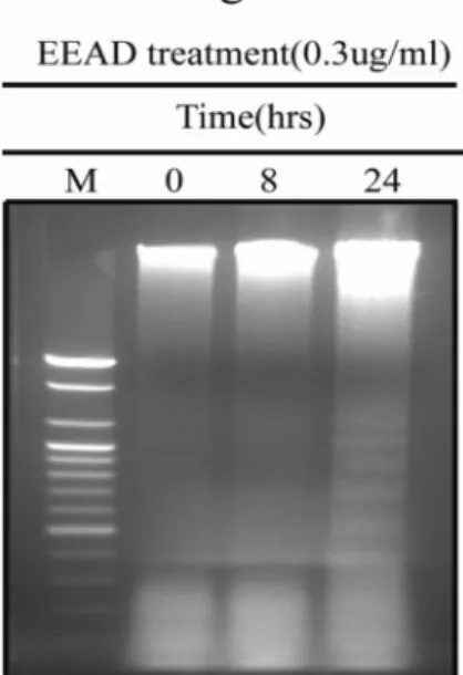
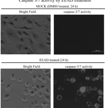
Angelica decursiva has been used in Korean traditional medicine as an antitussive, an analgesic, an antipyretic and a cough remedy. However, its anti-cancer properties have not yet been well defined. In our current study, we report the cytotoxic activity and the mechanism of cell death induced by ethanol extracts of Angelica decursiva (EEAD) against the human oral cancer cell line, KB. Treatment of KB cells with EEAD induced apoptotic cell death in both a dose- and time-dependent manner as determined by MTT assay and DNA fragmentation. However, no cytotoxic effects of EEAD against human normal oral keratinocytes (HNOK) were evident. By western blot analysis, we found that apoptosis in KB cells is associated with a decrease in procaspase-7 and -9.

In addition, the activation of caspase-7 was detectable in living KB cells by fluorescence microscopy. These results suggest that EEAD exhibits anti-cancer activity in KB cells via apoptosis and thus has potential as an anticancer agent in future drug development strategies.

Key words: Angelica decursiva, apoptosis, human oral cancer cell, anti-cancer activity

Introduction

In recent years, there has been a global trend toward the use of natural substances present in fruits, vegetable, oilseeds, and herbs as medicine and functional food. Several of these substances are shown to have potential values as cancer chemo-preventive or therapeutic agents within the human body. In instance, some vitamins and their derivatives have important biological roles related to can prevention and free radical scavenging (Poppel & Berg, 1997). Some phyto- chemicals, such as Taxol, Oncovin, and captothecin, are spot- lighted in current clinical use for cancer treatment (Mukherje et al., 2001; Christou et al., 2001; Pezutto et al., 1997). Most of these bioactive substances exert their cancer chemother- apeutic activity by blocking cell cycle progression and triggering apoptotic cell death. Therefore, induction of apoptosis in tumor cells has become an important indicator of the tumor treatment response in employing a plant- bioactive substance to reduce and control human mortality due to cancer (Smets, 1994; Pas, 1998).

Apoptosis, which is a major way of programmed cell death, plays an important role in the regulation of tissue development and homeostasis in eukaryotes (Green & Reed, 1998; Hen, 2000; Kaufmann & Hengartner, 2001). During past two decades, the molecular mechanism of apoptosis has been extensively studied. Apoptosis may occur via a death receptor-dependent extrinsic or a mitochondria-dependent intrinsic pathway. Apoptosis is induced by treatment of chemotherapeutic agents (Havrilesky et al., 1995; Haschtscha et al., 1996; Kaufmann & Earnshaw, 2000; Reed, 2001;

Contributed equally

*Corresponding author: Dr. Chun Sung Kim, Department of Oral Biochemistry, School of Dentistry, Chosun University, 375 Seosuk- Dong, Dong-Gu, Gwangju 501-759, Korea

Tel: 82-62-230-7088, Fax: 82-62-224-3706

E-mail: [email protected]

(2)

Shinomiya et al., 1994; Walker et al., 1991; Young et al., 2010;

Seo et al., 2007). Caspase, a family of cytosolic cysteine proteases, plays an essential role in the execution of apoptosis. The procaspase-9 is activated via a mitochondrial pathway (Denecker et al., 2001). Mitochondria pathway is mediated by Bcl-2 family proteins. Antiapoptotic Bcl-2 protein and proapoptotic Bax protein of the Bcl-2 family regulate the passage of cytochrome C from the mitochondria (Kelekar & Thompson, 1998). Caspase-9 activates executioner caspases such as caspases-3, -6 and -7 (Salvesen & Dixit, 1997).

There are several medicinal plants that are considered to possess significant anti-cancer activity and also recently, scientific attentions increased to oriental medicine for the discovery of novel drugs including anticancer agents (Cheng et al., 2005; Hu et al., 2002; Lee et al., 2002; Park et al., 2005;

Tan et al., 2005). One of traditional Korean medicine, Angelica decursiva has been used mainly as a folk remedy for treatment of antitussive, analgesic, antipyretic, and a cough.

However, biological studies of apoptosis induction with this herb were reported not at all.

Therefore, this study was carried out to evaluate the cytotoxic effect and to determine the possible mechanisms of cell death elicited by the extract of Angelica decursiva on human oral cancer cell line KB. Furthermore, these results generated from this study will help to support the development of human clinical trials in the future.

Materials and methods

Materials

3-[4,5-dimethylthiazol-2-yl]-2,5-diphenyltetrazolium brom- ide (MTT) was purchased from Sigma (St Louis, MO, USA).

Anti-caspase-7 and anti-caspase-9 antibodies were purchased from Cell Signaling Technology, Inc. (Danvers, MA, USA).

Cell-permeable fluorogenic substrate PhiPhiLux-G

1

D

2

was purchased from OncoImmunin, Inc. (Gaithersburg, MD, USA).

Plant material and extract preparation

Dried root parts of Angelica decursiva were purchased from Jeonnam herbal medicine farmer's cooperative, Korea. The botanical identification was made by Prof. Su-In Cho, School of Oriental Medicine, Pusan National University, Korea. The roots were ground with a Wiley mill to pass a 1-mm screen and were extracted with 95% ethyl alcohol (EtOH, Merck, Germany) at 40

o

C for 5 hours. The extract was then filtered through a Advantec No. 1 filter paper. The collected filtrate was dried by evaporation under vacuum at 40

o

C using a rotary evaporator (N-1000V-W, Eyela, Japan). After evaporation, the concentrated extract was freeze-dried at −40

o

C for 3 days and stored in a refrigerator at 2

o

C until used.

Cell Cultures

The KB cells were grown in MEM containing NEAA (non-essential amino acids) at a ratio of 100:1, and the media

was supplemented with 5% FBS. Cells were maintained as monolayers in plastic culture plates at 37

o

C in a humidified atmosphere containing 5% CO

2

.

Healthy oral mucosa was obtained with proper informed consent from healthy human subjects who were patients undergoing dental surgery for crown lengthening at the Dental Hospital, Chosun University, Gwangju, Korea. The surgical materials were approved by the Chosun University Institutional Research Board to be used for medical study. In this study, 15 tissue specimens came from healthy subjects aged between 15 and 40 years. Occasionally, more than one sample of tissue came from a single subject. After disinfecting in povidone iodine solution, the tissue samples, which had a size of approximately 5 × 10 mm, were immersed for 2 h at 4

o

C in 0.25% trypsin/Hank's balanced salt solution (HBSS), supplemented with 100 U/ml streptomycin and 1% ampho- tericin B, to facilitate the removal of connective tissue. The tissue samples were kept at room temperature for 30 min and the epithelium was separated using fine forceps. The epithelium was dissociated with 0.025% trypsin in EDTA for 10 min and centrifuged at 400 × g. The cells were collected and fed with keratinocyte growth medium (KGM-SFM) containing 50 µg/ml of gentamycin, 5 ng/ml of EGF (epidermal growth factor) and 50 µg/ml of pituitary gland extract as culture media. The seeding density of cells was at least 0.5 × 10

3

cells/

cm

2

. The human normal oral keratinocytes (HNOK) from passages 1 to 3 were used for this study.

Cell viability by extract of Angelica decursiva

The cell viability test was performed according to the previously-described method, (Kim et al., 2004; Keum et al., 2002) with minor modifications. The cells were seeded at a concentration of 5 × 10

3

cells/well in 24-well plates. After 24 h growth, the cells were treated with EEAD at various concentrations for 24 h. The cell viability was assessed using MTT assay. Three separate experiments were performed for each concentration.

DNA Fragmentation Analysis

Following treatment with 0.3 ug/ml EEAD for various incubation times (0 h, 8 h, 24 h) approximately 5 × 10

6

cells were collected and transferred to lysis buffer containing 100 mM NaCl, 10 mM EDTA, 300 mM Tris-HCl, pH 7.5, 200 mM sucrose, 0.5% SDS and 0.5 mg/ml proteinase K and incubated at 65

o

C. DNA was extracted with an equal volume of phenol/chloroform/isoamylalcohol (25:24:1, v/v) and pre- cipitated with ethanol. The DNA was resuspended in Tris- EDTA buffer, pH 8.0 containing 5 g/ml DNase-free RNase and incubated at 37

o

C for 1 h. The DNA was visualized on 2.0%

agarose gel in the presence of 0.5 g/ml ethidium bromide.

Immunoblotting

The cells were treated with 0.3 ug/ml Ethanol Extract of

Angelica decursiva (EEAD) for various incubation times (0

h, 8 h, 24 h). For western blot analysis, cells were washed

(3)

twice with PBS at 4

o

C, and 0.1 ml of lysis buffer (50 mM Tris-HCl [pH 7.4], 150 mM NaCl, 0.25% sodium deoxyc- holate, 0.1% Nonidet P-40, 0.1% Triton X-100, 50 mM NaF, 1 mM dithiothreitol, 0.5 mM phenylmethylsulfonyl fluoride, 50 mM sodium pyrophosphate, 10 mM sodium vanadate and 1 x protease inhibitor cocktail (Roche; Indianapolis, IN)) was added. After centrifugation, the supernatant was transferred to a new tube and SDS-PAGE sample buffer was added to the supernatant. Approximately 30 µg of protein from each lysate was resolved by SDS-PAGE and transferred to a polyvinylidene difluride membrane for immunoblotting.

The anti-caspase-7 or anti-caspase-9 antibody (1:1000 dilutions, Cell Signaling Technology, Inc., Danvers, MA, USA) was used as the primary antibody. Membranes were blocked one hour with 10% milk, followed by incubation for one hour with the anti-caspase-7, anti-caspase-9 antibody (1:1000 dilution, Cell Signaling Technology, Inc., Danvers, MA, USA), and anti- β-actin (sc-47778, Santa Cruz Biotech- nology) as primary antibodies. After washing 2 times with TTBS (0.1% Tween 20, 50 mM Tris-HCl [pH 7.4], 150 mM NaCl), secondary antibodies conjugated with a horseradish peroxidase-conjugated goat antirabbit or antimouse antibody were added. They were then incubated for 1 h before develop- ment with ECL Western blotting reagents (Amersham, NJ, USA). The intensity of individual bands was determined with ImageQuant analysis software (Amersham).

Determination of caspase activation

The activity of caspase-7 was determined using the cell- permeable fluorogenic substrate PhiPhiLux-G

1

D

2

(Onco- Immunin, Inc. Gaithersburg, MD, USA), which was used according to the manufacturer’s instructions. The KB cells were treated with 0.3 ug/ml of EEAD for 24 h and incubated with PhiPhiLux-G

1

D

2

. The activity of caspase-7 was visualized by Laser Confocal Scanning Microscope System (Leica TCS SP5 AOBS/Tendem, Germany) in Korea Basic Science institute, Kwangju center.

Data Analysis

All experiments were performed in triplicate. Results are presented as mean ± S.E.M. Statistical significance was analyzed by using Student's t-test for two groups and one way analysis of variance for multi-group comparisons. P < 0.05 is considered statistically significant.

Results

Cytotoxic activity of EEAD on KB cells and HNOK To analyze the effect of EEAD on the viability of cancer cells, the cells were treated with EEAD at various concen- trations for 24 h, and then the MTT assay was performed. As shown in Fig 1, cytotoxicity of EEAD on KB cells was shown in a dose dependent manner. The IC

50

value for each time point of EEAD treatment was 0.21 ± 0.032 ug/ml. In contrast,

the cytotoxocity of EEAD in HNOK was not detected. These results suggest that EEAD has a specific effect for inhibition of cancer cell growth.

DNA fragmentation assay

Increased cellular apoptosis is only one among several possible mechanisms involved in reduced cell proliferation. To determine if apoptosis is indeed the underlying mechanism for the reduced cell proliferation we had observed, the KB cells treated with EEAD were subjected to DNA fragmentation. As shown in Fig. 2, the formation of a DNA ladder in the KB cells treated with 0.3 ug/ml of EEAD was observed in a time- dependent manner.

Activation of caspases by EEAD in KB cells

The levels of procaspase-7 and procaspase-9 were examined by Western blot analysis and detected by fluorescence micro- scopy using a selective fluorogenic substrate since caspase-7 and caspase-9 are effector caspases of apoptotic cell death.

Treatment (0.3 ug/ml) of EEAD significantly promoted prote- olytic cleavage of procaspase-7 and -9 with a decrease in the amount of procaspase-7 and -9 in the KB (Fig. 3). Furthermore, activation of caspase-7 in EEAD treated KB cells was con- firmed by fluorescence microscopy using fluorogenic sub- strate. As shown in Fig. 4, EEAD treatment led to activate the caspase-7 in the KB cells.

Discussion

Chemotherapeutic drugs are known to induce cytotoxicity Fig. 1. Cytotoxic effects of EEAD on KB cells and HNOK. Cyto- toxic activity was detected by MTT assay. The cells were exposed to various concentrations of EEAD for 24 h. The KB ( ) and HNOK ( ) were treated with 0, 0.01, 0.03, 0.1, 0.3, 1, 3, 10, and 30 ug/ml EEAD for 24 h. Cell viabilities were determined by the MTT assay.

The percentage of cell viability was calculated as a ratio of A570 nm of EEAD treated cells and untreated control cells. Each data point represents the mean ± SEM for three experiments.

***p < 0.001 vs. control (the control cells were measured in the

absence of EEAD treatment).

(4)

in tumor cells through diverse mechanisms, in which signaling events play an important role depending upon the cell type and stimulus (Hoshino et al., 1991; Tian et al., 2006). There is a need to find new anti-cancer drugs that can kill cancerous cells with minimal toxicity. The cytostatic effect of whole plant extracts on cancer cells is often much better than the effect of their particular biologically active compounds (Yano et al., 1994; Vickers et al., 2002). The main goal of this study was to investigate the effect of ethanol extract of Angelica decursiva on cell growth and apoptosis induction in human oral cancer cell line. Results have shown that EEAD remarkably reduced proliferation of KB cells at 1 ug/ml. However, EEAD has not shown such anti-proliferative effects in HNOK. Even in high concentration (30 ug/ml), EEAD is not cytotoxic as

judged by MTT assay (Fig. 1).

In the MTT assay, EEAD inhibited cell growth in a dose- dependent manner in the KB cells, but not in normal cell line (Fig. 1). This corresponded with the results of several extracts (Echinacea root, toona sinensis, willow bark) that have anticancer effects via the suppression of cancer cell growth in a dose-dependent manner (Chicca et al., 2007; Yang et al., 2006). As shown in Fig. 1, relatively low concentration (1 ug/

ml) of EEAD was enough to suppress the cell growth and apoptosis compared with other extracts (Chicca et al., 2007;

Yang et al., 2006; Hostanska et al., 2007). We speculated that EEAD has specific cytotocity for only cancer cells and potential value for anticancer drug discovery.

Apoptosis is an important way to maintain cellular homeo- stasis between cell division and cell death (Green & Reed, 1998; Hen, 2000; Kaufmann & Hengartner, 2001). So, induc- tion of apoptosis in cancer cells is one of useful strategies for anticancer drug development (Hu & Kavanagh, 2003). In this respect, many studies were performed for screening of apoptosis including compounds from plants.

In this study, treatment with EEAD induced internucle- osomal DNA fragmentation in a time-dependent manner in KB cells, suggesting apoptotic cell death (Fig. 2). These results indicate that EEAD inhibited the growth of this cell by activating cell apoptosis. Furthermore, the activation of a family of intracellular cysteine proteases, called caspases, is known to play an important role in the initiation and execution of apoptosis induced by various stimuli (Datta et al., 1997; Liu et al., 1997). Among the caspases identified in mammalian cells, caspase-7 and caspase-9 may serve as effector caspases of apoptotic cell death (Datta et al., 1997; Liu et al., 1997).

Caspase-7 and caspase-9 are synthesized as inactive proen- zymes (of sizes 35 kDa and 47 kDa, respectively), which require proteolytic activation (Datta et al., 1997; Liu et al., 1997; Cohen, 1997). Our results showed that high levels of procapase-7 and low level of procaspase-9 were present in EEAD-untreated cancer cells, and the amount of procaspase- 7 and -9 was decreased after EEAD treatment in the KB cells (Fig. 3). In addition, the activity of caspase-7 was increased Fig. 2. Analysis of DNA fragmentation using agarose gel electro-

phoresis. KB cells were incubated in the presence of 0.3 ug/ml of EEAD for 0 h, 8 h, and 24 h. Genomic DNA was prepared as described in Materials and methods and analyzed by 2% agarose electrophoresis followed by ethidium bromide staining. The figure is a representative of results from three independent experiments.

Fig. 3. EEAD treatment induces apoptosis via proteolytic cleavage of procaspase-7 and -9. (A) Western blot analysis of procaspase-7 and -9

in KB cells. The cells were treated with 0.3 ug/ml of EEAD for the indicated time periods. The cell lysate was prepared and analyzed by west-

ern blot analysis as described in “MATERIALS AND METHODS”. (B) Quantitative analyses of the blots in (A).

(5)

by EEAD treatment in the KB cells compared with DMSO treatment as a control (Fig. 4).

In conclusion, this study clearly demonstrates that the ethanol extract of Angelica decursiva strongly inhibits cell proliferation and induces apoptosis in KB cells. EEAD induced apoptosis through activation of caspase-7 and -9 and degradation of chromosomal DNA. Because apoptosis was regarded as a new target in discovery of anticancer drugs, these results confirm the potential of Angelica decursiva as an agent of chemotherapeutic and cytostatic activity in human oral cancer cell. However, to elaborate this nascent possibility, further investigation of its activity including in vivo and purification of bioactive compounds is now in progress.

Acknowledgements

This study was supported by a grant of the Korea Healthcare technology R&D project, Ministry for Health, Welfare &

Family Affairs, Republic of Korea (A084461). We thank Prof.

Su-In Cho, School of Oriental Medicine, Pusan National University, Korea, for his kind support. We thank Su Young Kim for her helpful technical support.

References

Cheng YS, Lee SC, Lin SZ, Chang WL, Chen YL, Tsai NM, Liu YC, Tzao C, Yu DS, Harn HJ. Anti-proliferative activity of Bupleurum scrozonerifolium in A549 human lung cancer cells

in vitro and in vivo. Cancer Lett. 2005;222:183-193.

Chicca A, Adinolfi B, Martinotti E, Fogli S, Breschi MC, Pellati F, Benvenuti S, Nieri PJ. Cytotoxic effects of Echinacea root hehanic extracts on human cancer lines. J Ethnopharmacol.

2007;110:148-153.

Christou L, Hatzimichael E, Chaidos A, Tsiara S, Bourantas KL.

Treatment of plasma cell leukemia with vincristine, liposomal doxorubicin and dexamethasone. Eur J Haematol. 2001;67:

51-53.

Cohen GM. Caspases: the executioners of apoptosis. Biochem J. 1997;326:1-16.

Datta R, Kojima H, Yoshida K, Kufe D. Caspase-3-mediated cleavage of protein kinase C theta in induction of apoptosis.

J Biol Chem. 1997;272:20317-20320.

Denecker G, Vercammen D, Declercq W, Vandenabeele P.

Apoptotic and necrotic cell death induced by death domain receptors. Cell Mol Life Sci. 2001;58:356-370.

Green DR, Reed JC. Mitochondria and apoptosis. Science.

1998;281:1308-1312.

Havrilesky LJ, Elbendary A, Hurteau JA, Whitaker RS, Rodriguez GC, Berchuck A. Rodriguez and A. Berchuck, Chemotherapy- induced apoptosis in epithelial ovarian cancers. Obstet Gynecol.

1995;85:1007-1010.

Hengartner MO. The biochemistry of apoptosis. Nature. 2000;

407:770-776.

Hoshino T, Hara A, Inoue M, Honda J, Imai Y, Oizumi K, Yokoyama MM. Flow cytometric measurement of NK cell cytotoxicity. J Clin Lab Immunol. 1991;36:39-43.

Hostanska K, Jurgenliemk G, Abel G, Nahrstedt A, Saller R.

Willow bark extract (BNO1455) and its fractions suppress growth and induce apoptosis in human colon and lung cancer cells. Cancer Detect Prev. 2007;31:129-139.

Hu H, Ahn NS, Yang X, Lee YS, Kang KS. Ganoderma lucidum extract induces cell cycle arrest and apoptosis in MCF-7 human breast cancer cell. Int J Cancer. 2002;102:250-253.

Huschtscha WA, Bartier CEA, Ross CEA, Tattersall MH.

Characteristics in cancer death after exposure to cytotoxic drugs in vitro. Br J Cancer. 1996;73:54-60.

Hu W, Kavanagh JJ. Anticancer therapy targeting the apoptotic pathway. Lancet Oncol. 2003;4:721-729.

Kaufmann SH, Earnshaw WC. Induction of apoptosis by cancer chemotherapy. Exp Cell Res. 2000;256:42-49.

Kaufmann SH, Hengartner MO. Programmed cell death: alive and well in the new millennium. Trends Cell Biol. 2001;

11:526-534.

Kelekar A, Thompson CB. Bcl-2 family proteins: the role of the BH3 domain in apoptosis. Trends Cell Biol. 1998;8:324-330.

Keum YS, Kim J, Lee KH, Park KK, Surh YJ, Lee JM, Lee SS, Yoon JH, Joo SY, Cha IH, Yook JI. Induction of apoptosis and caspase-3 activation by chemopreventive [6]-paradol and struc- turally related compounds in KB cells. Cancer Lett. 2002;177:

41-47.

Kim DK, Kim IJ, Hwang S, Kook JH, Lee MC, Shin BA, Bae CS, Yoon JH, Ahn SG, Kim SA, Kanai Y, Endou H, Kim JK.

System L-amino acid transporters are differently expressed in rat astrocyte and C6 glioma cells. Neurosci Res. 2004;50:437- 446.

Lee SM, Li ML, Tse YC, Leung SC, Lee MM, Tsui SK.

Fig. 4. Activation of caspase-7 by EEAD treatment in living KB

cells. The cells were treated with 0.3 ug/ml of EEAD for 24 hours

and added specific cell-permeable substrate Phiphilux G

1

D

2

. Active

of caspase-7 was visualized by fluorescence microscopy.

(6)

Paeoniae Radix, a chinese herbal extract, inhibit hepatoma cells growth by inducing apoptosis in a p53 independent pathway. Life Sci. 2002;71:2267-2277.

Liu X, Zou H, Slaughter C, Wang X. DFF, a heterodimeric protein that functions downstream of caspase-3 to trigger DNA fragmentation during apoptosis. Cell.1997;89:175-184.

Mukherjee AK, Basu S, Sarkar N, Ghosh AC. Advances in cancer therapy with plant based natural products. Curr Med Chem. 2001;8:1467-1486.

Park DI, Lee JH, Moon SK, Kim CH, Lee YT, Cheong JH, Choi BT, Choi YH. Induction of apoptosis and inhibition of telomerase activity by aqueous extract from Platycodon grandiforum in human lung carcinama cells. Pharmacol Res.

2005;51:437-443.

Paschka AG, Butler R, Young CYF. Induction of apoptosis in prostate cancer cell lines by the green tea component, (−)- epigallocatechin-3-gallate. Cancer Lett. 1998;130:1-7.

Pezutto JM. Plant-derived anticancer agents. Bioche Pharmacol.

1997;53:121-133.

Reed JC. Apoptosis-regulating proteins as targets for drug discovery. Trends Mol Med. 2001;7:314-319.

Salvesen GS, Dixit VM. Caspases: intracellular signaling by proteolysis. Cell. 1997;91:443-446.

Seo KS, Kim JH, Kim YG. Anti-proliferative and apoptosis inducing effect of momordin I on oral carcinoma (KB) cells.

Int J Oral Biol. 2007;32:113-118.

Shinomiya N, Shinomiya M, Wakiyama H. Enhancement of CDDP cytotoxicity by caffeine is characterized by apoptotic cell death. Exp. Cell Res. 1994;210:236-242.

Smets LA. Programmed cell death (apoptosis) and response to anti-cancer drugs. Anti-Cancer Drugs.1994;5:3-9.

Tan ML, Suaiman SF, Najimuddin N, Smian MR, Tengku Muhammad TS. Methanolic extract of Pereskia bleo (Kunth) DC. (Cactaceae) induces apoptosis in breast carcinama, T47- D cell line. J Ethnopharmacol. 2005;96:287-294.

Tian Z, Chen S, Zhang Y, Huang M, Shi L, Huang F, Fong C, Yang M, Xiao P, The cytotoxicity of naturally occurring styryl lactones. Phytomedicine. 2006;13:181-186.

van Poppel G, van den Berg H. Vitamins and cancer. Cancer Lett. 1997;114:195-202.

Vickers A. Botanical medicines for the treatment of cancer:

rationale, overview of current data, and methodological con- siderations for phase I and II trials. Cancer Invest. 2002;20:

1069-1079.

Walker PR, Smith C, Youdale T. Topoisomerase II-reactive chemotherapeutic drugs induce apoptosis in thymocytes.

Cancer Res. 1991;51:1078-1085.

Yang HL, Chang WH, Chia YC, Huang CJ, Lu FJ, Hsu HK, Hseu YC. Toona sinensis extracts induces apoptosis via reactive oxygen species in human premyelocytic leukemia cells. Food Chem Toxicol. 2006;44:1978-1988.

Yano H, Mizoguchi A, Fukuda K, Haramaki M, Ogasawara S, Momoski S, Kojiro M. The herbal medicine Sho-saiko-to inhibits proliferation of cancer cell lines by inducing apoptosis and arrest at G0/G1 phase. Cancer Res. 1994;54:448-454.

Young JH, Kim DK, Lee SE, Kim IR, Jeong NY, Kim JY, Park

BS. Mechanism underlying NaF-induced apoptosis in human

oral squamous cell carcinoma. Int J Oral Biol. 2010;35:51-60.

수치

Fig. 3. EEAD treatment induces apoptosis via proteolytic cleavage of procaspase-7 and -9
Fig. 4. Activation of caspase-7 by EEAD treatment in living KB cells. The cells were treated with 0.3 ug/ml of EEAD for 24 hours and added specific cell-permeable substrate Phiphilux G 1 D 2

참조

관련 문서

To investigate whether or not piceatannol has effects on cancer cell viability, human oral YD-15 cells were treated with piceatannol (0, 50, and 100 μM).. Piceatannol treatment

This paper investigates the effects of Compositae extracts on the inhibition of proliferation and apoptosis of human gastric cancer AGS cells, human breast cancer MDA-MB-231 cells,

Abstract - In this study, we evaluated the anti-cancer activity and potential molecular mechanism of 70% ethanol extracts of leaves from Rodgersia podophylla against human

In this study, we investigated the anticancer effect of methanol extracts of Ficus carica (MeFC) and the mechanism of cell death in human FaDu hypopharyngeal

In our present study, we examined the expression profile of miRNAs related to oral cancer cell growth inhibition using normal human oral keratinocytes (NHOK) and YD-38 human

Taken together, we suggest that apop- tosis of prostate cancer cell line PC-3 induced by silibinin is associated with cell cycle arrest through decrease of cell cycle check

Therefore, we investigated the antican- cer effect of CAPE in human ovarian cancer cell line, OVCAR3 and demonstrated for the first time that CAPE suppressed cell

the combined treatment of Compound K and radiation enhanced the cell death in human lung cancer cells [22]; and that Compound K induced apoptosis in MCF-7 breast cancer