피세아타놀에 의한 YD-15 구강암세포의 세포자가사멸 유도 효과
이해님․장혜연․김형진․신성아․추강식․박병권․김병수․정지윤 공주대학교 특수동물학과
Induction of Apoptosis by Piceatannol in YD-15 Human Oral Cancer Cells
Hae-Nim Lee, Hye-Yeon Jang, Hyeong-Jin Kim, Seong-Ah Shin, Gang-Sik Choo, Byung-Kwon Park, Byeong-Soo Kim, and Ji-Youn Jung
Department of Companion and Laboratory Animal Science, Kongju National University
ABSTRACT Piceatannol (trans-3,4,3',5'-trihydroxystilbene), a natural stilbene, is an analogue of resveratrol. In the present study, possible mechanisms by which piceatannol exerts its pro-apoptotic action in cultured human oral cancer YD-15 cells were investigated. To investigate whether or not piceatannol has effects on cancer cell viability, human oral YD-15 cells were treated with piceatannol (0, 50, and 100 μM). Piceatannol treatment (100 μM) showed the strongest inhibition of cell proliferation and reduced cell viability in a dose-dependent manner. Chromatin condensation detected by DAPI staining significantly increased in a concentration-dependent manner, indicating apoptosis. Piceatannol treatment activated initiator Bax (pro-apoptotic) and cPARP in a concentration-dependent manner. Further, piceatannol induced down-regulation of Bcl-2 (anti-apoptotic). We also evaluated the activity of piceatannol against oral cavity cancer tumors in mice. Piceatannol-treated nude mice bearing YD-15 xenograft tumors exhibited significantly reduced tumor volume and weight due to the potent effect of piceatannol on tumor cell apoptosis, as determined by terminal deoxynucleotidyl transferase-mediated dUTP nick end labeling assay. Immunohistochemistry staining showed elevated expression of cleaved-caspase-3 as well as reduced expression of Ki-67 in the piceatannol-treated group. Therefore, piceatannol can be developed as a cancer preventive medicine due to its growth inhibitory effects and induction of apoptosis in human oral cancer cells.
Key words: piceatannol, oral cancer cell, apoptosis, anticancer
Received 19 March 2015; Accepted 21 April 2015
Corresponding author: Ji-Youn Jung, Department of Companion and Laboratory Animal Science, Kongju National University, Yesan, Chungnam 340-702, Korea
E-mail: [email protected], Phone: +82-41-330-1526
서 론
의학의 발달로 기대수명은 늘어났지만 생활습관의 변화 로 인해 암, 심장질환 등 퇴행성 질환은 오히려 증가하고 있다. 암에 대한 기존의 치료방법으로는 항암요법과 함께 외과적 수술요법, 방사선요법, 화학요법 등이 있지만 심각한 부작용과 더불어 재발의 위험도 있는 것으로 알려져 있어, 최근에는 인체에 무해하고 부작용이 나타나지 않는 천연물 에서 항암물질을 찾는 연구가 활발히 진행되고 있다(1-4).
2013년에 발표된 보건복지부 중앙암등록본부 자료에 의하 면 2011년에 우리나라 암발생자는 218,017명으로 2010년 에 총 205,680명 대비 6.0% 증가하였다. 그중 구강암은 전 체 암 발생률 중에 약 2%로 매우 적은 비중을 차지하나(5), 치료 후에도 저작, 발음 등의 구강 내 기능의 저하로 정상적 인 음식물 섭취가 어렵고 말하는 기능이 떨어지며 얼굴 외형
의 변형 등 만성적인 후유증을 유발하여 환자의 삶에 치명적 인 영향을 주는 것으로 보고되고 있다(6). 구강암은 생존율이 50%인 악성종양으로 구강암 중에서는 편평상피암이 가장 흔하게 발병하며, 이외에도 구강점막의 작은 침샘에서 발생 하는 타액선암, 턱뼈나 안면부의 근육 등의 연조직에서 발생 하는 육종, 구강점막의 입천장, 볼점막, 잇몸 등에서 발생하 는 악성흑색종, 드물게는 림프종 등이 발생한다(7). 최근 들 어 과거에 비해 젊은 층의 환자가 증가하는 것으로 보고되고 있는데 이는 서구화된 식습관과 흡연 및 음주의 증가가 그 원인으로 보고되고 있다(8). Resveratrol은 레드와인, 포도, 땅콩, 블루베리 등에 많이 함유되어 있고 항암작용, 항산화, 항염증작용 등의 효과가 있는 것으로 보고되고 있다(9-11).
또한 심장질환, 신경퇴행성질환을 예방하는 것으로도 보고 되고 있다(12,13). Resveratrol은 다양한 유형의 암세포에 존재하는 CYP1BA1 효소에 의해 활성을 나타내기 시작하 며, phenol 화합물인 piceatannol로 전환된다(14). Picea- tannol(3',4',3,5-tetrahydroxy-trans-stilbene)은 등대풀 속 식물인
Euphorbia lagascae
에서 처음 분리 동정되어 (15) 포도, 대황, 사탕수수 등에 풍부하게 들어 있는 resver- atrol의 유도체의 일종인 stilbene 계열 물질의 한 종류이다Fig. 1. Chemical structure of piceatannol.
(16,17). 이전의 연구에 의하면 piceatannol은 resveratrol 보다 강력한 항산화제, 항부정맥제로 알려져 있으며(18,19), nuclear factor kappa B(NF-κB)의 활성 억제를 통해 항염 증 활성이 보고된 바 있다(20). 항산화, 항염증 효과 이외에 도 폐암, 위암, 전립선암, 유방암, 혈액암 등 여러 암세포에 서 항암 효과를 나타냈다(2,21,22). 하지만 다양한 암세포에 서의 항암 효과가 밝혀진 바에 비해 piceatannol의 구강암 세포에서의 항암 효능은 제한적으로 보고되고 있다. 이에 본 연구에서는 만성적인 후유증으로 제2의 수술이 필요한 것으로 보고되고 있는 구강암에 대한 piceatannol의 항암 효과를 알아보기 위해 구강암세포주인 YD-15에서 천연물 유래 물질인 piceatannol의 암세포 억제 효과를 apoptosis 관점에서 연구하고
in vivo
실험에서 종양성장 억제 효과에 대하여 연구하였다.재료 및 방법
세포 및 재료
본 연구에 사용된 구강암세포주(YD-15, human oral cancer cells)는 한국세포주은행(KCLB, Korea Cell Line Bank, Seoul, Korea)에서 구입하여 본 실험에 사용하였다.
Piceatannol(Fig. 1)은 Sigma-Aldrich Co.(St. Louis, MO, USA)에서 구입하여, dimethyl sulfoxide(DMSO)에 100 mM의 stock solution으로 만든 후 적정농도로 배지에 희석 하여 처리하였다. Anti-rabbit IgG와 Bcl-2 antibody, Bax antibody, PARP antibody, caspase-3 antibody, Ki-67 antibody, β-actin antibody는 Cell Signaling Technology (Danvers, MA, USA)에서 구입하였다.
실험동물
실험동물은 4주령의 nude mice(nu/nu)를 Orient-Bio Inc.(Seoul, Korea)에서 구입하여 공주대학교 특수동물학 과 실험동물실 내 실내온도 23±5°C, 습도 40±10%, 환기횟 수 10~12회/h, 조명시간 12시간(08:00 점등, 20:00 소등), 조도 150~200 lux로 유지되는 공간에서 1주일간 순화시키 면서 사육하고 물과 식이는 자유급여 하였다. 순화 기간 중 건강한 개체를 선별하여 piceatannol 복강투여량에 따라 군 당 4마리씩 3개군으로 나누어 군별로 한 cage에서 3주간 사육하였다. 동물의 개체식별법은 문신법을 사용하였으며, tag 표시법에 의해서 사육 상자를 구별하였다. 동물실험은
공주대학교 동물실험윤리위원회의 승인(KNU_2014-13) 을 받아 동물실험윤리위원회규정에 따라 수행되었다.
세포배양
YD-15 세포는 10% fetal bovine serum(FBS)에 1%의 streptomycin/penicillin이 포함된 90%의 RPMI-1640 배 지를 사용하여 37°C, 5% CO
2
가 유지되는 incubator에서 배 양하였다. 175-cm2
flask에 세포가 80~90% 정도 되었을 때 phosphate buffered saline solution(PBS)으로 2번 씻 어내고 trypsin-EDTA를 이용하여 계대배양 하고 배지는 2~3일마다 교환하였다.MTT assay
YD-15 세포의 증식에 미치는 piceatannol의 영향을 조 사하기 위해 세포배양용 96 well plate에 YD-15 세포를 2×10
4
cells/mL로 분주하고 24시간 동안 안정화시킨 다음 piceatannol을 0, 6.25, 12.5, 25, 50, 100, 200 μM을 각각 처리하여 24시간 관찰하였다. 24시간 후 배지를 제거하고 PBS에 녹인 MTT solution을 각 well당 40 μL씩 처리한 후 2시간 동안 CO2
incubator에서 배양한 다음, MTT sol- ution을 제거한 뒤 DMSO를 100 μL씩 처리하여 well에 형 성된 formazan을 모두 녹인 후 ELISA reader(Bio-Rad, Hercules, CA, USA)로 595 nm에서 흡광도를 측정하였다.DAPI staining
Apoptosis가 유발되었을 때 특이적으로 나타나는 핵의 형태적 변화를 관찰하기 위해 DAPI 핵 염색을 수행하여 핵 의 형태적 변화를 측정하였다. YD-15 세포를 60 dish에 1×10
5
cells/mL로 분주하여 24시간 동안 안정화시키고 piceatannol을 0, 50, 100 μM 처리하여 24시간 incubator 에서 배양한 다음 PBS로 두 번 세척 후 4% paraformalde- hyde solution으로 15분간 고정시켰다. 그 후 PBS로 다시 세척하고 PBS에 10배 희석한 DAPI solution을 2 mL씩 처 치하여 암실에서 형광현미경(Zeiss fluorescence micro- scope, Thornwood, NY, USA)으로 200배 시야로 관찰하 였다.Western blotting
175-cm
2
flask에 YD-15 구강암세포를 24시간 배양한 후 piceatannol을 0, 50, 100 μM 농도로 24시간 동안 처리 하였다. 그 후 trypsin-EDTA를 처리하여 세포를 부유 상태 로 만들어 원심분리(1,200 rpm, 5 min, 4°C) 하였다. PBS 로 세포를 세척하고 원심분리 하여 cell pellet에 cell lysis buffer(Invitrogen, Carlsbad, CA, USA)를 첨가하여 4°C 에서 30분간 반응하였다. Lysate는 13,000 rpm에서 5분 동안 원심분리 하여 상등액을 취해 cell lysate로 사용하였 다. 추출한 단백질의 농도는 Bradford protein assay를 이 용해 측정하였다. 단백질을 12% sodium dodecyl sulfatepolyacrylamide gel electrophoresis(SDS-PAGE)로 크기 별로 분리한 후 nitrocellulose membrane(Bio-Rad)에 이 동시켰다. Membrane은 5% skim milk로 2시간 동안 blocking 한 후 anti-Bcl-2, anti-Bax, anti-PARP, anti-β- actin의 1차 항체를 각각 첨가하여 4°C에서 overnight 하였 다. 그 후 anti-rabbit IgG를 첨가하여 2시간 반응하였다. 각 protein band는 ECL detection reagents(Pierce, Rock- ford, IL, USA)를 이용하여 실험 결과를 보았다.
YD-15 oral tumor xenograft
37°C, 5% CO
2
가 유지되는 incubator에서 5% FBS를 첨 가한 RPMI-1640 배지에 YD-15 구강암세포를 배양하였 다. 175-cm2
flask에 80~90%의 세포가 배양되었을 때 trypsin-EDTA를 첨가하여 세포를 부유시켜 원심분리(1,200 rpm, 5 min, 4°C) 하였다. 그 후 PBS로 세척 후 다시 한 번 원심분리(1,200 rpm, 3 min, 4°C) 하여 cell pellet을 얻어 1×10⁷
cells/mL가 되도록 하여 배지에 분주하였다. 1주일 의 순화기간을 보낸 nude mice 등에 200 μL씩 YD-15 구강 암세포를 주입하여 종양을 유발시켰다. 그다음 종양 사이즈 가 군별로 일정하게 나오도록 군 분류를 한 후 고농도군 3마 리는 piceatannol을 DMSO에 희석시켜 매주 5번 20 mg/kg 을 같은 시간에 3주간 복강투여 하였고, 저농도군 3마리는 매주 5번 10 mg/kg을 같은 시간에 3주간 복강투여 하였다.대조군은 DMSO와 PBS를 섞어 매주 5번 같은 시간에 3주 간 복강투여 하였다. 투여기간 동안 마우스의 일반적인 상태 를 살피고 종양사이즈는 주 2회 측정하였다.
TUNEL assay
Dead End
TM
Colorimetric TUNEL System(Promega, Madison, WI, USA)을 이용한 TUNEL assay를 통해 apo- ptosis 세포를 확인하였다. 먼저 xylene을 이용해 탈파라핀 화를 거친 후 ethanol을 이용하여 100, 95, 85, 70, 50% 순서 로 함수시켰다. 그 후 PBS로 세척 후 Proteinase K를 각 슬라이드에 떨어트려 실온에서 15분간 반응시키고, equili- bration buffer와 Biotinylated Nucl. Mix, rTdT를 섞어 각 슬라이드에 처리하여 37°C에서 1시간 반응시켰다. 그 후 0.3% hydrogen peroxide를 PBS와 섞어 5분간 반응 후 Streptavidin HRP를 각 슬라이드에 처리하였고, 각 슬라이 드에 3,3'-diaminobenzidine tetrahydrochloride(DAB) solution을 10분간 반응시킨 후 microscope(×200)로 pos- itive cell을 관찰하였다.Immunohistochemistry
TUNEL assay를 통해 apoptotic cell을 확인한 후 apoptosis와 관련된 target protein을 확인하기 위해 im- munohistochemistry(IHC)를 실시하였다. 5 μm 두께로 절 단된 파라핀 조직을 먼저 xylene을 이용해 탈파라핀화를 한 후, ethanol을 이용해 rehydration을 시켰다(100, 90,
80, 70%). 그 후 PBS로 세척하고 0.3% H
2
O2
를 이용하여 내인성 peroxidase 불활성화시켰다. PBS로 세척하고 skin milk를 이용하여 내인성 biotin을 불활성화시킨 후 1차 항체 를 반응시켰다. Washing 후 2차 항체를 반응시키고 DAB에 H2
O2
를 첨가하여 반응시켰다. 그 후 microscope(×200)로 target protein을 관찰하였다.통계학적 분석
모든 실험 결과는 평균치와 표준편차와 표준오차를 사용 하여 나타내고 각 군 간 비교는 one-way ANOVA에 이은
t
-test 분석을 실시하였다. 대조군과 비교하여P
<0.05일 때 를 통계학적으로 유의성이 있다고 판정하였다.결과 및 고찰
Piceatannol에 의한 YD-15 세포의 생존율 억제 및 ap- optosis 효과
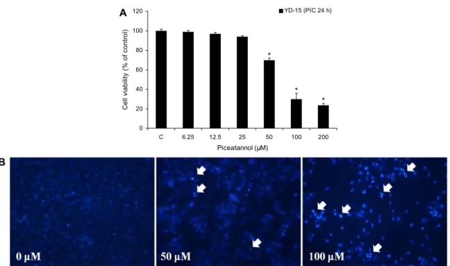
YD-15 구강암세포의 생존율에 미치는 piceatannol의 영 향을 알아보기 위하여 piceatannol을 농도(0, 6.25, 12.5, 25, 50, 100, 200 μM)별로 24시간 처리한 후 MTT assay 를 통해 관찰하였다(Fig. 2A). 각 농도별 세포 생존율은 처 리된 농도가 증가될수록 감소하였는데 50 μM 처리군의 경 우 대조군에 비해 약 30% 정도 세포 생존율 감소가 관찰되 었으며, 100 μM 처리군은 대조군에 비해 약 60% 이상 세포 생존율이 감소한 것을 관찰할 수 있었다. Choi(22)의 연구에 의하면 AGS 위암세포에 piceatannol을 0, 25, 50, 75, 100 μM로 48시간 동안 처리한 후 MTT assay를 통해 cell via- bility를 측정한 결과, 75 μM 및 100 μM의 농도에서는 대조 군에 비하여 약 50% 및 75%의 억제율을 보였다. 이러한 결과는 본 연구에서 piceatannol이 YD-15 구강암세포에서 농도 의존적으로 세포 증식을 억제시킨 것과 같은 결과로 나타나 piceatannol이 암세포 증식 억제에 효과를 보이는 것으로 사료된다. 이러한 piceatannol 처리에 의한 YD-15 세포의 생존율 저하가 apoptosis 유발에 의한 것인지 조사 하기 위하여 DAPI stain을 실시하였다. YD-15 구강암세포 에 piceatannol을 농도(0, 50, 100 μM)별로 24시간 처리한 후 DAPI 염색을 이용하여 형광현미경으로 관찰하였다(Fig.
2B). 그 결과 piceatannol을 100 μM 처치한 군에서 apop- tosis 발현세포의 증가가 관찰되었으며 MTT assay 결과와 동일하게 DAPI 염색에서도 대조군에 비해 piceatannol을 처리한 군에서 세포 수가 줄어들었고 세포질의 위축, 염색사 의 응축, apoptotic body와 같은 apoptosis 형태가 관찰되 었다. Kim 등(23)의 연구에 의하면 U937 백혈병 세포에 piceatannol을 0, 10, 20, 40, 60 μM로 48시간 동안 처리한 후 DAPI 염색을 수행한 결과 농도 의존적으로 세포의 생존 율이 감소하였으며 piceatannol을 처리한 군에서 핵과 염색 질 응축 및 apoptotic body가 관찰되었다. 이러한 결과를 종합해볼 때 piceatannol 처리에 의한 YD-15 구강암세포
0 20 40 60 80 100 120
C 6.25 12.5 25 50 100 200
Piceatannol (μM)
C e ll vi abil ity ( % of c ont ro l) .
YD-15 (PIC 24 h)
A
*
*
*
B
Fig. 2. The effect of piceatannol on cell viability and DAPI staining. (A) YD-15 cells were treated with piceatannol (0, 6.25, 12.5,
25, 50, 100, and 200 μM) for 24 h, and cell viability determined by MTT assay. The results are shown as means±standard deviation (SD) of two independent experiments performed in triplicate. Significance was determined by Dunnett's t-test with *P<0.05 considered
as statistically significant compared with non-treated controls. (B) YD-15 cells were treated with piceatannol (0, 50, and 100 μM) for 24 h, and apoptotic bodies stained with DAPI. The arrows indicate chromatin condensation in the YD-15 cells. Cleaved nuclei were examined using a fluorescence microscope (×200).의 세포생존율 감소는 apoptosis 유도에 의해 이루어지는 것으로 여겨진다.
Piceatannol이 apoptosis 경로 단백질 발현에 미치는 영향
Apoptosis는 다양한 단백질들에 의해 세포 내외 경로를 통해 조절되며, 이 중 Bcl-2 family 단백질들은 미토콘드리 아 막 투과성을 조절하는 단백질로 미토콘드리아 막에 존재 하거나 세포사멸 유도 신호에 의해 미토콘드리아 막으로 이 동하여 세포사멸을 조절하는 중요한 역할을 담당한다(24).Bcl-2 family에 속하는 몇 가지 단백질들은 apoptosis 조절 에 가장 대표적인 유전자로 알려져 있는데, 그중 Bcl-2는 anti-apoptotic 분자로서 apoptosis의 유발을 억제하는 기 능을 가지며, Bax는 대표적인 pro-apoptotic 분자로 apo- ptosis의 유발과 관계가 있다(25). YD-15 세포에서 picea- tannol에 의한 apoptosis 유도 기전을 확인하기 위해 west- ern blotting을 통하여 apoptosis를 조절하는 Bcl-2 family 단백질의 발현 양상을 확인하였다. Fig. 3에서 나타난 바와 같이 piceatannol을 처리한 군은 pro-apoptotic 인자인 Bax 단백질의 발현이 증가하였으며, anti-apoptotic 인자 인 Bcl-2 단백질은 감소하는 경향을 보였다. Zhang 등(26) 의 연구 결과 HCT116과 HT29 대장암세포에 piceatannol 을 30 μM로 48시간 동안 처리했을 때 Bcl-2의 감소와 Bax 의 증가를 확인하였다. 이러한 결과는 piceatannol이 YD- 15 구강암세포의 Bcl-2 family 단백질의 수준을 변화시킨
것과 동일한 결과로 나타나 piceatannol의 apoptosis 유도 기전은 Bcl-2 family 단백질과 연관이 있음을 확인하였다.
PARP 단백질은 손상된 DNA를 복구하는 단백질로 PARP 의 절단은 apoptosis의 대표적인 특징 중 하나이다(27).
YD-15 세포에 piceatannol을 0, 100 μM 처리하여 west- ern blotting을 통해 PARP 단백질의 발현 양상을 확인한 결과 PARP 단백질은 대조군에 비해 100 μM에서 PARP의 분절이 증가하였다(Fig. 4). 이러한 결과를 종합했을 때 pi- ceatannol은 YD-15 구강암세포의 Bcl-2 및 Bax 단백질의 발현 수준을 변화시키고 PARP의 분절을 유도하여 apopto- sis를 유도시키는 것으로 사료된다.
Xenograft를 이용하여 이식한 종양에 piceatannol이 미치 는 영향
Xenograft를 실시한 마우스의 YD-15 구강암세포 증식 에 piceatannol이 어떠한 영향을 미치는지 관찰하였다. 종 양 사이즈는 주 2회 측정하였고 piceatannol(10, 20 mg/
kg)은 DMSO에 희석시켜 주 5회 같은 시간에 3주간 복강투 여 하였으며, 대조군은 DMSO와 PBS를 희석시켜 주 5회 같은 시간에 복강투여 하였다. 그 결과 복강투여 8일째부터 대조군과 비교하였을 때 종양 사이즈가 감소하는 경향을 보 였다. 22일째에는 대조군에 비해 piceatannol을 20 mg/kg 처리한 군에서 약 50%의 억제율을 보였다(Fig. 5A). 최종 종양 사이즈는 대조군이 640 mm
3
, piceatannol 10 mg/kg0 50 100 (μM) Bax
Bcl-2
β-Actin
A
0 0.1 0.2 0.3 0.4 0.5 0.6 0.7 0.8 0.9 1
0 50 100
Piceatannol (μM)
Ba x/ β -a ct in .
**
B *
0
0.2 0.4 0.6 0.8 1 1.2
0 50 100
Piceatannol (μM)
Bc l- 2 /β -a ct in .
*
Fig. 3. The effect of piceatannol on Bcl-2 family protein in YD-15 cells. (A) YD-15 cells were treated with piceatannol (0, 50,
and 100 μM) for 24 h and cells harvested to measure protein levels of Bax and Bcl-2 by western blotting. The blots were also probed with anti-β-actin antibodies to confirm equal sample loading. (B) Each bar represents the mean±SD calculated from three independent experiments. Significance was determined by Dunnett's t-test with *P<0.05 considered as statistically significant compared
with non-treated controls.0 100 (μM) PARP
p-PARP β-Actin
A
0 0.2 0.4 0.6 0.8 1 1.2 1.4 1.6
0 100
Piceatannol (μM)
c- PARP/ PARP .
B *
Fig. 4. The effect of piceatannol on PARP protein in YD-15 cells. (A) YD-15 cells were treated with piceatannol (0 and 100
μM) for 24 h and cells harvested to measure protein levels of PARP by western blotting. The blots were also probed with anti-β-actin antibodies to confirm equal sample loading. (B) Each bar represents the mean±SD calculated from three independent experiments.Significance was determined by Dunnett's t-test with *
P<0.05 considered as statistically significant compared with non-treated controls.
을 투여한 군에서는 482 mm
3
였으며, 20 mg/kg을 투여한 군에서는 366 mm3
였다. 최종 종양의 무게는 대조군이 0.25 g, piceatannol 20 mg/kg 투여군이 0.21 g으로 picea- tannol 투여군의 종양성장을 억제하는 경향을 보였다(Fig.5B). TUNEL assay를 이용하여 적출한 종양에서 apopto- sis가 일어나는 세포를 확인한 결과 piceatannol을 투여하 지 않은 대조군과 비교 시 piceatannol을 20 mg/kg 투여한 군에서 apoptosis가 일어난 세포가 많이 관찰되었다(Fig.
5C, 5D). 이러한 결과에 따라 piceatannol은 YD-15 세포에 apoptosis를 유발하여 종양의 증식을 억제시키는 것으로 사 료된다.
Piceatannol이 종양조직에서 apoptosis 관련 단백질의 발현 및 종양성장에 미치는 영향
Xenograft를 실시한 마우스의 종양조직에서 picea- tannol이 apoptosis 관련 단백질 및 종양성장의 증식 속도 를 평가하는 단백질의 발현을 관찰하기 위해 immunohis- tochemistry를 수행하였다. Caspase는 세포사멸을 조절하 는 주요한 조절인자로 다양한 apoptosis signal의 공통적인 경로로서, initiator caspase에 의해 활성화된 caspase-3는 손상된 DNA를 복구하는 단백질인 PARP를 절단할 수 있다 (28). Ki-67은 증식하는 세포에 존재하는 핵을 검출하는 antibody로 증식하는 세포의 G1, S, G2, M기에는 핵에 존
0 100 200 300 400 500 600 700 800 900 1000
1 4 8 11 15 18 22
Days of treatment T u mo r vo lu me ( m m
3) .
Control 10 mg/kg 20 mg/kg
A
0 0.05 0.1 0.15 0.2 0.25 0.3 0.35
Control 10 20
Piceatannol (mg/kg)
T u m o r w e ig h t (g ) .
B
*
Piceatannol (mg/kg) 0 20
C
0 5 10 15 20 25 30
Control 20
Piceatannol (mg/kg)
% of apopt ot ic c e lls .
D *
Fig. 5. Piceatannol inhibits YD-15 oral cavity tumor growth and increases apoptosis. Male BALB/c nude mice received an injection
of YD-15 cells and were divided into three groups. Piceatannol was administered at a dose of 10 and 20 mg/kg five times per week, for a total 15 injections. On day 22, mice were sacrificed and tumors excised. (A) Piceatannol significantly reduced tumor volume beginning at day 8 of treatment. (B) The mean tumor weight in the piceatannol group was less than that of the control group. (C) Nude mice were treated with piceatannol (0 and 20 mg/kg) for 22 days and tumors analyzed by TUNEL assay. The slides were assessed under a microscope and photographed (×200). Paraffin-embedded tumors were sectioned to a thickness of 5 μm. (D) The percentage of labeled with TUNEL-positive apoptotic cells was calculated from 1,000 scored cells. Data presented as mean±standard error. Significance was determined by Dunnett's t-test with *P<0.05 considered as statistically significant compared
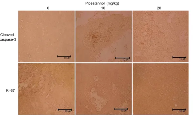
with non-treated controls.재하나 휴식세포의 핵에는 존재하지 않는다(29). 본 연구에 서는 실험동물을 희생하여 적출한 종양에서 apoptosis를 조 절하는 주요 인자인 caspase-3의 활성과 종양세포의 증식 속도를 평가하는 Ki-67을 확인하기 위해 immunohisto- chemistry를 수행하여 확인한 결과, 대조군에 비해 picea- tannol을 투여한 군에서 cleaved-caspase-3의 발현이 증 가하였으며, Ki-67의 발현은 감소한 것을 확인하였다(Fig 6). 이러한 결과에 따라 piceatannol은 YD-15 구강암세포 에 apoptosis를 유발하여 종양의 증식을 억제시키는 것으로 효과를 확인하였다.
요 약
Resveratrol 유도체의 일종으로 stilbene 계열 물질인 pi- ceatannol은 암세포의 증식을 억제하고 apoptosis를 유발 하는 것으로 알려져 있다. 본 연구에서는 YD-15 인체 구강 암세포를 대상으로 piceatannol에 의한 암세포 증식 억제와 연관된 부가적인 기전연구를 실시하였다. Piceatannol이 암 세포 성장에 미치는 영향을 확인하기 위하여 구강암세포주
YD-15에 piceatannol을 0, 6.25, 12.5, 25, 50, 100, 200 μM의 농도로 처리하고 MTT assay를 수행한 결과 picea- tannol은 농도 의존적으로 세포 성장을 억제하였다. Picea- tannol에 의한 암세포 증식 억제 효과가 apoptosis에 의한 것인지 확인하기 위해 DAPI 염색을 수행한 결과 apoptotic body와 세포질 응축이 농도 의존적으로 증가하는 것을 확인 하였다. Western blotting의 결과 piceatannol은 Bax와 cleaved-PARP 단백질의 발현을 농도 의존적으로 증가시 키고 Bcl-2 단백질 발현을 감소시켰다.
In vivo
실험에서는 누드마우스에 YD-15 구강암세포를 이종이식한 후 3주간의 piceatannol 복강투여를 통해 이식된 종양의 크기를 측정한 결과 piceatannol 처치군이 대조군에 비해 유의적으로 종양 부피가 감소하였고, 종양조직을 이용해 TUNEL assay를 수 행한 결과 piceatannol 처치군에서 TUNEL-positive cell 이 더 많이 관찰되었다. 또한 종양조직을 이용해 IHC stain 을 수행하여 cleaved-caspase-3와 Ki-67을 확인한 결과 piceatannol 처치군에서 cleaved-caspase-3가 증가하는 경향을 보였고 Ki-67은 감소하는 경향을 보였다. 따라서 piceatannol은 인체 구강암세포에서 apoptosis를 유도시키Piceatannol (mg/kg)
0 10 20
Cleaved- caspase-3
Ki-67
Fig. 6. Effect of piceatannol on expressions of caspase protein and cell proliferation markers. Male BALB/c nude mice received
an injection of YD-15 cells and were divided into three groups. Piceatannol was administered at a dose of 10 and 20 mg/kg five times per week, for a total 15 injections. On day 22, mice were sacrificed and tumors excised. To identify target protein (cleaved-cas- pase-3, Ki-67), immunohistochemistry assay was used as described in material and methods. The slides were assessed under a microscope and photographed (×200). Paraffin-embedded tumors were sectioned to a thickness of 5 μm.고 종양 형성을 억제함으로써 구강암 치료제로의 개발 가능 성이 있는 것으로 확인하였다.
감사의 글
이 논문은 2013년도 정부(교육과학기술부)의 재원으로 한 국연구재단의 지원을 받아 수행된 기초연구사업임(NRF- 2013R1A1A4A01012315)
REFERENCES
1. Lee SH, Park SY, Kim IS, Park OJ, Kim YM. 2012. Effects of resveratrol on migration and proliferation in HT-29 colon cancer cells. KSBB J 27: 289-294.
2. Choi YH. 2012. Induction of apoptosis and inhibition of NO production by piceatannol in human lung cancer A549 cells. J Life Sci 22: 815-822.
3. Kim EJ, Moon JS, Kang JI, Lee YK, Koh YS, Yoo ES, Kang HK, Yim DS. 2013. The effect of rice bran extract on the apoptosis induction of HL-60 leukemia cells. Kor J Pharma-
cogn 44: 269-274.
4. Chen K, Zhang S, Ji Y, Li J, An P, Ren H, Liang R, Yang J, Li Z. 2013. Baicalein inhibits the invasion and metastatic capabilities of hepatocellular carcinoma cells via down-reg- ulation of the ERK pathway. PLoS One 8: e72927.
5. NCC. Annual report of cancer statistics in Korea in 2011.
http://www.cancer.go.kr/ebook/84/PC/84.html (accessed Dec 2013).
6. Chandu A, Sun KC, DeSilva RN, Smith AC. 2005. The assessment of quality of life in patients who have undergone
surgery for oral cancer: a preliminary report. J Oral Maxil-
lofac Surg 63: 1606-1612.
7. NCIC. Oral cavity cancer cell. http://www.cancer.go.kr/mbs/
cancer/jsp/cancer/cancer.jsp?cancerSeq=3461&menuSeq=
3465&viewType=all&id=cancer_020112000000 (accessed Feb 2013).
8. La Vecchia C, Tavani A, Franceschi S, Levi F, Corrao G, Negri E. 1997. Epidemiology and prevention of oral cancer.
Oral Oncol 33: 302-312.
9. Kim YY. 2004. Resveratrol induces cell cycle arrest and apoptosis in cancer cells and inhibits cytotoxicity by LPS and amyloid-β in glial cells. PhD Dissertation. Pusan Nation- al University, Busan, Korea.
10. Kwon JK, Park YS, Park BK, Kim BS, Kim SK, Jung JY.
2012. Resveratrol induces apoptosis through PI3K/Akt and p53 signal pathway in MDA-MB-231 breast cancer cells.
Korean J Food Sci Technol 44: 452-459.
11. Jang M, Cai L, Udeani GO, Slowing KV, Thomas CF, Beecher CW, Fong HH, Farnsworth NR, Kinghorn AD, Mehta RG, Moon RC, Pezzuto JM. 1997. Cancer chemo- preventive activity of resveratrol, a natural product derived from grapes. Science 275: 218-220.
12. Lee JK. 2012. Neuroprotective effect of capsaicin and/or resveratrol against glutamate-induced toxicity in mouse cer- ebral cortical neurons. MS Thesis. Chungbuk National Uni- versity, Chungbuk, Korea.
13. Park HJ. 2007. The cardioprotective effects of resveratrol in hypoxic injury of myocardial cells. MS Thesis. Daegu Catholic University, Daegu, Korea.
14. Potter GA, Patterson LH, Wanogho E, Perry PJ, Butler PC, Ijaz T, Ruparelia KC, Lamb JH, Farmer PB, Stanley LA, Burke MD. 2002. The cancer preventative agent resveratrol is converted to the anticancer agent piceatannol by the cyto-
chrome P450 enzyme CYP1B1. Br J Cancer 86: 774-778.
15. Ferrigni NR, McLaughlin JL, Powell RG, Smith CR Jr.
1984. Use of potato disc and brine shrimp bioassays to de- tect activity and isolate piceatannol as the antileukemic prin- ciple from the seeds of Euphorbia lagascae. J Nat Prod 47: 347-352.
16. Matsuda H, Kageura T, Morikawa T, Toguchida I, Harima S, Yoshikawa M. 2000. Effects of stilbene constituents from rhubarb on nitric oxide production in lipopolysaccharide- activated macrophages. Bioorg Med Chem Lett 10: 323-327.
17. Rimando AM, Kalt W, Magee JB, Dewey J, Ballington JR.
2004. Resveratrol, pterostilbene, and piceatannol in vacci- nium berries. J Agric Food Chem 52: 4713-4719.
18. Hung LM, Chen JK, Lee RS, Liang HC, Su MJ. 2001.
Beneficial effects of astringinin, a resveratrol analogue, on the ischemia and reperfusion damage in rat heart. Free
Radic Biol Med 30: 877-883.
19. Lee SK, Mbwambo ZH, Chung H, Luyengi L, Gamez EJ, Mehta RG, Kinghorn AD, Pezzuto JM. 1998. Evaluation of the antioxidant potential of natural products. Comb Chem
High Throughput Screen 1: 35-46.
20. Ashikawa K, Majumdar S, Banerjee S, Bharti AC, Shishodia S, Aggarwal BB. 2002. Piceatannol inhibits TNF-induced NF-κB activation and NF-κB-mediated gene expression through suppression of IκBα kinase and p65 phosphoryla- tion. J Immunol 169: 6490-6497.
21. Choi YH. 2008. Induction of S phase arrest of the cell cycle by piceatannol is associated with inhibition of telomerase activity in human leukemic U937 cells. J Life Sci 18: 96- 102.
22. Choi YH. 2012. Piceatannol-induced G1 arrest of the cell
cycle is associated with inhibition of prostaglandin E
2
pro- duction in human gastric cancer AGS cells. J Korean SocFood Sci Nutr 41: 907-913.
23. Kim YH, Park C, Lee JO, Kim GY, Lee WH, Choi YH, Ryu CH. 2008. Induction of apoptosis by piceatannol in human leukemic U937 cells through down-regulation of Bcl-2 and activation of caspases. Oncol Rep 19: 961-967.
24. Kim EJ, Park SY, Hong JE, Shin MJ, Lim SS, Shin HK, Yoon Park JH. 2007. Inhibitory effect of the methanolic extract of Symphyocladia latiuscula on the growth of HT-29 human colon cancer cells. J Korean Soc Food Sci Nutr 36:
431-438
25. Donovan M, Cotter TG. 2004. Control of mitochondrial in- tegrity by Bcl-2 family members and caspase-independent cell death. Biochim Biophys Acta 1644: 133-147.
26. Zhang H, Jia R, Wang C, Hu T, Wang F. 2014. Piceatannol promotes apoptosis via up-regulation of microRNA-129 ex- pression in colorectal cancer cell lines. Biochem Biophys
Res Commun 452: 775-781.
27. Galluzzi L, Kepp O, Trojel-Hansen C, Kroemer G. 2012.
Mitochondrial control of cellular life, stress, and death. Circ
Res 111: 1198-1207.
28. Kim EJ, Lee YJ, Shin HK, Yoon JH. 2006. A study on the mechanisms by which the aqueous extract of Inonotus obli-
quus induces apoptosis and inhibits proliferation in HT-29
human colon cancer cells. J Korean Soc Food Sci Nutr 35:516-523.
29. Gerdes J, Lemke H, Baisch H, Wacker HH, Schwab U, Stein H. 1984. Cell cycle analysis of a cell proliferation-asso- ciated human nuclear antigen defined by the monoclonal antibody Ki-67. J Immunol 133: 1710-1715.