J. Exp. Biomed. Sci. 2012, 18(1): 16~21 pISSN : 1738-3226
The Extract of Pseudomonas aeruginosa Induces the Apoptosis of the Human Colorectal Cancer Cell Line, HCT 116 Cells, via Mitochondrial Pathway
Eun Ju Yang and Jeong Hyun Chang†
Department of Clinical Laboratory Science, Daegu Haany University, Gyeongsan 712-715, Korea
Although there are many potential cytotoxic molecules released from bacteria, the role of these molecules on the apoptosis of various cancer cells is not well understood. Pseudomonas aeruginosa (P. aeruginosa) is a Gram-negative, aerobic and rod-shaped bacterium, and has a number of virulence factors. To understand the cytotoxic effect of bacterial extracts on the colorectal cancer cell line, HCT 116 cells, we examined alteration of the cell viability, proliferation, cell cycle and apoptosis of HCT 116 cells after treatment with extract of P. aeruginosa (PaE). These cytotoxicity of PaE occurred in a time- and a dose-dependent manners. In addition, PaE arrested the cell cycle of HCT 116 cell in a time- dependent manner. PaE inhibited the protein levels of Bcl-2 and induced the release of cytochrome c from mitochondria of HCT 116 cells. The decrease of procaspase-3 was induced by the treatment of PaE. These results indicate that PaE has a cytotoxicity in HCT 116 cells via the induction of apoptosis associated with mitochondrial pathway. Therefore, PaE may used as the potential target for the treatment of colorectal cancer.
Key Words: Pseudomonas aeruginosa, HCT 116 cells, Cytotoxicity, Anti-cancer effect
서 론
매년 증가하는 암환자의 치료를 위해 꾸준히 새로운 치료제가 개발되고 방사선 치료 및 다양한 치료방법이 증가하고 있지만 여전히 항암 치료의 부작용에 대해서는 계속 언급되고 있다. 이를 극복하기 위해 부작용을 최소 로 할 수 있는 물질 개발이 필요하다. 특히 암 발생의 기 전을 파악하여 치료제를 개발하고자 연구가 진행되고 있 는데 그 중 세포자멸사를 이용한 연구가 있다. 세포자멸 사는 수명이 다한 세포를 제거하기 위해 세포 생존기간 이 일정시간 지나면 일어나는 세포 죽음의 한 기전이다.
이러한 세포자멸사를 이용하여 특정세포에 작용하면 다 양한 암 치료 및 염증 치료에 이용되기도 한다 (Johnstone et al., 2002; Kroemer and pouyssegur, 2008; Melet et al., 2008).
특히, 세균에서 세포자멸사를 유도하는 연구들이 보고되 고 있다 (Parkin, 2005). 그 중, Clostridium hystolyticum에서 추출한 물질을 암세포에 처리하였더니 추출물 중 효소성 분이 암세포의 성장을 억제함을 관찰하였다 (Chakrabarty, 2003). 세균들은 숙주에 침입한 다음 직접적으로 균체 가 항원으로 작용하거나 다양한 독성인자들을 분비하여 항원으로 작용해 질병들을 유도하는데 이 중 일부가 암세포에 세포독성 효과를 보이는 것이다. Pseudomonas aeruginosa (P. aeruginosa, 녹농균)도 exotoxin A라고 하 는 immunotoxin을 함유하고 있는데 이 물질이 세포 내 caspase 활성을 유도하여 편평상피암종의 악화를 억제한 다는 보고가 있다 (Keppler-Hafkemeyer et al., 1998; Wei et al., 2008). Caspase는 cystinyl aspartate-specific protease의 약 자로 활성부위에 cysteine을 가지고 있으며, 현재까지 알 려진 14종의 caspase들 모두가 공통적으로 자르는 위치에 서 N-terminus 방향으로 aspartate가 존재한다. 이들은 발 현이 되면 전구체로 존재하다가 세포자멸사 유도 자극 이 오면 잘려지게 되어 활성을 가진 형태로 바뀌게 된다 (Wilson et al., 1994). 이러한 caspase 활성 분자들이 암세포 에서 발현이 증가하게 되면 세포자멸사를 유도하여 암 치료 효과를 기대할 수 있다. 그러나 이전의 보고를 보면
*Received: 30 March, 2012 / Revised: 31 March, 2012 Accepted: 31 March, 2012
†Corresponding author: Jeong Hyun Chang. Department of Clinical Laboratory Science, Daegu Haany University, Gyeongsan, 712-715, Korea.
Tel: 82-53-819-1352, Fax: 82-53-819-1269 e-mail: [email protected]
○CThe Korean Society for Biomedical Laboratory Sciences. All rights reserved.
Original Article
Pseudomonas aeruginosa exotoxin A에 대한 연구가 대부분 이어서 그 외 물질들, 특히 균체 성분에 대한 연구가 미 비한 실정이다. 또한 다양한 암세포에서의 효과를 확인하 지 못하였다. 그래서 본 연구에서 여러 암유발 세포 중 대장암세포주인 HCT 116 세포에서의 증식억제 효과를 관찰하고자 한다.
대장암은 최근 꾸준히 증가하고 있는 암의 일종으로 암으로 인해 사망하는 환자의 대부분이 대장암으로 알려 져 있다 (Rychahou et al., 2008). 대장암은 급격한 세포 증 식으로 인해 발생하고 대장에서 직장을 구성하고 있는 외피세포로부터 유래된다. 비록 대장암 치료를 위해 외과 적 수술이나 다양한 화학요법을 실시하고 있지만 대부분 의 치료 효과가 극히 적거나 일부 부작용을 보이는 경우 가 있다. 그러므로 대장암 치료에 대한 새로운 물질 개발 이 시급한 실정이다. 그러기 위해 기전연구가 활발히 진 행되고 있는데, 암세포는 세포 내 신호전달 과정에서 생 존 관련 단백질들의 발현이 증가하고 세포 죽음 관련 단 백질들의 발현이 현저히 줄어드는 것이 보고되어져 왔다 (Markowitz and Bertagnolli, 2009). 특히 anti-apoptotic Bcl-2 단백질은 미토콘드리아에서 cytochrome c의 유출을 막고 MMP을 조절하여 세포자멸사를 억제한다 (Groww et al., 1999). Bcl-2에 의해 억제되는 cytochrome c는 미토콘드리 아의 전자전달계의 구성성분으로 세포 내 칼슘의 급격한 증가에 의해 세포자멸사의 초기에 관여한다 (Liu et al., 1996). 세포질로 cytochrome c이 분비되어 세포자멸사 활 성인자인 caspase 9을 활성화 시키고 활성화된 caspase 9 은 caspase 7과 caspase 3을 활성화시켜서 DNA 손상 및 세포자멸사를 일으킨다 (Liu et al., 1996).
본 연구에서는 P. aeruginosa이 함유하고 있는 물질을 HCT 116 세포에 처리하여 세포 생존 및 증식에 미치는 효과를 보고자 하였다. 세포억제 효과는 P. aeruginosa의 물질을 농도별로 처리하여 세포 생존을 확인하였고 세포 주기 및 자멸사에 미치는 효과를 관찰하고자 하였다. 또 한 세포자멸사 유도 시에 진행되는 세포 내 신호전달 과 정 중 mitochondrial pathway와 procaspase-3의 발현 변화를 확인하였다.
재료 및 방법
세포 배양
사람 대장암세포주인 HCT 116 세포 (American Type Culture Collection, Rockville, MD, USA)는 10% heat-
inactivated fetal bovine serum (FBS), 100 U/ml의 penicillin, 100 μg/ml의 streptomycin (Life technologies, Inc., Gaithersburg, MD)이 포함된 DMEM 배지 (Life technologies, Inc.)에서 배양하였다. 세포는 37℃, 5% CO2 조건으로 배양하였다.
P. aeruginosa
의 추출물 획득
P. aeruginosa는 Trypticase soy broth (BBl microbiology systems, cockeysvile, MD)에 접종하여 37℃ 배양기에서 3일 동안 배양하였다. 세균이 함유하고 있는 물질들을 얻 기 위해 배양된 세균을 모은 다음 Dulbecco's phosphate- buffered saline (DPBS)에 부유시킨 다음 50~60%의 진폭 을 설정하여 30초간 sonication한 다음 1분 동안 얼음에서 방치하고, 이 과정을 5번 반복하였다. 그 다음 원심분리 를 통해 침전물을 제거하고 다음 실험에 사용하기 전까 지 4℃에 보관하였다. 추출물은 Lowry assay를 통해 단백 질을 정량하였다.
Trypan blue exclusion test
세포 생존율은 측정하기 위해 trypan blue exclusion test를 실시하였다. 세포는 1 × 10⁵ cells/100 μl의 농도 로 배양액에 부유하여 96 well plate에 분주한 다음 P.
aeruginosa 추출물 (P. aeruginosa extract, PaE)를 첨가하여 24시간, 48시간 동안 처리하였다. 배양 후 trypsin-EDTA (Life technologies, Inc.)를 이용하여 세포를 떼어낸 다음 세포부유액을 trypan blue 염색액과 동량으로 희석하여 hemocytometer를 이용하여 세포수를 계측하였다. 살아있 는 세포는 세포질이 투명하게 보일 것이고, 반면, 죽은 세포는 trypan blue 염색약이 흡수되어 파란색으로 보일 것이다. 세포 생존율을 계산하는 방법은 아래와 같다.; 세 포 생존율 (%)은 전체 세포 중 살아있는 세포를 전체세 포수로 나눈 값에 100을 곱하여 계산하였다.
MTT assay
세포독성을 확인하기 위해 MTT assay kit (Roche, Penzberg, Germany)를 이용하여 HCT 116 세포의 생존율을 측정하였다. 세포는 1 × 10⁵ cells/50 μl의 농도로 부유한 다음 96 well plate에 분주하여 PaE를 50 μl 첨가하여 24 시간, 48시간 동안 처리하였다. 각 배양 시간이 지난 다 음, MTT (3-(4,5-Dimethylthiazol-2-yl)-2,5-diphenyltetrazolium bromide) solution을 10 μl 첨가하여 37℃, 5% CO2 배양기 에서 4시간을 배양한 다음 100 μl의 solubilization solution 을 추가로 첨가하였다. 37℃, 5% CO2 배양기에서 하루 동
안 방치한 다음 550 nm에서 흡광도를 측정하였다.
유세포 분석기를 이용한 세포주기 측정
HCT 116 세포를 trypsin-EDTA를 이용하여 떼어내고 DPBS에 부유시킨 다음 원심분리하여 상층액을 제거하는 과정을 통해 세포를 2번 세척한 다음 다시 DPBS에 부유 시켜 PI를 처리하여 15분간 염색한다. 그 후 유세포 분 석기를 통해 측정하였다. Sub G0/G1기의 세포 수를 측정 하여 세포주기를 파악하였다. Sub G0/G1기가 증가한 만큼 세포주기가 멈춘 세포의 증가로 정의하고 각 샘플 당 총 10,000개의 세포를 분석하였다.
유세포 분석기를 이용한 세포자멸사 측정
PaE를 처리한 후 trypsin-EDTA를 이용하여 세포를 떼어낸 다음 세포부유액에 annexin V-FITC와 propidium iodide (PI) (BD bioscience, San Diego, CA)를 첨가하여 실온 에서 15분간 방치한 후 염색된 세포는 CellQuest software 를 이용하여 유세포 분석기 (BD bioscience)를 통해 측정 하였다. Annexin V-FITC가 염색된 모든 세포를 세포자멸 사가 일어난 세포로 정의하고 각 샘플 당 총 10,000개의 세포를 분석하였다.
Western blotting
Trypsin-EDTA 처리하여 떼어 낸 세포를 lysis buffer (10 mM HEPES, 10 mM NaCl, 0.1 mM EDTA, 0.1 mM EGTA, 1% NP-40, 0.5 mM PMSF, 0.1 mM DTT, 0.1 mM Na3VO4, protease inhibitors)에 부유시켜 cell lysate를 만들었 다. Cell lysate는 12% sodium dodecyl sulfate-polyacrylamide gel electrophoresis (SDS-PAGE)을 통해 분리한 다음 nitrocellulose (NC) membrane으로 이동시켰다. 이동된 NC membrane은 anti-cytochrome c, anti-Bcl-2, anti-caspase-3 antibody (Santa Cruz Biotechnology, Santa Cruz, CA)와 반 응시킨 다음 enhanced chemiluminescence detection (ECL) system을 이용하여 현상하였다. 현상 후 TBST (tris- buffered saline-Tween 20)로 세척한 NC membrane에 anti- ERK2 antibody (Santa Cruz Biotechnology)을 다시 반응 시 켜 단백질 발현 대조 단백질로 사용하였다.
통계처리
모든 실험 결과를 mean ± S.D로 표현하였다. 각 대조 군과 실험군간의 통계학적의 유의성은 SPSS statistical software package (Version 10.0, Chicago, IL)을 이용하여
student's t-test로 분석하였고 P value가 0.05 이하면 통계 학적인 유의성이 있다고 판단하였다.
결 과
P. aeruginosa
추출물 (PaE)에 의한 세포독성 효과
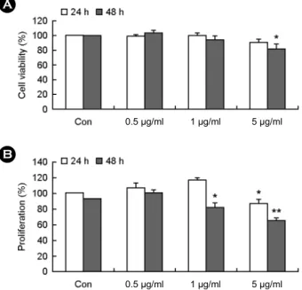
HCT 116 세포에 PaE를 각각 24 시간, 48시간 동안 처 리하고 trypan blue exclusion test로 세포 생존율을 측정하 였다. 그 결과, Fig. 1A와 같이 PaE를 처리한 다음 PaE 농도 의존적으로 HCT 116 세포의 생존율이 감소함을 보 였다. 특히, PaE을 48시간 처리 후 유의성 있게 생존율이 감소하였다. 그 다음 MTT assay를 이용하여 HCT 116 세 포의 증식율을 측정하여 PaE의 작용을 관찰하였다. Fig.
1A와 마찬가지로 PaE 처리 후에 농도 및 시간 의존적으 로 HCT 116 세포의 증식이 억제되는 것을 확인하였다 (Fig. 1B). 이는 PaE가 HCT 116 세포에 세포 생존율 및 증식율을 억제하여 세포독성 효과가 있음을 보여준다.
A
B
0.5 μg/ml 1 μg/ml 5 μg/ml 0.5 μg/ml 1 μg/ml 5 μg/ml
Fig. 1. The effect of Pseudomonas aeruginosa extract on the
viability and the proliferation of HCT 116 cells. HCT 116 cells
were incubated with 0.5~5 μg/ml of Pseudomonas aeruginosa extract (PaE) for the indicated times. (A) After treatment with PaE, viable cells were determined by trypan blue exclusion test as described in the materials and methods section. (B) After treatment with PaE, proliferation rate of the cells were determined by MTT assay as described in the materials and methods section. Data are presented in relation to the control, which was set at 100%. Data are expressed as the means ± S.D. *P<0.05 and **P<0.01 indicate a statistically significant difference between the control and PaE-treated groups at same incubation time.PaE에 의한 세포주기 조절 효과
HCT 116 세포가 PaE에 의해 세포자멸사가 유도됨을 확인하여 세포주기에도 영향을 미치는지 확인하였다. 세 포주기 중 Sub G0/G1기가 증가하면 cell cycle arrest가 진행 된 것으로 보이는데, Fig. 2에서 나타난 것과 같이 PaE를 처리하였을 때, Sub G0/G1기가 급격히 증가함을 관찰하였 다. Cell cycle arrest가 증가함으로써 PaE가 HCT 116 세포 에서 세포 증식을 억제하고 세포의 죽음을 유도하는 것 으로 보여진다.
PaE에 의한 세포자멸사 유도 효과
PaE에 의한 HCT 116 세포의 죽음이 세포자멸사인지를 파악하기 위해 앞의 방법과 동일하게 HCT 116 세포에 PaE를 5 μg/ml로 처리하여 24시간, 48시간 동안 방치한 다음 annexin-V와 PI 염색을 시행하였다. Annexin-V는 세 포자멸사 초기에 세포막에 발현되는 phosphatidyl serin (PS)에 결합하는 물질로 annexin-V가 염색된 세포는 세포 자멸사가 진행된 세포로 정의한다. 염색된 세포는 유세포 분석기를 통해 측정하였다. Fig. 3에 나타난 바와 같이 PaE에 의해 HCT 116 세포의 세포자멸사가 증가하였고 각각 24시간, 48시간 처리한 결과가 유의성 있게 나타 났다.
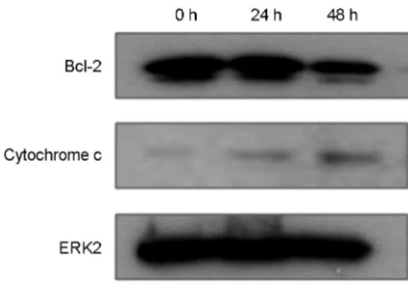
Fig. 4. The effect of Pseudomonas aeruginosa extract on the expression of cytochrome c and Bcl-2 proteins in HCT 116 cells.
HCT 116 cells were incubated for 24 h and 48 h in the absence (Con) and presence of PaE (5 μg/ml). Harvested cells were lysed and were performed Western blotting as described in the materials and methods section. The expression of cytochrome c and Bcl-2 were detected with anti-cytochrome c or anti-Bcl-2 antibody. The membrane was reprobed with anti-ERK2 as an internal control.
Fig. 5. The suppressive effect of Pseudomonas aeruginosa extract on the cleavage of procaspase-3 proteins in HCT 116 cells. HCT 116 cells were incubated for 24 h and 48 h in the
absence (Con) and presence of PaE (5 μg/ml). Harvested cells were lysed and were performed Western blotting as described in the materials and methods section. The expression of procaspase-3 were detected with anti-procaspase-3 antibody. The membrane was reprobed with anti-ERK2 as an internal control.Fig. 2. The effect of Pseudomonas aeruginosa extract on the cell cycle of HCT 116 cells. HCT 116 cells were incubated for 24 h
and 48 h in the absence (Con) and presence of PaE (5 μg/ml). Cell cycle analysis was carried out by fixation of cells in 70% ethanol, staining with PI and flow cytometry analysis of DNA content. In cell cycle, Sub G0/G1 phase were shown as means ± S.D in three individual experiments. **P<0.01 indicates a statistically significant difference between the control and PaE-treated groups at same incubation time.Fig. 3. The effect of Pseudomonas aeruginosa extract on apoptosis of HCT 116 cells. HCT 116 cells were incubated for 24 h
and 48 h in the absence (Con) and presence of PaE (5 μg/ml). The apoptosis of these cells was analyzed by measuring the binding of annexin V-FITC and PI using flow cytometry as described in the materials and methods section. The percentage of apoptotic cell in total cell population was included all annexin V binding cells. Data are expressed as the means ± SD in three individual experiments.*P<0.05 and **P<0.01 were considered a significant difference between the untreated group and PaE-treated group at same incubation time.
Mitochondrial pathway를 통한 PaE의 세포자멸사 유도
PaE에 의한 HCT 116 세포의 세포자멸사의 기전을 확 인하기 위해 mitochondrial pathway에 작용하는 세포자멸 사 관련 단백질인 cytochrome c와 Bcl-2 단백질 발현 변 화를 확인하고자 하였다. HCT 116 세포에 PaE 5 μg/ml을 각각 24시간, 48시간 처리한 다음 cytochrome c와 Bcl-2 단백질을 확인한 결과 cytochrome c의 발현이 증가한 반 면 Bcl-2의 발현은 감소하였다 (Fig. 4). 또한 세포자멸사 의 강력한 실행자인 procaspase-3의 활성을 확인해본 결 과 procaspase-3의 발현이 PaE 처리 24시간 때부터 서서 히 감소하기 시작하여 48시간 때에 급격히 감소하였다.
이러한 결과는 PaE가 HCT 116 세포의 미토콘드리아에서 cytochrome c와 Bcl-2의 발현을 조절하여 세포자멸사를 유도함을 보여준다 (Fig. 5).
고 찰
P. aeruginosa의 추출물인 PaE가 HCT 116 세포에서 세 포 생존율 및 증식율을 억제하고 세포주기를 비정상적으 로 차단함으로써 세포 증식 억제를 유도함을 관찰하였다.
또한 mitochondrial pathway인 Bcl-2와 cytochrome c를 조 절하여 세포고사를 유도함으로써 암세포 제거에 관여하 였다. P. aeruginosa의 독성인자들은 세포자멸사를 통해 세포 죽음을 유도한다 (Andersson et al., 2004; Adams and Cooper, 2007).
HCT 116 세포는 대장암세포로써, 이에 대한 발병기전 및 항암 효과 연구가 꾸준히 진행되어 왔지만 아직 부족 한 실정이다 (Brattain et al., 1981; Santoro and Groden, 1997;
Bender et al., 1998). 그러나, Fig. 3과 Fig. 5에서처럼 PaE가 HCT 116 세포에서 caspase 3 pathway를 거쳐 세포자멸사 를 유도함을 확인하였다. 이러한 세포자멸사는 프로그램 된 세포의 죽음을 표현하는 용어로, 죽는 세포가 면역반 응을 일으켜서 주위세포 및 조직에 악영향을 주는 부작 용 없이 phagocyte에 의해 청소가 될 수 있는 형태로 진 행되게 된다. 그 후 핵의 분해 및 세포소기관의 변화가 발생하여 세포자멸사체를 형성하는 등의 특징을 가지게 된다. 특히 caspase-3는 세포자멸사 실행자로 잘 알려져 있는 물질로 세포 내 다양한 신호전달 과정으로 조절된 다. Caspase는 불활성 형태의 효소로 합성되어 있다가 세 포자멸사 신호에 의하여 단백질이 분해됨으로써 활성화 된다. 처음 세포자멸사 유도 물질이 세포를 자극하게 되
면 세포 내 신호전달 단백질들의 발현 및 활성이 조절되 어 결국 caspase cascade를 작동시켜 마지막엔 procaspase- 3가 활성 형태로 분해되어 급속도로 세포자멸사가 일어 난다. 특히, Bcl-2나 Bax 등의 세포자멸사 관련 단백질 들의 작용에 의해 미토콘드리아 내에서 cytochrome c가 분비되어 세포자멸사를 촉진시킨 것으로 알려져 있다 (Yang and Chang, 2011). PaE 역시 HCT 116 세포의 세포자 멸사 유도 시 Bcl-2 단백질 발현을 억제하고 cytochrome c의 발현을 증가시켜 procaspase-3의 분해를 촉진하였다 (Fig. 4, Fig. 5). 또한 PaE는 HCT 116 세포의 세포주기 중 Sub G0/G1기를 증가시켰다 (Fig. 2). 세포주기는 세포분열 기인 M기와 DNA 합성기인 S기가 반복적으로 일어나면 서 세포분열이 지속되고 있고, 이때 M기와 S기 사이에 휴지기인 G1 또는 G2기가 존재한다. 그러나 G1기로 진 행되지 못하고 그 전 단계인 Sub G0/G1기가 증가하면 세 포 증식이 진행되지 못하고 결국 세포죽음이 일어난다 (Peng et al., 2005). 결국 PaE가 cell cycle을 억제하여 세포 증식을 차단함으로써 세포죽음을 유도하는데 관여함을 나타낸다.
P. aeruginosa가 일으키는 세포자멸사 유도 물질로 예상 되는 것 중 Pseudomonas exotoxin A (PE)이 있는데 이 독 성인자는 fibroblast에서 세포 죽음을 억제하고 생존을 지 속시키는데 관여하는 단백질인 Mcl-1의 분해를 촉진시키 고, 반대로 세포자멸사를 유도하는 단백질인 Bak의 발 현을 증가시켜서 세포자멸사를 유도한다고 보고되었다 (Du et al., 2010). 또한 pyocyanin은 P. aeruginosa가 분비 하는 색소로 선충류인 Caenorhabditis elegan에 산화적 스트레스를 일으켜 세포죽음을 유도하는 독성인자이다 (Cooper et al., 2003). Phospholipid를 분해하는 효소인 phospholipase C (PLC) 역시 P. aeruginosa에 존재하면서 다른 세포의 세포막에 손상을 주어 세포 죽음을 유도하 는 독성인자로 작용한다 (Songer, 1997).
결론적으로, PaE가 HCT 116 세포의 세포주기의 진행 을 차단하고, 세포 내에서 Bcl-2를 억제함으로써 미토 콘드리아 안으로부터 cytochrome c의 유출이 증가하여 procaspase-3의 활성화를 통해 세포자멸사를 유도하다. 이 를 통해 HCT 116 세포의 죽음을 유의하게 증가하였고, PaE가 암 발생 및 진행을 억제하는 잠재적인 효과가 있 을 것으로 보이고, 앞으로 구체적인 세포자멸사 기전을 파악하여 대장암 치료제 개발에 기여할 수 있다고 사료 된다.
REFERENCES
Adams KW, Cooper GM. Rapid turnover of mcl-1 couples translation to cell survival and apoptosis. J Biol Chem. 2007.
282: 6192-6200.
Andersson Y, Juell S, Fodstad Q. Downregulation of the anti- apoptotic MCL-1 protein and apoptosis in MA-11 breast cancer cells induced by an anti-epidermal growth factor receptor-Pseudomonas exotoxin A immunotoxin. Int J Cancer.
2004. 112: 475-483.
Bender CM, Pao MM, Jones PA. Inhibition of DNA methylation by 5-Aza-2'- deoxycytidine suppresses the growth of human tumor cell lines. Cancer Research. 1998. 58: 95-101.
Brattain MG, Fine WD, Khaled FM, Thompson J, Brattain DE.
Heterogeneity of malignant cells from a human colonic carcinoma. Cancer Research. 1981. 41: 1751-1756.
Chakrabarty AM. Microorganisms and Cancer: Quest for a therapy.
J Bacteriol. 2003. 185: 2683-2686.
Cooper M, Tavankar GR, Williams HD. Regulation of expression of the cyanide-insensitive terminal oxidase in Pseudomonas aeruginosa. Microbiology. 2003. 49. 1275-1284.
Du X, Youle RJ, FitzGerald DJ, Ira P. Pseudomonas Exotoxin A-Mediated apoptosis is Bak dependent and preceded by the degradation of Mcl-1. Molecular and Cellular Biology. 2010.
30: 3444-3452.
Groww A, McDonnell JM, Korsmeyer SJ. Bcl-2 family members and the mitochondria in apoptosis. Genes Dev. 1999. 13: 1899 -1911.
Johnstone RW, Ruefli AA, Lowe SW. Apoptosis: a link between cancer genetics and chemotherapy. Cell. 2002. 108: 153-164.
Keppler-Hafkemeyer A, Brinkmann U, Pasan I. Role of caspases in immunotoxin-induced apoptosis of cancer cells. Biochemistry.
1998. 37: 16934-16942.
Kroemer G, Pouyssegur J. Tumor cell metabolism: cancer's Achilles' heel. Cancer Cell. 2008. 13: 472-482.
Liu X, Kim C, Yang J, Jemmerson R, Wang X. Induction of apoptotic program in cell-free extracts: requirement for dATP and cytochrome c. Cell. 1996. 86: 147-157.
Markowitz SD, Bertagnolli MM. Molecular basis of colorectal cancer. N Engl J Med. 2009. 361: 2449-2460.
Melet A, Song K, Bucur O, Jagani Z, Grassian AR, Khosravifar R.
Apoptotic pathways in tumor progression and therapy. Adv Exp Med Biol. 2008. 615: 47-79.
Parkin DM, Bray J, Ferlay J, Pisani P. Global cancer statistics 2002. CA Cancer J Clin. 2005. 55: 74-108.
Peng CH, Huang CN, Wang CJ. The anti-tumor effect and mechanisms of action of penta-acetyl geniposide. Current Cancer Drug Targets. 2005. 5: 299-305.
Rychahou PG, Kang J, Gulhati P, Doan HQ, Chen LA, Xiao SY, Chung DH, Evers BM. Akt2 overexpression plays a critical role in the establishment of colorectal cancer metastasis.
Proc Nat Acad Sci USA. 2008. 105: 20315-20320.
Santoro IM, Groden J. Alternative splicing of the APC gene and its association with terminal differentiation. Cancer Research.
1997. 57: 488-494.
Songer JG. Bacterial phospholipases and their role in virulence.
Trends Microbiol. 1997. 5: 156-161.
Wei MQ, Mengesha A, Good D, Anné J. Bacterial targeted tumour therapy-dawn of a new era. Cancer Letters. 2008.
259: 16-27.
Wilson KP, Black JA, Thomson JA, Kim EE, James PG, Manuel AN, Murcko MA, Chambers PS, Aldape RA, Raybuck SA, Livingston DJ. Structure and mechanism of interleukin-1 beta converting enzyme. Nature. 1994. 370: 270-275.
Yang EJ, Chang JH. Potassium cyanate induces apoptosis of human colorectal cancer cell via mitochondrial pathway. J Exp Biomed Sci. 2011. 17: 177-184.