INTRODUCTION
Ovarian cancer is the fourth most common cancer in women and is the leading cause of death from all gynecolog- ical carcinomas (Zhang et al. 2004; Dundr 2010). Recently, pathogenesis of this disease remains to be elucidated and novel therapeutic agents are constantly being explored (Dundr 2010; Kandalaft et al. 2010). Moreover, the available exper- imental/human evidence on natural products intake and risk of ovarian cancer is limited. Thus, novel therapeutic appro- aches are direly needed. The phenethyl ester of caffeic acid (CAPE) is an active component of honeybee propolis extract and is used as a traditional medicine in the Far East (Grun- berger et al. 1988), and has been shown to acts as an antiviral (Ho et al. 2005; Fruehauf and Meyskens 2007), anti-inflam-
matory (Orban et al. 2000; Lee et al. 2010), antiatherosclero- tic agent (Hishikawa et al. 2005; Ho et al. 2009), and anti- tumoral (Nagaoka et al. 2003; Demestre et al. 2009; Szliszka et al. 2009) in diverse systems. Although these studies sug- gest that at least some of these effects of CAPE are due to its anticancer effects, it is not known whether CAPE can inhibits cell proliferation or induces apoptosis in human ovarian cancer cells. Therefore, we investigated the antican- cer effect of CAPE in human ovarian cancer cell line, OVCAR3 and demonstrated for the first time that CAPE suppressed cell proliferation and induced apoptosis in ovarian cancer cells.
MATERIALS AND METHODS
1. Cell culture
The OVCAR3 cells, human ovarian cancer cell line, were obtained from the American Type Culture Collection (Rock-
─
─ 196 ──
Caffeic Acid Phenethyl Ester Inhibits Cell Proliferation and Induces Apoptosis in Human Ovarian Cancer Cells
Hyung Joo Park†, Seung Joo Yang†, Jin Young Mo, Geun Chang Ryu1and Kyung Jin Lee*
Asan Institute for Life Sciences, Asan Medical Center, Seoul, Korea
1Department of Optometry and Optic Science, Dongshin University, Najoo, Korea
Abstract -- The phenethyl ester of caffeic acid (CAPE), an active component of honeybee propolis extract, is shown to inhibit cancer growth previously. However, studies on human ovarian cancer are largely obscure. This study evaluated the effects of CAPE as a potential anti-proliferative and pro-apoptotic agent in the human ovarian cancer line, OVCAR-3. CAPE treated OVCAR-3 cells showed inhibition of cell viability and proliferation in a dose-dependent manner by WST-1 assay, LDH assay and bromodeoxyuridine (BrdU) incorporation assay. Furthermore, CAPE-mediated OVCAR-3 cell growth inhibition was associated with apoptotic changes as evident by cell cycle arrest and accumulation of cells in the apoptotic phase and DNA fragmentation. Taken together, CAPE inhibits cell proliferation via DNA synthesis reduction and induces apoptotic cell death via DNA damage, thus elucidating a novel, plausible mechanism of CAPE anti-tumorigenic property in OVCAR-3 cells.
Key words : phenethyl ester of caffeic acid, DNA fragmentation, apoptosis, OVCAR-3
†This authors contributed equally to this work
* Corresponding author: Kyung Jin Lee, Tel. 02-3010-4147, Fax. 02-3010-8566, E-mail. [email protected]
ville, MD, USA). Tissue culture media and fetal bovine serum (FBS) were purchased from Gibco (Carlsbad, CA, USA).
The CAPE and all the other reagents, except where indicated, were purchased from sigma chemical company (St. Louis, Mo, USA).
CAPE was dissolved in DMSO and stored at -20�C. The OVCAR3 cell line was maintained in RPMI1640 medium supplemented with 10% FBS, penicillin (100 units mL-1), streptomycin (100μg mL-1) and 1 mM glutamine. The cells were cultured at 37�C in a humidified atmosphere under 5%
CO2. For all the experiments, unless otherwise stated, 1×10³ cells were seeded in 96-well plates and grown to confluence for 24 hrs, prior to being treated with CAPE or the carrier solvent DMSO. The final concentrations of CAPE used for all the experiments were prepared by diluting the stock with cell culture media. The cells were then treated with either the vehicle (DMSO) or CAPE at various concentrations for 48 hrs.
2. Cell viability
Cell viabilities were determined using WST-1 assays, and by measuring LDH release, which was quantified in super- natants using LDH kits. WST-1 and LDH assays were both performed according to the manufactures’ instructions des- cribed previously (Lee et al. 2008a).
3. Cell proliferation
Cell proliferation was measured BrdU incorporation in place of thymidine during DNA synthesis before cell division by DNA precursor analog incorporation assay kit (Roche Diagnostics, Indianapolis, USA.) In brief, both the vehicle and CAPE-treated cells were labeled with BrdU (10μM) for 6 h prior to incubation with anti-BrdU-peroxidase (10μL well-1) for 2 hrs. The immune complex was detected follow- ing the addition of trimethyl benzidine substrate and measur- ed at 450 nm using an ELISA reader. Cell proliferation was expressed as the % BrdU incorporation.
4. Cell cycle analysis
Flow cytometry was used for cell cycle analysis. In brief, 3×10⁵cells were cultured in 6-well plates and treated with CAPE or vehicle for 48 hrs. Both the floating and adherent cells were collected and washed twice with DMSO prior to staining using propidium iodide. Modfit software (BD Bio-
sciences, Mountain View, CA, USA) were used for data acquisition and analysis.
5. Caspase-3 activity and DNA fragmentation analysis
Caspase-3 assay and DNA Fragmentation were both per- formed according to the manufactures’ instructions as desc- ribed previously (Lee et al. 2008b). Briefly, cell lysates were incubated in reaction buffer in the presence of 50μM fluoro- genic substrates (Biomol, Germany), preferentially cleaved by caspase-3 (AC-DEVD-AMC). Cleavage of the fluoroge- nic substrates for 60 min at 37�C was measured using a fluorescence reader (Varioskan, Thermo Electon Co.) at an excitation wavelength of 380 nm and an emission wavelength of 460 nm. DNA samples for DNA laddering were extracted from lysates using a total DNA separator kit (Promega, USA) and electrophoretically separated in a 0.9% agarose gel, which were treated ethidium bromide and visualized and photographed under ultraviolet light.
6. Statistical analysis
All data were expressed as the mean±S.D. from three independent experiments, performed in triplicate. Statistical significance was determined by one-way analysis of variance (ANOVA) followed by the student t test, using p⁄0.05 as the level of significance.
RESULTS
1. Effect of CAPE on cell cytotoxicity and proliferation
The cytotoxicity effects of CAPE in OVCAR3 cells were quantified using LDH and WST-1 assays. CAPE fully induc- ed cell cytotoxicity in a dose-dependent manner based on WST-1 assay results (Fig. 1A). In particular, CAPE also induced the leakage of LDH from OVCAR3 cells (Fig. 1B).
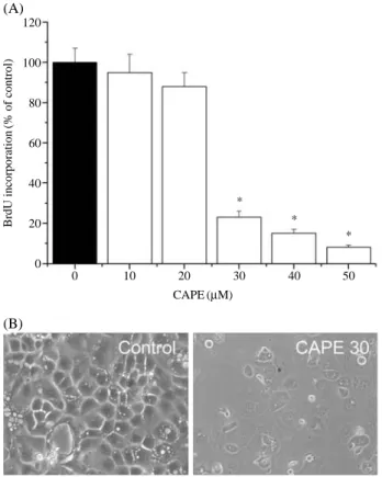
Moreover, CAPE inhibited DNA synthesis in a dose-depen- dent manner as evident by the observed decrease in the incor- poration of BrdU into DNA in the CAPE-treated cells com- pared to the controls (Fig. 2A).
2. Effects of CAPE on cell cycle
To explore CAPE on cell cycle arrest in ovarian cancer
cells, we further analyzed FACS analysis. As the results, CAPE increased the percentage of OCVAR-3 cells in the
G1-phase (Fig. 3A) with an associated decrease in the per- centage of cells in S-phase (DNA synthesis) (Fig. 3B) and G2-phase (Fig. 3C) of the cell cycle when compared to the untreated controls. Furthermore, the percentage of cells in the apoptosis phase (G0 arrest) was increased when compared to the controls (Fig. 3D).
3. Effects of CAPE on apoptosis
The CAPE-mediated OVCAR-3 inhibition of cell prolifera- tion/survival was associated with apoptotic changes. At CAPE concentrations where inhibition of cell proliferation/
survival was observed (10~50μM), the cells were shrunken, rounded and detached from the cell culture substratum (Fig.
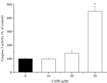
2B). Furthermore, DNA fragmentation/laddering (character- istic of apoptosis) was observed when the cells were treated with CAPE, indicating cell death by apoptosis in these cells while no DNA laddering was observed in the untreated cells (controls) (Fig. 4). The apoptotic cells were identified by the caspase-3 assay kit. As the result, CAPE-treated cells increased in a does dependent manner, when compared to control cells (Fig. 5).
DISCUSSION
The flavonoids and phenolic components found in propolis are known to affect the apoptosis of various cancer cells and may play an important role in cancer chemoprevention (Shukla and Gupta 2006; Syed et al. 2008; Demestre et al.
Fig. 1. Effect of CAPE on cytotoxicity of OVCAR-3 cells. The cells were seeded at an initial density of 1×10³cells well-1. Cells were treated with various concentrations of CAPE were treated for 48 hrs. Cell viability were estimated by the WST-1 assay (A) and LDH leakage assay (B), as described in Materials and Methods. The cytotoxicity was determined by relative absorbance normalized to the control cells. Each bar represents the mean±S.D. calculated from three independent experiments. *p⁄0.05 Compared to control.
Cell viability (% of control) LDH leakage (fold of control)
120
100
80
60
40
20
0
4
3
2
1
0
0 10 20 30 40 50 0 10 20 30 40 50
CAPE (μM) CAPE (μM)
* *
*
*
*
*
(A) (B)
BrdU incorporation(% of control)
(B) (A)
CAPE (μM)
0 10 20 30 40 50
120
100
80
60
40
20
0
*
*
*
Fig. 2. Effect of CAPE on proliferation of OVCAR-3 cells. Cells were treated with various concentrations of CAPE were treat- ed for 48 hrs. (A) DNA synthesis was measured by BrdU incorporation assay as described in Materials and Methods.
(B) Cellular morphology of OVCAR-3 cells. Each bar repre- sents the mean±S.D. calculated from three independent experiments. *p⁄0.05 Compared to control.
2009; Szliszka et al. 2009). The present study showed that CAPE inhibited the viability of OVCAR-3 cells. This study Fig. 4. DNA fragmentation analysis of OVCAR-3 cells treated with
CAPE for 48 hrs. Lane 1: Protein marker (M); lane 2: vehicle- treated cells; lane 3; CAPE (10μM)-treated cells; lane 4:
CAPE (20μM)-treated cells; lane 5: CAPE (30 μM)-treated cells.
CAPE (μM)
0 10 20 30
Fig. 3. Effect of CAPE on cell cycle progression. After 48 hrs of CAPE- treatment, the cell cycle phase distribution was determined by FACS analysis as described in Materials and Methods. Each bar represents the mean±S.D. calculated from three independent experiments.
*p⁄0.05 Compared to control.
G1 phase(%)G2 phase(%) S phase(%)G0 arrest(%)
80
70
60
50
40
30
20
70
60
50
40
30
20
20
15
10
5
10
8
6
4
2
0
0 30 0 30
0 30 0 30
CAPE (μM) CAPE (μM)
CAPE (μM) CAPE (μM)
*
*
*
* (A)
(C)
(B)
(D)
Caspase-3 activity (% of control)
500
400
300
200
100
0
0 10 20 30
CAPE (μM)
Fig. 5. Effect of CAPE on caspase-3 activation of OVCAR-3 cells.
After 48 hrs of CAPE-treatment, the caspase-3 activation was determined as described in Materials and Methods.
Each bar represents the mean±S.D. calculated from three independent experiments. *p⁄0.05 Compared to control.
*
had the advantage that the MTT and LDH assay was used, which measures the mitochondrial activity and cell membrane damage of cells. CAPE-treated cells lost the ability to provi- de and maintain energy for metabolic activity and survival indicating that CAPE caused cellular damage. DNA replica- tion occurs before cell division/doubling, thus the measure- ment of DNA synthesis, is an attractive marker for cell pro- liferation. A decrease in BrdU-labeled DNA was noted in the CAPE treated cells, suggesting that CAPE is an inhibitor of DNA synthesis and cell proliferation in OVCAR-3 cells.
Moreover, the percentage of OVCAR-3-treated cells in the apoptotic phase was significantly increased as compared to the untreated cells, suggesting a block in cell cycle progres- sion leading to programmed cell death. This was in agreement with previous studies which established that apoptosis is the likely mechanism of action of honeybee propolis extract for inhibiting cell growth (Kamatou et al. 2008). Thus, the activa- tion of apoptosis is believed to play a critical role in both the chemo-prevention and treatment of human carcinomas.
The biochemical hallmark of apoptosis is genomic DNA fragmentation and caspase-3 activity increasing that commits the cell to die. The nuclear endonuclease enzyme cleaves DNA resulting in DNA fragments through some kind of caspase activites. In the previous study, CAPE has the potent prooxidant potential in DNA cleavage, and ortho-dihydroxyl functionality of CAPE may be a major determination of its prooxidant activity (Wang et al. 2008). In present study, CAPE confirmed by the labeling the DNA fragments and Caspase-3 assay, and indicating that CAPE induced apopto- sis in the OVCAR-3 cells by causing DNA damage. In recent study, ethanolic extract of propolis augments TRAIL- induced apoptotic death in prostate cancer cells (Szliszka et al.
2009). Because its phenolic components may be one of the mechanisms responsible for their cancer preventive effects, more studies are needed to further explore this mecanisms.
Taken together, the anti-proliferative and proapoptotic properties of CAPE in the human ovarian cancer cell line, OVCAR-3, were supported by the observed reduction in cell survival and DNA synthesis, as well as cell cycle arrest and the accumulation of cells in the apoptotic phase caused by DNA damage.
ACKNOWLEDGEMENT
This study was supported by Technology Development
Program for Agriculture and Forestry and a grant (2010- 505) from the Asan Institute for Life Sciences, Seoul, Korea.
REFERENCES
Demestre M, SM Messerli, N Celli, M Shahhossini, L Kluwe, V Mautner and H Maruta. 2009. CAPE (caffeic acid phene- thyl ester)-based propolis extract (Bio 30) suppresses the growth of human neurofibromatosis (NF) tumor xenografts in mice. Phytother. Res. 23:226-230.
Dundr P. 2010. Ovarian carcinoma: current diagnostic principles.
Cesk. Patol. 46:53-61.
Fruehauf JP and FL Meyskens Jr. 2007. Reactive oxygen species:
a breath of life or death? Clin. Cancer. Res. 13:789-794.
Grunberger D, R Banerjee, K Eisinger, EM Oltz, L Efros, M Caldwell, V Estevez and K Nakanishi. 1988. Preferential cytotoxicity on tumor cells by caffeic acid phenethyl ester isolated from propolis. Experientia 44:230-232.
Hishikawa K, T Nakaki and T Fujita. 2005. Oral flavonoid supplementation attenuates atherosclerosis development in apolipoprotein E-deficient mice. Arterioscler Thromb. Vasc.
Biol. 25:442-446.
Ho CC, SS Lin, MY Chou, FL Chen, CC Hu, CS Chen, GY Lu and CC Yang. 2005. Effects of CAPE-like compounds on HIV replication in vitro and modulation of cytokines in vivo. J. Antimicrob. Chemother. 56:372-379.
Ho HC, SL Hsu, CT Ting, CY Kuo and VC Yang. 2009. Caffeic acid phenethyl ester inhibits arterial smooth muscle cell proliferation and migration in vitro and in vivo using a local delivery system. Cell Mol. Biol. (Noisy-le-grand) 55 Suppl:
OL1161-1167.
Kamatou GP, NP Makunga, WP Ramogola and AM Viljoen.
2008. South African Salvia species: a review of biological activities and phytochemistry. J. Ethnopharmacol. 119:664- 672.
Kandalaft LE, DJ Powell Jr., N Singh and G Coukos. 2010.
Immunotherapy for Ovarian Cancer: What’s Next? J. Clin.
Oncol. In press.
Lee KJ, JH Choi, YP Hwang, YC Chung and HG Jeong. 2008a.
Protective effect of caffeic acid phenethyl ester on tert-butyl hydroperoxide-induced oxidative hepatotoxicity and DNA damage. Food Chem. Toxicol. 46:2445-2450.
Lee KJ, JH Choi, HG Kim, EH Han, YP Hwang, YC Lee, YC Chung and HG Jeong. 2008b. Protective effect of saponins derived from the roots of Platycodon grandiflorum against carbon tetrachloride induced hepatotoxicity in mice. Food Chem. Toxicol. 46:1778-1785.
Lee Y, DH Shin, JH Kim, S Hong, D Choi, YJ Kim, MK Kwak
and Y Jung. 2010. Caffeic acid phenethyl ester-mediated Nrf2 activation and IkappaB kinase inhibition are involved in NFkappaB inhibitory effect: structural analysis for NFkap- paB inhibition. Eur. J. Pharmacol. 643:21-28.
Nagaoka T, AH Banskota, Y Tezuka, K Midorikawa, K Mat- sushige and S Kadota. 2003. Caffeic acid phenethyl ester (CAPE) analogues: potent nitric oxide inhibitors from the Netherlands propolis. Biol. Pharm. Bull. 26:487-491.
Orban Z, N Mitsiades, TR Burke Jr., M Tsokos and GP Chousos.
2000. Caffeic acid phenethyl ester induces leukocyte apop- tosis, modulates nuclear factor-kappa B and suppresses acute inflammation. Neuroimmunomodulation 7:99-105.
Shukla S and S Gupta. 2006. Molecular targets for apigenin- induced cell cycle arrest and apoptosis in prostate cancer cell xenograft. Mol. Cancer Ther. 5:843-852.
Syed DN, Y Suh, F Afaq and H Mukhtar. 2008. Dietary agents
for chemoprevention of prostate cancer. Cancer Lett. 265:
167-176.
Szliszka E, ZP Czuba, J Bronikowska, A Mertas, A Paradysz and W Krol. 2009. Ethanolic Extract of Propolis Augments TRAIL-Induced Apoptotic Death in Prostate Cancer Cells.
Evid Based Complement Alternat. Med.
Wang T, LX Chen, Y Long, WM Wu and R Wang. 2008. DNA damage induced by caffeic acid phenyl ester in the presence of Cu (II) ions: potential mechanism of its anticancer proper- ties. Cancer Lett. 263:77-88.
Zhang M, AH Lee, CW Binns and X Xie. 2004. Green tea con- sumption enhances survival of epithelial ovarian cancer.
Int. J. Cancer 112:465-469.
Manuscript Received: October 25, 2010 Revision Accepted: November 10, 2010 Responsible Editor: Hak Young Lee