ISSN 2233-4084
Introduction
Symphysis in which the mandibular incisor is located becomes the anatomical limits1 of tooth movement. When excessive force is applied during orthodontic treatment, teeth can touch the cortical plate of alveolus which causes resorption of corti- cal bone, fenestration, dehiscence, root exposure, external root resorption, and gingival recession.2,3 It has been reported that they have occurred in almost all adults that underwent orthodontic treatment.4 In general, Class I bi-dentoalveolar protrusive patients who need premolar extraction with maximum retrac- tion of the incisors, bone defects must be observed
carefully before orthodontic treatment and anatomi- cal characteristics must be considered when planning tooth movements. Particularly for symphysis which is a thin narrow part, special care must be taken for the lingual movement of mandibular incisor.5 Excessive lingual movement of mandibular incisors bring about irreversible resorption of the lingual alveolar bone, which results in permanent recession of the lingual alveolar bone.3,6 After 4-month retention period with penetrated cortical plate, it was insufficient to com- pletely cover the root, and the perforation site of the bone could be repaired only by its relapse to original position.7
Because the conventional two-dimensional lateral
*Correspondence to: Min-Ji Kim, DDS, MSD, PhD
Ewha Womans University, Mokdong Hospital, 911-1 Mok-dong, Yangcheon-gu, Seoul, 158-710, Republic of Korea
Tel: +82-2-2650-5112, Fax: +82-2-2650-5764, E-mail: [email protected] Received: June 23, 2014/Last Revision: August 11, 2014/Accepted: August 15, 2014
Morphological difference of symphysis according to various skeletal types using cone-beam computed tomography
Hyun-Jin Kwon, Youn-Sic Chun, Min-Ji Kim*
Graduate School of Clinical Dentistry, Ewha Womans University, Seoul, Korea
Purpose: The aim of this study was to investigate differences between the morphology of the mandibular symphysis and four facial skeletal types. Materials and Methods: 40 cone-beam computed tomographies were selected and classified in to 4 groups according to their vertical and anterior-posterior skeletal patterns. The bone volume (mm3) of the symphysis, the cross sectional area corresponding to the 4 mandibular incisors’ axis: the cross sectional area of total bone (mm2), the area of the cancellous bone (mm2) and the thickness (mm) of labial and lingual alveolar bone at 2 mm, 3 mm under the cemento-enamel junction (CEJ) were measured. General linear model (GLM), Kruskal-Wallis test and Tukey honestly significant difference (HSD) test were subsequently used for statistical analysis. Results: The lingual cortical bone thickness of the lateral incisors at 2, 3 mm under CEJ was greater in the Class I low angle group than the other 3 groups (P < 0.05). There were no statistically significant differences in the volume of the mandibular incisor bony support, cross-sectional area of total bone and cancellous bone at the mandibular incisor’ axis. Conclusion:
Patients in Class I, low angle group have a thicker lingual mandibular symphysis than Class I, high angle patients. (J Dent Rehabil Appl Sci 2014;30(3):215-22)
Key words: facial skeletal type; mandibular symphysis; CBCT; volume of mandibular symphysis
Copyright© 2014 The Korean Academy of Stomatognathic Function and Occlusion.
It is identical to Creative Commons Non-Commercial License.
cc
cephalometric images cannot accurately provide the thickness of the alveolar bone and bone density, ex- presses at least 5% enlargement,8 and does not show the concave surface of symphysis properly, the ac- tual symphysis is narrower than the image.3 To solve these limits of 2D images, the use of Cone-beam CT (Cone-beam computed tomography, CBCT) is increasing in dental treatment these days.9,10
Previous studies on shapes of symphysis according to skeletal pattern were mostly done by 2D cepha- lometrcis having limitations. Therefore, this study intended to examine any differences in mandibular shapes by measuring the sectional area, thickness, and volume of symphysis according to Class I Class II skeletal patterns and vertical (high, low) facial skel- eton types.
Materials and Methods
1. Materials
The subjects of this study were 658 patients who visited the orthodontic department of Ewha Wom- ans University Mokdong Hospital between Febru- ary 2011 and April 2012 and took CBCT. Patients who were 18 years old or younger, who had received orthodontic treatment, who had severe crowding in the mandibular incisor (Little’s irregularity ≥ 6),11 whose number of teeth was abnormal (supernumer- ary or missing teeth), who had abnormal shapes of teeth (giant or dwarf teeth), or whose IMPA was out of the average range (90 < IMPA < 102)12 were
excluded. The subjects were divided into Class I (0˚- 4˚) or Class II (4˚ or greater) based on ANB angle13 and into low angle (25 - 32˚) or high angle (38 - 49˚) based on the SN-MP angle.14 Thus, they were classi- fied into four groups (Class I low angle, Class I high angle, Class II low angle and Class II high angle). As a result, a total of 40 subjects (7 males, 33 females) were chosen (Table 1). This study was approved by the Institutional Review Board of Ewha Womans University Medical School Mokdong Hospital (ECT 11-24-01).
2. Methods 1) CBCT data
The images taken with CBCT (DinnovaTM, Willmed, Seoul, Korea, 9 mA, 80 kV, 24 s, 20 cm × 15 cm field of view) were saved as DICOM (digital imaging and communication in medicine) files. Lateral cephalo- metric images were obtained using the X-ray Genera- tion Module of OnDemand 3DTM (Cybermed Inc., Seoul, Korea). They were saved as DICOM files and the facial skeletons were analyzed through cephalo- metric radiography analysis program (V-cephTM 5.5, OSSTEM Inc., Seoul, Korea).
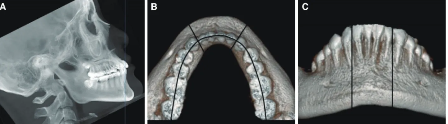
The volume (mm3), sectional areas (total sectional area and the sectional area of cancellous bone; mm2), and thickness (mm) of the alveolar bone were mea- sured. The DICOM file was reoriented so that the mandibular central incisor would be perpendicular to the horizontal plane (Fig. 1A). The mandible was cut in such a way that it was perpendicular to the
Table 1. General characteristics of subjects in four subgroups (Mean ± SD)
Class I Class II
Low High Low High
Number of subjects (M/F) 10 (2/8) 10 (2/8) 10 (1/9) 10 (2/8)
Age (year) 25.30 ± 5.42 24.60 ± 8.76 30.90 ± 7.25 23.50 ± 5.50
IMPA (˚) 98.21 ± 4.37 93.13 ± 2.64 99.67 ± 3.00 95.92 ± 4.11
ANB (˚) 2.17 ± 1.18 3.00 ± 0.73 5.67 ± 1.56 6.74 ± 1.66
SN-MP (˚) 28.21 ± 2.15 41.26 ± 1.66 28.65 ± 2.06 43.15 ± 2.96
Class I, angle’s Class I (0˚ < ANB <4˚); Class II, angle’s Class II (ANB > 4˚), Low, low angle (25˚ < sella-nasion to mandibular plan angle < 32˚);
High, high angle (38 < sella-nasion to mandibular plan angle < 49˚).
SD, standard deviation; IMPA, lower incisor to mandibular plane angle; ANB, A point - Nasion - B point angle; SN-MP, angle between sella-nasion line and mandibular plane.
reoriented plane and the mandibular dental arch and it would pass through the center of both mandibular lateral incisors, and the volume (mm3) of the alveolar bone of the mandibular incisor was measured (Fig.
1B, 1C) The total sectional area (mm2) of the alveolar bone was measured from a plane based on the axis of each of the 4 mandibular incisors, and the sec- tional area of the cancellous bone (mm2) excluding the cortical bone was measured. Furthermore, the thicknesses of the labial and lingual alveolar bones at 2 mm and 3 mm below CEJ (cemento-enamel junc- tion) were measured (Fig. 2).
3. Intra-examiner reliability
To evaluate intra-examiner reliability, 10 of the 40 CBCT data were chosen and measured again with a week interval. An evaluation using ICC (Intraclass Correlation Coefficient) showed that the data were reliable at the confidence level of 0.994 (volume), 0.968 (total sectional area), 0.978 (sectional area of cancellous bone), and 0.939 (thickness).15
4. Statistical analysis
The collected data were analyzed for computer sta- tistics with the PASW Statics 18 (SPSS Inc., Chicago, IL, USA) application. General linear model (GLM), Kruskal-Wallis test, and Tukey posteriori test were performed (P < 0.05). To examine differences of the Fig. 1. Measuring volume of mandibular symphysis. (A) CT image was re-oriented to make the axis of the mandibular central incisor perpendicular to the horizontal plane, (B) Symphysis was sectioned in the direction that was vertical to the re-oriented horizontal plane and to the mandibular dental arch and passing by the distal side of lateral incisors, (C) Frontal view.
A B C
Fig. 2. Measurement of the alveolar bone thickness (mm) and dimension (mm2) on the sagittal section. La (labial side, thick black lines): thickness of mandibular alveolar bone at 2, 3 mm under CEJ, Li (lingual side, thick red dotted lines): thickness of mandibular alveolar bone at 2, 3 mm under CEJ. Area of total alveolar bone, cancellous bone.
La, labial side of incisor; CEJ, cemento-enamel junction;
Li, lingual side of incisor.
La
Li
CEJ
2 mm 3 mm 2 mm
3 mm
four groups in symphysis volume, the sectional area of sagittal plane based on the axes of 4 mandibular incisors (total sectional area and sectional area of the cancellous bone), alveolar bone thicknesses (labial and lingual at 2 mm and 3 mm below CEJ), GLM was performed to adjust age, sex, and IMPA. To determine correlations, two-way ANOVA, Kruskal- Wallis test and Tukey post-hoc test were performed.
Results
The thicknesses of lingual alveolar bone at 2 mm and 3 mm below CEJ of the lateral incisor were significantly different, and the ClassⅠlow angle was thicker than other three groups (P < 0.05). The dif- ferences between the four groups in volume, section- al areas (total sectional area and sectional area of the
cancellous bone), and the thicknesses of the buccal alveolar bone at 2 mm and 3 mm below CEJ were statistically insignificant (P > 0.05, Table 2, 3).
It was found that the differences in thicknesses of the lingual alveolar bone at 2 mm below CEJ of the left and right mandibular lateral incisors were signifi- cant (P < 0.05). The lingual alveolar bone at 3 mm below CEJ showed different significancy with differ- ent statistical methods, which could be interpreted as to have weaker effect by vertical or horizontal skeletal pattern. As a result of the post-hoc test, the thickness of the lingual alveolar bone at 2 mm below CEJ of the Class I low angle was thicker than other three groups (P < 0.05, Table 4), and the thickness of the lingual alveolar bone at 3 mm below CEJ of the Class I low angle was thicker than Class I high angle and Class II high angle (P < 0.05, Table 4).
Table 2. Volume (mm3), cross sectional area of total bone (mm2) and cancellous bone (mm2) at the incisor’s axis of mandibular symphysis according to facial types
Class I Class II
P value
Low High Low High
Volume (mm3) 8996.37 ± 2033.94 8006.37 ± 1951.2 9887.71 ± 2959.31 7498.47 ± 1615.61 0.119 Total area (mm2) 262.66 ± 39.45 252.24 ± 47.49 285.85 ± 47.52 247.51 ± 34.21 0.300 Cancellous area (mm2) 124.59 ± 25.33 115.27 ± 28.05 139.44 ± 41.57 110.02 ± 26.68 0.218
Gender, age and IMPA adjusted.
Class I, angle’s Class I (0˚ < ANB < 4˚); Class II, angle’s Class II (ANB > 4˚), Low, low angle (25˚ < sella-nasion to mandibular plan angle < 32˚);
High, high angle (38 < sella-nasion to mandibular plan angle < 49˚).
Table 3. Means and standard deviations of the alveolar bone thickness of mandibualr incisors under CEJ 2, 3 mm
Alveolar bone thickness Class I Class II
P value
Low High Low High
Central incisor La 2 mm 0.58 ± 0.38 0.50 ± 0.4 0.47 ± 0.52 0.57 ± 0.39 0.332 3 mm 0.48 ± 0.30 0.57 ± 0.44 0.64 ± 0.51 0.56 ± 0.35 0.364 Li 2 mm 0.67 ± 0.58 0.53 ± 0.39 0.36 ± 0.42 0.46 ± 0.30 0.210 3 mm 0.90 ± 0.69 0.76 ± 0.37 0.64 ± 0.48 0.61 ± 0.46 0.636 Lateral incisor La 2 mm 0.47 ± 0.32 0.50 ± 0.44 0.64 ± 0.57 0.44 ± 0.43 0.251 3 mm 0.35 ± 0.28 0.65 ± 0.49 0.63 ± 0.45 0.56 ± 0.43 0.051 Li 2 mm 1.33 ± 0.61b 0.43 ± 0.34a 0.48 ± 0.46a 0.56 ± 0.38a 0.000*
3 mm 1.70 ±0.87b 0.73 ± 0.33a 0.98 ± 0.63a 0.72 ± 0.54a 0.012*
Gender, age and IMPA adjusted. *P < 0.05, Kruskal-Wallis test, Tukey post hoc test (a < b), Two-way anova, Values with the same alphabetical superscript are not statistically different.
Class I, angle’s Class I (0˚ < ANB < 4˚); Class II, angle’s Class II (ANB > 4˚), Low, low angle (25˚ < sella-nasion to mandibular plan angle < 32˚);
High, high angle (38˚ < sella-nasion to mandibular plan angle < 49˚); La, labial surface of incisor; Li, lingual surface of incisor.
Discussion
This study investigated the differences in the shape of mandibular symphysis by Class I Class II skeletal patterns and the vertical facial skeletons in adult pa- tients. There were no differences in the volume and sectional area of the alveolar bone of mandibular in- cisors, but the thickness of the lingual alveolar bone near CEJ varied. Among the 4 incisors, the lateral in- cisor showed a difference. The lingual alveolar bone of the low angle group was thicker than that of the high angle, which indicated that the symphysis thick- ness was more affected by the vertical skeleton rather than the horizontal skeleton in Class I Class II. The symphysis volume and sectional areas showed no significant difference of facial skeleton. It seems that the thin, long symphysis and the thick, short sym- physis only changed their shapes maintaining their volume during growth. And seems that the alveolar
bone thickness is more affected by vertical skeletal pattern in the more coronal part.
According to a study by Swasty et al.,16 there were no differences in mandibular shape and alveolar bone thickness between genders, except for mandibular body height. However, in this study, the gender dif- ferences between the four groups were not statisti- cally significant (P > 0.05).
Previous studies reported conflicting results of dif- ferent alveolar bone thickness according to different age groups.17,18 But because only adults were included in this study, no statistically differences in alveolar bone thickness were found according to ages (P >
0.05).
The differences in IMPA between groups were detected in this study. Since the plane for measuring volume was determined with mandibular incisors, the volume could be affected by IMPA. Thus, in this study, the IMPA values were controlled to certain Table 4. Means and standard deviations of the alveolar bone thickness at 4 incisors (detailed by parts)
Class I Class II
P value
Low High Low High
Central incisor Rt La 2 mm 0.61 ± 0.36 0.44 ± 0.43 0.36 ± 0.49 0.59 ± 0.35 0.131 3 mm 0.55 ± 0.31 0.59 ± 0.48 0.68 ± 0.48 0.59 ± 0.36 0.651 Li 2 mm 0.71 ± 0.49 0.54 ± 0.43 0.36 ± 0.41 0.54 ± 0.29 0.190 3 mm 1.00 ± 0.61 0.75 ± 0.44 0.63 ± 0.46 0.66 ± 0.24 0.346 Lt La 2 mm 0.56 ± 0.41 0.57 ± 0.39 0.58 ± 0.56 0.56 ± 0.44 0.920 3 mm 0.41 ± 0.30 0.55 ± 0.42 0.58 ± 0.57 0.53 ± 0.36 0.463 Li 2 mm 0.63 ± 0.68 0.52 ± 0.38 0.35 ± 0.47 0.39 ± 0.32 0.630 3 mm 0.81 ± 0.79 0.77 ± 0.31 0.63 ± 0.56 0.56 ± 0.61 0.947 Lateral incisor Rt La 2 mm 0.45 ± 0.35 0.55 ± 0.48 0.59 ± 0.56 0.43 ± 0.39 0.359 3 mm 0.36 ± 0.31 0.58 ± 0.55 0.61 ± 0.37 0.54 ± 0.37 0.270 Li 2 mm 1.41 ± 0.67b 0.47 ± 0.36a 0.57 ± 0.53a 0.50 ± 0.40a 0.009**
3 mm 1.88 ± 1.08b 0.75 ± 0.31a 1.03 ± 0.66ab 0.76 ± 0.61a 0.071 Lt La 2 mm 0.49 ± 0.31 0.45 ± 0.41 0.69 ± 0.61 0.46 ± 0.49 0.505 3 mm 0.34 ± 0.26 0.66 ± 0.49 0.78 ± 0.51 0.58 ± 0.50 0.082 Li 2 mm 1.25 ± 0.58b 0.48 ± 0.37a 0.44 ± 0.36a 0.61 ± 0.38a 0.001**
3 mm 1.53 ± 0.58b 0.72 ± 0.37a 0.97 ± 0.62ab 0.69 ± 0.50a 0.120
Gender, age and IMPA adjusted.
*P < 0.05, **P < 0.01 , Kruskal-Wallis test, Tukey post hoc test (a < b), Two-way anova, Values with the same alphabetical superscript are not statistically different. ab: There is no statistically difference with a and b.
Class I, angle’s Class I (0˚ < ANB < 4˚); Class II, angle’s Class II (ANB > 4˚), Low, low angle (25˚ < sella-nasion to mandibular plan angle < 32˚);
High, high angle (38˚ < sella-nasion to mandibular plan angle < 49˚); Rt, right side; Lt, left side; La, labial surface of incisor; Li, lingual surface of incisor.
levels and the results were analyzed with statistical adjustment.
Even though a few researchers insisted that there was no correlation between orthodontic tooth move- ment and gingival recession,19,20 many studies re- ported that narrow symphysis was a major cause of fenestration and dehiscence.6,21 Due to the compact labio-lingual alveolar bone around the roots of man- dibular incisors, the movement of mandibular teeth are limited.22,23 The alveolar bone condition must be evaluated when moving mandibular incisors to prevent gingival recession and bone fenestration.23,24 Due to such anatomical limitations of the symphysis, the positions of mandibular incisors and the quantity of tooth movement planned are important factors.
Unlike other sites, symphysis has thicker lingual alveolar bone than the labial alveolar bone.25 Handel- man1 investigated the correlations between facial form and symphysis thickness, and reported that the symphysis was thin in the long face for Class I and II and in all facial forms in Class III. Tsunori et al.26 re- ported that patients with low angle exhibited thicker buccal alveolar bone, which was in contrast to this study showing differences in lingual alveolar bone thickness. This could be explained by including pos- terior teeth in investigating alveolar bone thickness which was not included in this study.
Cephalometric x-ray which is two-dimensional im- age is difficult to identify the adverse reactions of al- veolar bone during or after tooth movement, and the symphysis thickness appears thicker than the actual thickness because the sagittal plane projected.1 While conventional x-ray cannot evaluate the actual bone defects such as dehiscence and conventional CT has the burden of high radiologic dose and high price, Cone-beam CT can provide precise results for hard tissues in the craniofacial area without high exposure to radiation.27
Retraction of mandibular incisors has the risk of adverse reactions. More care should be taken in pa- tients with narrow symphysis. Therefore, the volume and thickness of alveolar bone are important, but it is difficult to identify its shape through cephalo- metric x-ray or clinical intra-oral examination, CT can reveal them more clearly. The possibility of the
resorption of alveolar bone must be explained to the patient, and the mandibular incisor retraction must be performed while paying attention to the thin sym- physis for facial skeletons of Class II, high angle.
Conclusion
The differences in morphology of symphysis were examined according to vertical skeletal types and Class I and Class II facial patterns. It was found that Class I group and low angle had thicker lingual al- veolar bone of mandibular incisor than that of Class II and high angle, respectively. Therefore, high angle patients are likely to have thin symphysis, extra care must be taken during retraction of the mandibular teeth.
References
1. Handelman CS. The anterior alveolus: its impor- tance in limiting orthodontic treatment and its influence on the occurrence of iatrogenic sequelae.
Angle Orthod 1996;66:95-109.
2. Edwards JG. A study of the anterior portion of the palate as it relates to orthodontic therapy. Am J Or- thod 1976;69:249-73.
3. Mulie RM, Hoeve AT. The limitations of tooth movement within the symphysis, studied with lami- nagraphy and standardized occlusal films. J Clin Orthod 1976;10:882-93, 886-9.
4. Lupi JE, Handelman CS, Sadowsky C. Prevalence and severity of apical root resorption and alveolar bone loss in orthodontically treated adults. Am J Orthod Dentofacial Orthop 1996;109:28-37.
5. Nahm KY, Kang JH, Moon SC, Choi YS, Kook YA, Kim SH, Huang J. Alveolar bone loss around incisors in Class I bidentoalveolar protrusion pa- tients: a retrospective three-dimensional cone beam CT study. Dentomaxillofac Radiol 2012;41:481-8.
6. Wehrbein H, Bauer W, Diedrich P. Mandibular inci- sors, alveolar bone, and symphysis after orthodon- tic treatment. A retrospective study. Am J Orthod Dentofacial Orthop 1996;110:239-46.
7. Wainwright WM. Faciolingual tooth movement: its influence on the root and cortical plate. Am J Or-
thod 1973;64:278-302.
8. Adams GL, Gansky SA, Miller AJ, Harrell WE Jr, Hatcher DC. Comparison between traditional 2-dimensional cephalometry and a 3-dimensional approach on human dry skulls. Am J Orthod Den- tofacial Orthop 2004;126:397-409.
9. Mozzo P, Procacci C, Tacconi A, Martini PT, An- dreis IA. A new volumetric CT machine for dental imaging based on the cone-beam technique: pre- liminary results. Eur Radiol 1998;8:1558-64.
10. Baumrind S, Carlson S, Beers A, Curry S, Norris K, Boyd RL. Using three-dimensional imaging to assess treatment outcomes in orthodontics: a prog- ress report from the University of the Pacific. Or- thod Craniofac Res 2003;6 Suppl 1:132-42.
11. Little RM. The irregularity index: a quantitative score of mandibular anterior alignment. Am J Or- thod 1975;68:554-63.
12. Kim JH, Gansukh O, Amarsaikhan B, Lee SJ, Kim TW. Comparison of cephalometric norms between Mongolian and Korean adults with normal occlu- sions and well-balanced profiles. Korean J Orthod 2011;41:42-50.
13. Erbay EF, Caniklioğlu CM. Soft tissue profile in Anatolian Turkish adults: Part II. Comparison of different soft tissue analyses in the evaluation of beauty. Am J Orthod Dentofacial Orthop 2002;121:
65-72.
14. Bae KW, Ryu YK. A cephalometric study on the vertical and anteroposterior dysplasia of the cranio- facial skeleton. Korean J Orthod 1988;18:175-88.
15. Landis JR, Koch GG. The measurement of observ- er agreement for categorical data. Biometrics 1977;
33:159-74.
16. Swasty D, Lee J, Huang JC, Maki K, Gansky SA, Hatcher D, Miller AJ. Cross-sectional human man- dibular morphology as assessed in vivo by cone- beam computed tomography in patients with different vertical facial dimensions. Am J Orthod Dentofacial Orthop 2011;139:e377-89.
17. Choe HY, Park W, Jeon JK, Kim YH, Shon BW.
Differences in mandibular anterior alveolar bone
thickness according to age in a normal skeletal group. Korean J Orthod 2007;37:220-30.
18. Swasty D, Lee JS, Huang JC, Maki K, Gansky SA, Hatcher D, Miller AJ. Anthropometric analysis of the human mandibular cortical bone as assessed by cone-beam computed tomography. J Oral Maxillo- fac Surg 2009;67:491-500.
19. Ruf S, Hansen K, Pancherz H. Does orthodontic proclination of lower incisors in children and ado- lescents cause gingival recession? Am J Orthod Dentofacial Orthop 1998;114:100-6.
20. Djeu G, Hayes C, Zawaideh S. Correlation between mandibular central incisor proclination and gingival recession during fixed appliance therapy. Angle Or- thod 2002;72:238-45.
21. Artun J, Krogstad O. Periodontal status of man- dibular incisors following excessive proclination. A study in adults with surgically treated mandibular prognathism. Am J Orthod Dentofacial Orthop 1987;91:225-32.
22. Sarikaya S, Haydar B, Ciğer S, Ariyürek M. Changes in alveolar bone thickness due to retraction of anterior teeth. Am J Orthod Dentofacial Orthop 2002;122:15-26.
23. Dorfman HS. Mucogingival changes resulting from mandibular incisor tooth movement. Am J Orthod 1978;74:286-97.
24. Wehrbein H, Fuhrmann RA, Diedrich PR. Peri- odontal conditions after facial root tipping and palatal root torque of incisors. Am J Orthod Den- tofacial Orthop 1994;106:455-62.
25. Schwartz-Dabney CL, Dechow PC. Variations in cortical material properties throughout the human dentate mandible. Am J Phys Anthropol 2003;120:
252-77.
26. Tsunori M, Mashita M, Kasai K. Relationship between facial types and tooth and bone charac- teristics of the mandible obtained by CT scanning.
Angle Orthod 1998;68:557-62.
27. Mah J, Hatcher D. Three-dimensional craniofacial imaging. Am J Orthod Dentofacial Orthop 2004;
126:308-9.
*교신저자: 김민지
(158-710) 서울시 양천구 목5동 이대목동병원 치과
Tel: 02-2650-5112|Fax: 02-2650-5764|E-mail: [email protected] 접수일: 2014년 6월 23일|수정일: 2014년 8월 11일|채택일: 2014년 8월 15일
안면골격 유형에 따른 하악 전치 치조골의 형태 차이: Cone-beam CT를 이용한 정량적 평가
권현진, 전윤식, 김민지*
이화여자대학교 임상치의학대학원
목적: 본 연구는 수평적, 수직적 안면 골격 유형에 따른 하악 전치부 치조골의 형태학적 차이를 알아보기 위하여 시행 하였다.
연구 재료 및 방법: 40명의 Cone-beam computed tomography (Cone-beam CT)를 선별하여, 4개 군으로 분류하였다.
Cone-beam CT 자료를 이용하여 하악 전치부 치조골의 부피(mm3), 하악 4절치 치축 기준 시상단면의 단면적(총 단면
적, 해면골 단면적: mm2), 백악법랑경계(cemento-enamel junction: CEJ) 2 mm, 3 mm 아래 순, 설측 치조골 두께를 측 정하였다. 통계분석은 GLM, Kruskal-Wallis test and Tukey HSD를 사용하였다.
결과: 측절치의 백악법랑경계 2 mm, 3 mm 하방 설측 치조골 두께가, Class I low angle군이 나머지 3군 보다 두꺼웠다 (P < 0.05). 하악 전치 치조골의 부피, 전체 치조골 및 해면골의 단면에서의 통계적으로 유의한 차이는 없었다.
결론: Class I low angle군은 Class II high angle군에 비해 하악 전치 치조골의 설측 부위가 더 두껍다.
(구강회복응용과학지 2014;30(3):215-22)
주요어: 안면골격형태; 하악전치 치조골; CBCT; 설측치조골 부피