DOI: 10.21851/obr.41.04.201712.221 ORAL BIOLOGY RESEARCH
Clinical study and statistical analysis of intrabony location of poste- rior superior alveolar artery using cone-beam computed tomography
In-Sook Park*
Department of Oral & Maxillofacial Surgery, Department of Dentistry School of Medicine, Daegu Catholic University, Daegu 42472, Republic of Korea
(Received Oct 23, 2017; Revised version received Nov 13, 2017; Accepted Nov 13, 2017)
ABSTRACT ···
The posterior superior alveolar artery (PSAA) is the branch of the maxillary artery that supplies the lateral sinus wall and overlying membrane. This artery has the potential to cause bleeding complications in approximately 20% of normally positioned lateral window osteotomies. The aim of this clinical study was to evaluate prevalence, diameter, and location of the PSAA before sinus augmentation using cone-beam computed tomography (CBCT). Sixty-nine CBCT scans from 18 patients undergoing lateral sinus augmentation and implant placement using a thin saw insert with a piezoelectric surgical device were included. Distance of the arterial inferior border from the alveolar crest, diameter, and horizontal position of the artery were statistically analyzed. Sinus membrane perforation and arterial damage were recorded at the creation of the lateral bony window. The artery was identified in 63.9% and was mostly intraosseous (53.62%). The average distance between the alveolar crest and the inferior border of the PSAA was 16.34±5.67 mm; the lowest border was located at maxillary first molar (p = .056). The mean diameter of the PSAA was 1.14±0.45 mm. Significant positive correlation between diameter and age was observed (p = .015*). The results from this study suggest that evaluation of the PSAA is needed before sinus augmentation, especially in elderly edentulous patients. Use of a piezoelectric surgical device can reduce the incidence of potential vascular damage and sinus membrane perforation due to micro-vibration. For these reasons, the creation of a lateral bony window during sinus augmentation is recommended.
KEY WORDS: Cone-beam computed tomography, Posterior superior alveolar artery, Sinus floor augmentation
서 론
상악 구치부의 치아가 상실되면 치조골의 위축과 상악 동의 함기화에 의해 임플란트의 성공에 필수적인 초기고 정을 얻기 어려우며, 치관 대 치근비가 증가하여 임플란트 의 실패율도 높아지게 된다[1-3]. 따라서 부족한 수직 골량 을 증가시키기 위한 상악동 골 이식술이 필요하다[4-6].
1970년대 Tatum에 의해 소개된 상악동 골 이식술의 측방 접근법은 상악 무치악 구치부의 수직적 한계를 해결할 수 있는 성공적이며 예지성이 있는 술식으로 널리 사용되어 왔다[7-10].
상악동 골 이식술 시 사용된 골 이식재는 거상된 상악
동 막과 상악동 내측벽으로부터 공급되는 혈액에 의해 신 생골로 치유된다. 상악동의 혈류공급은 상악동맥으로부터 기인하며, 상악동맥은 후상치조동맥(Posterior Superior Alveolar Artery: PSAA) 과 안와하동맥(infraorbital artery) 으로 분지되어 상악동의 외측벽과 점막에 분포한다. 이 두 가지 동맥이 문합하여 골내분지와 골외분지로 나뉘며 특 히 이 골내분지는 위턱뼈융기(maxillary tuberosity)와 상악 동 외측벽에 주행하고 있어 골창 형성 시 약 20%의 손상 가능성이 있다[11]. 후상치조동맥의 외과적 손상은 대량 출혈을 일으켜 수술 시 시야를 방해하므로 상악동막의 천 공 가능성을 증가시키고 골 이식재의 치유를 어렵게 한다 [12, 13].
콘빔전산화단층장치(Cone-Beam Computed Tomography:
CBCT) 는 공간 해상능(spatial resolution)과 영상 재구성능 (image reconstruction)이 우수하여 상악동 내부의 병소, 격 벽과 후상치조동맥의 존재여부, 상악동저와 치조정간의 관계를 평가하기에 적합하다[14, 15]. 이러한 이유로 CBCT를 이용한 상악동의 해부학적 구조물에 대한 분석 연구가 선 Original Article
*Corresponding author: In-Sook Park
Department of Oral & Maxillofacial Surgery, Department of Dentistry School of Medicine, Daegu Catholic University, 33 Duryugongwon-ro 17-gil, Nam-gu, Daegu 42472, Republic of Korea
Tel.: +82-10-2008-1512, Fax: +82-53-622-7067
E-mail: [email protected]
행되었다.
따라서 본 연구에서는 측방 접근법으로 상악동 거상술 을 시행한 환자의 CBCT 영상을 이용하여 후상치조동맥의 유병률(prevalence rate), 분포 양상, 직경과 평균 위치를 분 석하였다. 상악동 거상술 측방 접근법 시 골창의 형성부의 위치와 형성 방법에 대해 다시 한 번 알아보고, 초음파 기 구를 이용한 골창 형성 시 상악동막의 천공률과 천공 양 상, 후상치조동맥의 손상 여부를 비롯한 초음파 수술 기구 의 장점에 대하여 상기하고자 본 연구를 시행하였다.
재료 및 방법
연구 대상
본 연구는 2010년 1월부터 2011년 12월 동안 상악 무치 악 구치부에 임플란트 식립을 위해 내원한 환자 중 임플 란트 식립과 동시에 측방 접근법으로 상악동 거상술을 시 행한 환자 18명을 대상으로 하였다. 총 24개의 상악동(편 측 상악동 증대술 12명, 양측 상악동 증대술 6명)에 임플 란트를 식립하였고, 환자 18명의 좌, 우측 상악동에서 제 2 소구치, 제 1 대구치, 제 2 대구치의 총 108 부위의 영 상을 얻었다. 남자가 12명, 여자가 6명으로 평균 나이는
52.8 세(±12.4, 35~75세)이며 술 전 환자의 의학적 병력을 조사하였다.
연구 방법
CBCT 영상의 평가 방법 및 내용
술 전에 수술 부위의 잔존 치조골의 높이와 상악동 내 부의 점막 병변을 알아보기 위해 CBCT (Combi
®; Pointnix, Seoul, Korea) 를 촬영하였다. 이에 연동된 컴퓨터 프로그램 (Realscan2.0, pointnix, Seoul, Korea)상의 축 평면영상 (axial view)에 cross-sectional guide curve를 설정하고, 이 를 통해 파노라마 영상(panoramic view)을 획득하였다. 파 노라마 영상에서 단면을 이동시켜가면서 상악 제 2 소구 치부, 제 1 대구치, 제 2 대구치부의 절단면 영상(cross- sectional view)을 연속적으로 관찰하여 상악동 외측벽에 후상치조동맥이 발현되는 69개의 영상을 획득하였다.
CBCT 상 확인되지 않는 경우는 제외되었다. 프로그램의
‘measure tool’의 ruler로 구조물 간의 거리 및 길이를 측정 하였다(Fig. 1).
아래와 같은 사항을 측정하였으며 측정값은 소수점 둘 째 자리까지 구하였다.
1) 동맥의 유병률(prevalence rate) – 상악동 외측벽의 원 형 또는 타원형 방사선 투과상을 관찰하여 동맥의 존재유
Fig. 1. Reformatted Cross-sectional image using RealScan 2.0 (pointnix, Seoul, Korea).
무를 확인
2) 동맥의 분포 양상(후상치조동맥과 상악동 외측벽과의 수평적 위치) – 골내형 (intraosseous - IO), 상악동내형 (intrasinusal - IS), 상악동외형(extrasinusal - ES) (Fig. 2)
3) 동맥의 직경 – 1 mm이내, 1~2 mm 사이, 2 mm 이상 4) 치조정으로부터 동맥 하연까지의 수직거리
5) 치조정으로부터 상악동저까지의 수직거리 (잔존 치조 골 높이)
6) 상악동저에서 동맥 하연까지의 수직거리 [4) - 5)]
수술 과정 및 골창 형성 방법
예방적 항생제로 Cefditoren pivoxil (Meiact; Boryung Parm., Seoul, Korea) 300 mg을 수술 하루 전에 3번 복용 하고 수술 이후 총 7일간 복용하였다. 수술 부위에 1:100,000 epinephrine 을 포함한 2% lidocaine으로 침윤 마 취 후 점막을 거상하고 상악동 외측벽으로 접근하여 상악 동 거상술을 시행하였다. 얇은 piezoelectric saw insert (S- Saw, Bukboo Dental Co., Daegu, Korea) 를 초음파 수술 기 구(Surgybone, Silfradent srl, Sofia, Italy)에 연결하여 상악 동 외측벽에 골창을 형성하였다. 골창은 상악동의 전방 경 계선의 2~3 mm 후방에서 상악동 내측으로 경사지게 saw tip 을 기울인 후 약 10 mm 높이의 수직 골 삭제선을 형성
하였고, 상악동 하연의 2~3 mm 상방에 동일하게 약 20 mm 길이로 골 삭제선을 형성하였다(Fig. 3). 형성한 골편을 떼 어내고 상악동막을 골창의 상층과 평행해 질 때까지 거상 하였다. 이는 상악동 내측벽을 노출하여 이로부터 혈류 공 급을 받아 신생골 형성을 유도하기 위함이다.
임플란트 식립 후 연조직이 상악동 내부로 자라 들어가 는 것을 막고 상악동 내 혈병을 유지시키며 치유 후에는 환자의 상악동 외측벽이 그대로 유지되도록 골편을 재위 치 시켰다.
봉합은 PTFE suture (Cytoplast
®, Osteogenic Biomedical, Texas, USA)를 사용하였고 수술 직 후 panorama와 CBCT 를 촬영하였다. 수술 후 주의 사항을 설명하고 수술 하루 후 환부의 소독을 시행하였으며 발사는 술 후 2주째 시행 하였다.
골창 형성 시 후상치조동맥의 손상 유무와 확인 여부, 천공이 일어난 증례에서는 천공이 일어난 시기, 부위 및 크기를 측정하였다.
통계학적 분석
SPSS (SPSS for windows 14.0, SPSS Inc., Chicago, IL, USA) 프로그램을 이용하여 통계처리를 실시하였다. 후상 치조동맥의 유병률이 성별, 좌측과 우측, 치아의 유무에 Fig. 2. Distribution types of PSAA. (a) Intraosseous type, (b) Intrasinusal type, (c) Extrasinusal type.
Fig. 3. (a) Lateral bony window osteotomies using the piezoelectric saw insert. (b) Unperforated sinus membrane was observed after bony
window removal.
따라 차이가 있는지, 동맥의 분포 형태가 부위에 따라 차 이가 있는지 알아보기 위하여 chi-square 검정을 시행하였 다. 동일 환자에게서 상악 좌, 우 같은 치아의 위치에서 후 상치조동맥의 직경차이를 알아보기 위하여 정규성 검증을 거쳐 paired t-test를 시행하였다. 치조정에서 동맥 하연까 지의 수직 거리를 치아의 유무에 따라 비교하기 위하여 t- test를 시행하였고, 치아의 위치에 따른 차이가 있는지 알 아보기 위하여 one-way analysis of variance (ANOVA) test 를 시행하였다. 나이에 따른 동맥 직경의 증가 추세를 알 아보기 위하여 피어슨 상관분석을 시행하여 양의 상관관 계가 있는지 검증하였다. 모든 통계학적 유의수준은 5%
미만에 두었다.
결 과
후상치조동맥의 유병률(prevalence rate)
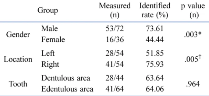
총 108개 중 69개의 절단면 영상에서 후상치조동맥의 관찰이 가능하였고 63.9%의 유병률을 나타내었다. 후상치 조동맥은 남성은 73.61%, 여성은 44.44% (p = .003
*), 우측 상악동에서는 75.93%, 좌측 상악동에서는 51.85% (p = .005
†), 유치악 부위에서는 63.64%, 무치악 부위에서는
64.06% 의 유병률을 나타내었다(p = .964) (Table 1).
후상치조동맥의 분포 양상
동맥의 분포 양상은 상악동 외측벽의 내부를 관통하는 골내형은 53.62%, 상악동막 하방과 상악동 외측벽의 내측 에 존재하는 상악동내형은 42.03%, 외측벽의 외측과 치은 하방에 존재하는 상악동외형은 4.35%의 비율로 나타났다.
치아의 부위에 따른 동맥의 분포 양상과 비율은 제 2 소 구치부에서 골내형 54. 55%, 상악동내형 56.25%, 상악동 외형 9.09%로 나타났다. 제 1 대구치부에서는 골내형, 상 악동내형 각각 31.82%, 68.18%로 나타났고 상악동외형은 나타나지 않았다. 제 2 대구치부에서는 골내형, 상악동내 형, 상악동외형 각각 69.23%, 42.03%, 4.35%로 나타났다 (p = .023
*) (Table 2).
후상치조동맥의 직경
후상치조동맥의 평균 직경은 1.14±0.45 mm 였으며 좌 측은 1.24±0.48 mm, 우측은 1.04±0.41 mm로 측정되었다.
동일 환자의 상악 좌, 우 같은 치아의 위치에서 동맥의 직 경차이를 알아보기 위하여 정규성 검증을 거쳐 paired t- test 를 시행한 결과, 통계학적으로 유의한 차이를 보이지 않았으며 이는 좌, 우 동맥의 직경 차이가 없음을 시사하 였다(p = .351). 52.17%에서 1 mm 이내의 직경을 보였으 며, 42.03%는 1~2 mm, 5.8%에서 2 mm 이상의 직경을 가 진 것으로 나타났다. 나이에 따라서는 30대에서 평균 1.92 mm, 40대에서 1.98 mm, 50대에서 2.19 mm, 60대에 서 2.8 mm, 70대 환자는 1명으로 평균 직경 1.35 mm로 Table 1. Prevalence of PSAA according to gender, location and
tooth
Group Measured
(n)
Identified rate (%)
p value (n)
Gender Male 53/72 73.61
.003*
Female 16/36 44.44
Location Left 28/54 51.85
.005
†Right 41/54 75.93
Tooth Dentulous area 28/44 63.64 Edentulous area 41/64 64.06 .964 PSAA: posterior superior alveolar artery.
This table was created by performing chi-square tests.
*P<0.05: Gender group showed statistically significant difference.
†
P<0.05: Location group did not show statistically significant difference.
Table 2. The distribution type of PSAA according to the tooth region
Second premolar First molar Second molar Total p value
(n) (%) (n) (%) (n) (%) (%) (n)
IO 12/22 54.55 7/22 31.82 18/25 72.00 53.62
.023*
IS 8/22 36.36 15/22 68.18 6/25 23.07 42.03
ES 2/22 9.09 - - 1/25 3.84 4.35
IO: intraosseous, IS: intrasinusal, ES: extrasinusal, PSAA: posterior superior alveolar artery. This table was created by performing chi- square tests.
*P<0.05: All measurements showed statistically significant differences.
Table 3. Arterial diameter related to location
N Minimum
(mm)
Maximum (mm)
Mean
(mm) SD
Left 28 0.3 2.2 1.24 0.48
Right 41 0.4 2.3 1.04 0.41
Total 69 0.35 2.25 1.14 0.45
SD: standard deviation.
This table was created by performing a paired t-test. (p = .351)
Arterial diameter of left and right sides did not show statistically
significant differences.
나타나 고령일수록 동맥 직경도 증가하는 양상을 보였으 며 통계적으로 유의성이 있었다(p = .015
*)(Table 3, 4).
후상치조동맥의 수직적 위치
치조정으로부터 동맥 하연까지의 수직거리는 평균거리 16.34±5.67 mm로 나타났다. 치아가 존재할 경우 치조정에 서 동맥 하연까지의 평균 수직거리는 18.95±1.16 mm, 무 치악 부위에서는 14.54±1.45 mm로 나타나 무치악일 경우 치조정과 동맥이 더 가까운 거리에 위치함을 알 수 있었 다(p = .001
*). 부위에 따라 치조정에서 동맥 하연까지의 평 균 수직거리는 제 2 소구치 부위, 제 1 대구치 부위, 제 2 대구치 부위에서 각각 18.29 mm, 14.34 mm, 16.34 mm로 나타나 상악 제 1 대구치 부위에서 가장 낮은 위치에 존 재함을 알 수 있었으나 통계적으로 유의한 차이는 없었다 (p = .056) (Table 5).
합병증
골창의 형성 시 후상치조동맥의 파열이나 손상이 일어 난 증례는 없었다. 1 증례에서는 골창의 제거 후 후상치조 동맥을 눈으로 확인할 수 있었다(Fig. 4).
골창의 형성 시 2 개의 상악동에서 평균 직경 3 mm의 천 공이 발생하여 8.33%의 천공률을 보였다. 이 증례에서는 상 악동막의 거상 후 천공부위를 흡수성 collagen membrane (CollaTape, Zimmer Dental, Carlsbad, CA, U.S.A.)으로 막은 후 통상의 임플란트 식립을 시행하였으며 치유기간 중 감염 소견을 보이지 않았다. 낭종을 제거한 2 개의 증례와 상악동 내부의 임플란트를 제거한 1개의 증례는 골창 형성 후 인위 적으로 막을 절개하였으므로 천공에서 제외하였다. 총 12 개 의 상악동에서 격벽이 11.11%의 비율로 관찰되었으며, 우측 상악동이 좌측 상악동에 비해 2배의 비율로 관찰되었다.
고 찰
상악동 거상술은 상악 구치부의 임플란트 식립 시 부족 한 골량을 증대시킬 수 있는 비교적 안전하고 예지력 있 는 술식으로 알려져 있다[7, 16]. 그러나 상악동 외측벽을 주행하는 후상치조동맥이 골창의 형성 시 외과적으로 손 상받으면 극심한 출혈이 생길 수 있다[17]. 후상치조동맥 의 손상은 출혈로 인한 시야의 방해와 더불어 상악동막의 천공 가능성을 증가시키며, 지혈로 수술 시간을 증가시켜 임플란의 성공률을 저하시키는 요인으로 작용한다[12, 17].
전산화 단층 촬영을 이용하여 Elian 등
[11]은 52.9%, Mardinger 등
[18]은 55%, Gϋncϋ 등
[19]은 64.5%의 후상치조 동맥 유병률을 보고 하였다. 본 연구에서는 비교적 높은 63.9% 의 발현율을 보였으며 성별과 좌, 우 상악동의 유병 률에 통계적으로 유의한 차이를 보였다. 그러나 모집단이 큰 다른 연구에서 성별과 좌, 우 상악동의 동맥 유병률에 통계적으로 유의한 차이를 보이지 않는 연구들이 보고되 었으므로 향후 추가적인 연구가 필요할 것이다[11, 18, 19].
Ella 와 Gϋncϋ 등은 모두 동맥의 평균 직경을 1.2~1.3 mm로 보고하여 본 연구의 1.14±0.45 mm와 비슷한 수치를 나타 내었다[19, 20]. 좌측 상악동의 동맥의 평균 직경은 1.24 Table 4. Arterial diameter related to age
30’s 40’s 50’s 60’s 70’s p value (n)
N 14 9 27 18 1
.015*
Average (mm) 1.92 1.98 2.19 2.8 1.35
SD 0.76 0.58 0.87 1.04 0.49
SD: standard deviation.
This table was created by performing a Pearson correlation test.
(r = .292, p = .015
*)
*P<0.05: All measurements showed statistically significant differences.
Table 5. Distance of the arterial inferior border from the alveolar crest
N Mean
(mm) SD p value
(n)
I
Total 69 16.34 5.67
.001*
Dentulous area 28 18.95 1.16
Edentulous area 41 14.54 1.45
II
Second premolar 22 18.29 0.03
First molar 22 14.34 0.37 .056
Second molar 25 16.34 2.79
SD: standard deviation.
Group I was created by performing a t-test. (p = .001)
*P<0.05: All measurements in group I showed statistically significant differences.
Group II was created by performing one-way ANOVA. (p = .056) Group II did not show statistically significant differences.
Fig. 4. Undamaged PSAA was observed after bony window
removal.
±0.48 mm, 우측은 1.04±0.41 mm로 나타났다. 52.17%에서 1 mm 이내의 직경을 보였으며, 42.03%는 1~2 mm, 5.8% 에서 2 mm 이상의 직경을 가진 것으로 나타나, Solar 등과 Mardinger 등과의 연구 결과와 유사하다 [13, 18]. 직경이 작은 동맥은 전산화 단층 영상에서 관찰 되지 않으며, 관찰되는 후상치조동맥의 대부분이 2 mm 이하의 직경을 가진다는 것을 알 수 있었다. 이들의 경 우 통상의 방법으로 골창 형성 후 상악동 거상술을 시행 하여도 출혈로 인한 합병증은 크지 않을 것이다[12]. 그 러나 환자의 나이가 증가할수록 동맥의 직경도 증가하는 양상을 보였고 통계적으로 유의성 있는 결과가 도출되었 다. 이는 성별이나 부위와는 관계없이 나이와 동맥의 직 경이 양의 상관관계를 가지므로 고령의 환자에서 후상치 조동맥의 손상 시 출혈의 가능성과 임플란트의 실패율이 증가함을 의미한다[18].
상악동 외측벽에 골창 형성 시 통상적으로 치조골정에 서 15~16 mm의 위치에 상방 골절제선을 형성하게 된다.
이 골절제선은 동맥의 위치와 근접하므로 후상치조동맥에 외과적 손상을 줄 가능성이 있다. 앞선 연구에서 후상치조 동맥의 수직적 위치는 치조골정에서 16.4~19.6 mm의 거 리를 가지는 것으로 보고되었다[11-13, 18, 21]. 본 연구에 서는 치조정으로부터 동맥 하연까지의 수직거리는 평균 16.34±5.67 mm 로 나타났다. 치아가 존재할 경우 치조정에 서 동맥 하연까지의 평균 수직거리는 18.95±1.16 mm, 무 치악 부위에서는 14.54±1.45 mm로 나타나 무치악일 경우 치조정과 동맥이 더 가까운 거리에 위치하며 통계적으로 유의성 있는 결과가 도출되었다. 치조정에서 15 mm 이하 에 존재하는 동맥은 39.13%, 15~20 mm의 높이에는 34.78%, 20 mm 이상의 높이에 위치하는 동맥은 26.09%로 나타났 다. 또한 부위별로 제 2 소구치 부위, 제 1 대구치 부위, 제 2 대구치 부위에서 각각 18.29 mm, 14.34 mm, 16.34 mm로 측정되어 이는 통계적으로 유의한 차이를 보이지 않았다. 이는 Hur 등의 후상치조동맥의 주행이 S type(직 선주행 – 78.1%)와 U type(곡선주행 – 21.9%) 나뉜다는 주장과 유사한 결론이다[22]. 그러나 선행된 많은 연구에 서 후상치조동맥의 주행이 완만한 곡선으로 상악 제 1 대 구치 부위에서 가장 낮은 위치에 존재함을 알 수 있었다.
이들 결과를 종합하여 보면 무치악 상태가 오래되어 잔존 치조골이 낮은 환자에서 동맥이 더 낮은 위치에 존재하는 양상을 나타내며, 나이가 증가할수록 동맥의 직경도 증가 하므로 상악 무치악 구치부에 측방 접근법을 이용한 상악 동 거상술 시 골창의 형성 위치를 변경하거나 연조직의 손 상이 적은 초음파 수술 기구의 사용을 고려하여야 할 것 이다.
상악동 외측벽과 동맥의 상대적인 위치 또한 동맥의 손 상에 중요한 요인으로 작용한다. 상악동외형의 경우 치은
에 절개만으로 손상될 가능성이 있으며 상악동내형은 골 창 형성 시 연조직에 손상을 주지 않는다면 손상되지 않 을 가능성이 크다.
상악동 골 이식술 중 측방 접근법의 또 다른 가장 큰 합 병증은 상악동막의 천공이다[23, 24]. 상악동막의 천공은 기존의 고속회전기구를 이용하여 골창의 형성 시 10~44%
의 빈도로 나타난다고 보고되고 있다[16, 25]. 뿐만 아니라 상악동막의 천공 시 형성된 크기와 위치에 따라 천공 폐 쇄 처치가 적절하게 시행되지 못한다면 골 이식술의 실패 로 인한 임플란트의 실패, 나아가 상악동염으로 인한 구강 농루형성 등의 술 후 합병증을 발생시킨다[31, 33, 34].
초음파 수술 기구는 25~29 kHz의 미세 진동 압전 효과 를 이용하여 경조직의 선택적 삭제를 가능하게 하여 연골, 신경, 혈관, 점막 등의 연조직을 보호하는 효과가 있으며 수술 중 진동과 소음이 적어 환자의 부담을 줄여준다는 연 구가 보고되었다[28-35]. 초음파 수술 기구를 사용한 상악 동 거상술은 상악동막의 천공과 후상치조동맥의 손상 가 능성을 줄이며 궁극적으로 수술의 안정성과 성공률을 높 이는데 기여한다.
본 연구에서 천공은 2개의 상악동에서 골창 형성 시 하
방 골 삭제선이나 전, 후방 골 삭제선의 형성 시 발생하였
으며 8.33%의 천공율을 보였다. 2 증례 중 1 증례에서는
만성 치주염이 만성 상악동염으로 발전한 증례로 상악동
내부를 관류 세척 후 상악동 거상술을 시행하였고, 나머지
1 증례는 75세의 여환으로 상악동막의 두께가 얇아 골창
형성 시 천공이 발생하였다. 초음파 기구와 얇은 saw
insert를 이용하여 골창 형성 시 무리한 힘을 가하는 경우
상악동막의 천공이 발생할 수 있으므로 기구의 절삭력을
이용한 골 삭제를 시행하여야 한다. 2 증례 모두 천공의
크기가 약 3 mm 정도로, 막의 거상 후 흡수성 콜라겐 차
단막을 사용하여 폐쇄하였다. 치조정 접근법의 시행 시 천
공이 일어나 측방 접근법으로 상악동 거상술을 시행한 3
증례에서는 천공의 크기가 작아 막의 거상 시에 중첩되어
저절로 폐쇄되었다. 상악동 내 병변을 제거 한 2개의 증례
와 15년전에 식립했던 임플란트가 상악동 내에 존재하여
제거한 1개의 증례에서는 인위적으로 막을 절개하였으므
로 천공의 경우에서 제외하였다. 1개의 증례에서 골창을
형성하고 제거 후에 손상되지 않은 후상치조 동맥의 확인
이 가능하였다. 또한 상악동내 격벽이 존재할 때에도 골창
을 나눠서 형성하였고 여러 개의 골창 모두 재위치 시킬
수 있었다. 얇은 piezoelectric saw insert를 사용 시 정밀한
골 삭제선의 형성이 가능하므로 골편이 상악동 내부로 밀
려들어가지 않고 골창 형성부위에 재위치 시킬 수 있었으
며 이는 연조직이 상악동내로 자라 들어가 신생골량의 감
소를 막고 골질이 불량해지는 것을 방지하는 차단막 역할
을 하였다.
본 연구에서 살펴본 바와 같이 상악동거상수술 시 CBCT를 이용하여 후상치조 동맥의 분포양상 및 위치를 파악하고 초음파 수술기구를 사용한다면 후상치조 동맥의 손상과 상악동 천공을 줄여 좀 더 안전하고 성공적인 상 악동 거상수술이 가능할 것이고 임플란트 식립의 성공률 를 높일 수 있을 것으로 사료된다.
Conflict of Interest
The authors declare that they have no competing interests.
ORCID
In-Sook Park 0000-0001-8360-8473
References