https://doi.org/10.5624/isd.2017.47.3.181
Introduction
The implant position is one of the most important deter- minants of the long-term maintenance of implant esthet- ics and function.1 If an implant could be placed into the extraction socket at the same angulation as the that of the root inside the alveolar bone, the prosthetic crown would have an ideal position.2,3 Although the position of an implant should be based on future restoration plans, the
placement of an implant axis in alignment with the crown is often limited by the morphology of the alveolar ridge.1
In addition to the root position in its alveolar socket, the thickness of the facial bone wall is also of crucial im- portance in selecting an appropriate treatment approach and determining the dimensions of the implant.4,5 In a previous study, the buccal bone wall of the anterior tooth was found to be very thin in most patients; therefore, the implant was positioned slightly on the palatal side to en- sure sufficient buccal bone thickness.3 Especially in the esthetic zone, the facial bone wall is often thin and is pre- dominantly composed of the bundle bone, such that its resorption results in a vertical reduction of the facial bony crest.6 Clinical guidelines suggest that a minimal buccal bone thickness of 1-2mm is required to maintain the tis-
Analysis of the root position of the maxillary incisors in the alveolar bone using cone-beam computed tomography
Yun-Hoa Jung1, Bong-Hae Cho1, Jae Joon Hwang2,*
1Department of Oral and Maxillofacial Radiology, School of Dentistry, Pusan National University, Yangsan, Korea
2Department of Oral and Maxillofacial Radiology, Yonsei University College of Dentistry, Seoul, Korea
ABSTRACT
Purpose: The purpose of this study was to measure the buccal bone thickness and angulation of the maxillary incisors and to analyze the correlation between these parameters and the root position in the alveolar bone using cone-beam computed tomography(CBCT).
Materials and Methods: CBCT images of 398 maxillary central and lateral incisors from 199 patients were retrospectively reviewed. The root position in the alveolar bone was classified as buccal, middle, or palatal, and the buccal type was further classified into subtypes I, II, and III. In addition, the buccolingual inclination of the tooth and buccal bone thickness were evaluated.
Results: A majority of the maxillary incisors were positioned more buccally within the alveolar bone, and only 2 lateral incisors(0.5%) were positioned more palatally. The angulation of buccal subtype III was the greatest and that of the middle type was the lowest. Most of the maxillary incisors exhibited a thin facial bone wall, and the lateral incisors had a significantly thinner buccal bone than the central incisors. The buccal bone of buccal subtypes II and III was significantly thinner than that of buccal subtype I.
Conclusion: A majority of the maxillary incisor roots were positioned close to the buccal cortical plate and had a thin buccal bone wall. Significant relationships were observed between the root position in the alveolar bone, the angulation of the tooth in the alveolar bone, and buccal bone thickness. CBCT analyses of the buccal bone and sagittal root position are recommended for the selection of the appropriate treatment approach.(Imaging Sci Dent 2017; 47: 181-7)
KEY WORDS: Maxilla; Incisor; Tooth Root; Cone-Beam Computed Tomography
Copyright ⓒ 2017 by Korean Academy of Oral and Maxillofacial Radiology
This is an Open Access article distributed under the terms of the Creative Commons Attribution Non-Commercial License(http://creativecommons.org/licenses/by-nc/3.0) which permits unrestricted non-commercial use, distribution, and reproduction in any medium, provided the original work is properly cited.
Imaging Science in Dentistry·pISSN 2233-7822 eISSN 2233-7830
*This study was supported by a 2016 Clinical Research Grant from Pusan National University Dental Hospital.
Received April 15, 2017; Revised May 16, 2017; Accepted June 2, 2017
*Correspondence to : Dr. Jae Joon Hwang
Department of Oral and Maxillofacial Radiology, Yonsei University College of Dentistry, 50-1 Yonsei-ro, Seodaemun-gu, Seoul 03722, Korea
Tel) 82-2-2228-8843, Fax) 82-2-363-5232, E-mail) [email protected]
sue architecture following tooth extraction and implant placement.7
The sagittal root position can have a significant impact on the occurrence of labial bone perforation, and cone- beam computed tomography(CBCT) images of the root position within the alveolar bone provide information that is useful for avoiding labial bone perforation.8 Therefore, preoperative diagnostic procedures are required to assess the risk factors for implant placement.4,9 Preoperative CBCT is now routine for implant placement, especially in the esthetic zone, both because of its accuracy and be- cause of its convenience and low radiation dosage.10-13
The objective of this study was to measure the buccal bone thickness and angulation of the maxillary incisors and to analyze the correlation between these parameters and the root position in the alveolar bone using CBCT.
Materials and Methods
This study was approved by the Institutional Review Board of Pusan National University Dental Hospital. The subjects of this retrospective study were randomly select- ed from patients who visited Pusan National University Dental Hospital and underwent CBCT imaging between 2013 and 2014. Each image was examined to identify fully formed, intact, and healthy maxillary incisors for analysis. The CBCT data were excluded if any radio- graphically detectable caries, periapical pathology, alveo- lar bone loss of more than 4mm from the cementoenamel junction, restoration, fracture, or history of orthodontic treatment was present. A total of 199 CBCT images met the inclusion criteria, resulting in a sample size of 398
maxillary central and lateral incisors. The patients com- prised 100 males and 99 females, with a mean age of 28.3 years(range, 20-50 years).
All images used in this study were obtained using a PaX-Zenith 3D apparatus(Vatech Co., Hwaseong, Ko- rea), with 5.7mA, 110kV, 24s exposure time, 0.2mm voxel size, and a field of view of 16×14cm or 12×9cm.
CBCT data were saved in the Digital Imaging and Com- munications in Medicine format, and the images were analyzed using Ez3D Plus Professional CBCT software (Vatech Co., Hwaseong, Korea).
The arch form selector tool was centered at the middle of the arch in the axial plane. The root position, angula- tion, and buccal bone thickness were evaluated by view- ing the cross-sectional images made at the midpoint of the tooth parallel to its long axis. The root position of the maxillary incisor in the alveolar bone was evaluated ac- cording to the position of the apex.14 The root position of the incisors was classified as follows: buccal type, the apical point of the incisor was within the buccal third of the alveolar bone and the root was closer to the buccal bone wall; middle type, the apical point of the incisor was within the middle third of the alveolar bone and the buc- cal and palatal bone walls were approximately equal in thickness; and palatal type, the apical point of the incisor was within the palatal third of the alveolar bone and the root was closer to the palatal bone wall(Fig. 1). The buc- cal type was further classified into subtypes I, II, and III.
In subtype I, the incisor root was covered by the buccal bone wall and the bone thickness increased toward the apex. In subtype II, the incisor root was covered by a thinner buccal bone wall than found in subtype I and the
Fig. 1. The root position of the incisors in the alveolar bone is classified as the buccal, middle, or palatal type. A. Buccal type: the apical point of the incisor is within the buccal third of the alveolar bone and the root is closer to the buccal bone wall. B. Middle type: the apical point of the incisor is within the middle third of the alveolar bone and the buccal and palatal bone walls are approximately equal in thick- ness. C. Palatal type: the apical point of the incisor is within the palatal third of the alveolar bone and the root is closer to the palatal bone wall.
A B C
bone thickness did not noticeably increase toward the apex that was covered by the bone tissue in the long axis of the tooth. In subtype III, the axis of the apex was angu- lated very buccally and the apex was not covered by the bone tissue in the long axis of the tooth(Fig. 2).
Furthermore, the angle between the long axis of the tooth and the long axis of the corresponding alveolar bone was determined. The long axis of the tooth was defined as the line through the lowest point of the crown to the high- est point of the apex in the cross-sectional image(Fig. 3).
The thickness of the buccal bone wall was assessed per- pendicular to the long axis of the tooth at the following 5 locations: at the crest; 2, 4, and 6mm apical to the crest;
and at the root apex. If the root apex was located anterior to the natural contour of the maxillary alveolar buccal bone and the extremely thin bony wall belonged to buccal subtype III, a value of 0mm was used to record the buc- cal bone thickness(Fig. 4).
All measurements were performed by a single examin- er. The Kolmogorov-Smirnov test was used to determine the normality of the data. To estimate the intraexaminer deviations in the measurements, assessments of the max- illary incisors of 30 patients were performed twice. Two sets of measurements were carried out at different times on the same CBCT scans. The Wilcoxon signed-rank test was used to compare the numeric values of the duplicate measurements, and no significant differences were noted between the 2 sets of measurements. Moreover, no sig- nificant differences were observed between the measure- ments on the right and left sides. The Wilcoxon signed- rank test was used to compare the angulation and buccal bone thickness between the central and lateral incisors because the samples did not follow a normal distribution.
The buccal bone thickness was presented as mean±stan- dard deviation. In addition, the buccal bone thickness
Fig. 2. The buccal type is further classified as follows. A. Subtype I: the incisor root is covered by the buccal bone wall, and the bone thickness increases toward the apex. B. Subtype II: the incisor root is covered by a thinner buccal bone wall than in subtype I and the bone thickness does not noticeably increase toward the apex that is covered by the bone tissue in the long axis of the tooth. C. Subtype III: the axis of the apex is angulated very buccally and the apex is not covered by the bone tissue in the long axis of the tooth.
A B C
Fig. 3. The angle between the long axis of the tooth and the long axis of the corresponding alveolar bone is measured.
Fig. 4. The thickness of the buccal bone is measured at the alveo- lar crest; 2, 4, and 6mm apical to the alveolar crest; and at the root apex.
was grouped into the following categories for descriptive analysis: missing bone wall, bone thickness <1mm, and bone thickness ≥1mm. The Kruskal-Wallis test was con- ducted to compare the buccal bone thickness and angula- tion according to the root position. P values <.05 were considered to indicate statistical significance. The statis- tical analyses were performed using SPSS version 23.0 (IBM Corp., Armonk, NY, USA).
Results
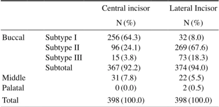
In this study, the root position within the alveolar bone was examined. A majority of the maxillary incisors were positioned more buccally within the alveolar bone. Among the buccal-type incisors, 64.3% of the central incisors were classified as subtype I and 67.6% of the lateral inci- sors were classified as subtype II. Only 2 lateral incisors (0.5%) were positioned more palatally(Table 1).
Furthermore, the angulation of the maxillary incisor within the alveolar bone was determined. The angulation of the maxillary lateral incisor was significantly greater than that of the central incisor(P<.05). The angulation of buccal subtype III was the greatest and that of the middle type was the lowest. A significant relationship between the root position within the alveolar bone and the angula- tion in the alveolar bone was found(P<.05; Table 2).
The mean thickness of the facial bone wall was 0.92mm at the central incisors and 0.57mm at the lateral incisors.
The maxillary lateral incisors demonstrated a significant- ly thinner buccal bone thickness than the central incisors (P<.05). In the lateral incisors, the lowest bone thickness was observed 6mm apical to the crest(Table 3).
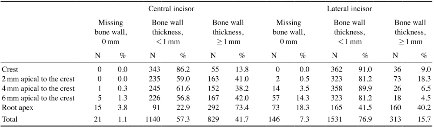
A majority of the maxillary incisors exhibited a thin buccal bone wall(<1mm). In addition, a thick buccal bone wall(≥1mm) was found in 41.7% of the central incisors and 15.7% of the lateral incisors. The lowest per- centage(4.5%) of thick walls was found 6mm apical to
the crest of the lateral incisors. At the root apex, the buc- cal bone was missing in 3.8% of the central incisors and 18.3% of the lateral incisors(Table 4).
Table 5 shows the buccal bone thickness of the maxil- lary incisors according to sagittal root position. The buc- cal bone thickness of buccal subtypes II and III was sig- nificantly thinner than that of buccal subtype I at 4mm, 6 mm, and the root apex of the central incisors and at 2mm, 4mm, 6mm, and the root apex of the lateral incisors, re- spectively(P<.05). The buccal bone thickness gradually decreased apically at 2, 4, and 6mm in buccal subtypes II and III.
Discussion
In this study, we investigated the root position in the al- veolar bone, the sagittal angulation between the long axes of the teeth and their respective alveolar bone, and the buccal bone thickness of the maxillary incisors on CBCT images. Several studies have investigated the root posi- tion of the maxillary central incisors in the alveolar bone and have reported that the buccal type was the predomi- nant incisor root position.2,5,14-16 In our study, the buccal type of root position was identified in more than 90% of cases and the palatal type was found in only 2 cases(5%)
Table 1. Root position of the maxillary incisors in the alveolar bone
Central incisor Lateral Incisor
N(%) N(%)
Buccal
Middle Palatal
Subtype I Subtype II Subtype III Subtotal
256(64.3) 96(24.1) 15(3.8) 367(92.2)
31(7.8) 0(0.0)
32(8.0) 269(67.6)
73(18.3) 374(94.0) 22(5.5)
2(0.5)
Total 398(100.0) 398(100.0)
Table 2. Angulation of the maxillary incisors with reference to the alveolar bone according to the root position (unit: degrees)
Central incisor* Lateral incisor*
Buccal subtype I Buccal subtype II Buccal subtype III Middle
Palatal
5.2±3.0 9.2±3.4 11.5±4.7 1.7±1.4
9.9±2.7 11.8±3.1 16.3±4.1 4.8±2.4 16.8±0.1
Total 6.1±3.9 12.1±4.2
*P<.05.
Table 3. Buccal bone thickness measurements at the alveolar crest; 2, 4, and 6mm apical to the alveolar crest; and at the root apex for the maxillary incisors (unit: mm)
Central incisors*
(n=398) Lateral incisors*
(n=398) Crest
2mm apical to the crest 4mm apical to the crest 6mm apical to the crest Root apex
0.79±0.16 0.91±0.24 0.86±0.27 0.87±0.35 1.18±0.53
0.70±0.26 0.67±0.42 0.40±0.38 0.26±0.31 0.81±0.63
Total 0.92±0.36 0.57±0.46
*P<.05.
of the lateral incisors, similar to the results of previous reports.5,14,16
We further classified the buccal type into 3 subtypes based on the thickness of the buccal bone wall at different root levels and the presence of an apical bone wall. Pa- tients with buccal subtype I had an adequate buccal bone thickness and bone wall around the root. During imme- diate placement, the direction of placement more or less coincided with the long axis of the tooth.14,17 In our study, buccal subtype I accounted for 64.3% of the central inci- sors and 8.0% of the lateral incisors. The buccal subtype II incisors had a thinner buccal bone wall than buccal sub- type I, and immediate placement was more challenging than in patients with subtype I in such cases.14 The buccal subtype III teeth not only had a very thin buccal plate, but the axis of the root apex was also angulated very buccal- ly, passing anterior to the natural contour of the maxillary alveolar bone.2 Therefore, patients with a subtype III root
position may not be suitable for immediate placement.2,14 In this study, subtypes II and III were more frequent in the lateral incisors than in the central incisors.
The buccolingual inclination of the tooth was critical in determining the appropriate dental implant treatment approach and implant size.3 In this study, the angle be- tween the long axis of the tooth and the long axis of the corresponding alveolar bone was less than 10° in buccal subtype I and the middle type. It may be relatively easy to insert implants into this kind of extraction socket. In our results, the angulation of buccal subtype III was the great- est, to the point that the implant position might be com- promised.2,3
Numerous studies have revealed that a thin facial al- veolar bone wall is usually present in the anterior maxil-
la.4,15,18-21 Hence, in most situations, adjunctive bone aug-
mentation has been recommended for immediate or early implant placement in the anterior esthetic zone.18,22,23 The
Table 4. Frequency distribution of the maxillary incisors according to the thickness of the buccal bone wall
Central incisor Lateral incisor
Missing bone wall,
0mm
Bone wall thickness,
<1mm
Bone wall thickness,
≥1mm
Missing bone wall,
0mm
Bone wall thickness,
<1mm
Bone wall thickness,
≥1mm
N % N % N % N % N % N %
Crest
2mm apical to the crest 4mm apical to the crest 6mm apical to the crest Root apex
0 0 1 5 15
0.00.0 0.31.3 3.8
343 235 245 226 91
86.259.0 61.656.8 22.9
55163 152167 292
13.841.0 38.242.0 73.4
0 2 14 57 73
0.00.5 14.33.5 18.3
362 323 358 323 165
91.081.2 89.981.2 41.5
36 73 26 18 160
18.39.0 6.54.5 40.2
Total 21 1.1 1140 57.3 829 41.7 146 7.3 1531 76.9 313 15.7
Table 5. Buccal bone thickness according to the sagittal root position at the crest; 2, 4, 6mm apical to the crest; and at the root apex (unit: mm) Buccal
Middle Palatal
Subtype I Subtype II Subtype III
Central incisor
Crest
2mm apical to the crest 4mm apical to the crest*
6mm apical to the crest*
Root apex*
0.78±0.15 0.91±0.18 0.93±0.18 1.00±0.21 1.33±0.32
0.82±0.20 0.92±0.34 0.68±0.32 0.53±0.29 0.70±0.42
0.79±0.14 0.90±0.32 0.50±0.29 0.25±0.24 0.00±0.00
0.81±0.19 0.97±0.26 1.03±0.29 1.18±0.34 1.91±0.40
Total 0.99±0.28 0.73±0.35 0.49±0.40 1.18±0.49
Lateral incisor
Crest
2mm apical to the crest*
4mm apical to the crest*
6mm apical to the crest*
Root apex*
0.68±0.26 0.76±0.32 0.74±0.20 0.79±0.17 1.51±0.40
0.69±0.25 0.63±0.37 0.35±0.30 0.22±0.18 0.86±0.42
0.70±0.24 0.67±0.41 0.30±0.31 0.03±0.06 0.00±0.00
0.65±0.18 0.74±0.35 0.65±0.35 0.65±0.43 1.76±0.79
1.85±0.21 3.20±0.28 3.20±0.28 2.25±0.07 2.40±0.42
Total 0.89±0.42 0.55±0.39 0.34±0.39 0.89±0.63 2.58±0.60
*P<.05.
vertical resorption of the buccal crest was 3 times greater at sites with a thin buccal bone(<1mm) than at sites with a thick buccal bone(≥1mm).24 In this study, the buccal bone was thin(<1mm) in 58.3% of the central incisors and 84.3% of the lateral incisors. Moreover, a statistically significant association was noted between the root posi- tion in its socket and the buccal bone thickness, and the buccal bone was significantly thinner in buccal subtypes II and III than in buccal subtype I. The buccal bone of the middle type was significantly thicker than that of buccal subtype I in the central incisors. However, there was no significant difference in buccal bone thickness between buccal subtype I and the middle type in the lateral incisors because the lateral incisors had thinner alveolar bones than the central incisors.
In conclusion, a majority of the maxillary incisors were positioned close to the buccal cortical plate and had a thin buccal bone wall. Significant relationships were noted be- tween the root position in the alveolar bone, angulation in the alveolar bone, and buccal bone thickness. CBCT anal- yses of the buccal bone and sagittal root position are rec- ommended for the selection of an appropriate treatment approach.
References
1. Chan HL, Garaicoa-Pazmino C, Suarez F, Monje A, Benavides E, Oh TJ, et al. Incidence of implant buccal plate fenestration in the esthetic zone: a cone beam computed tomography study.
Int J Oral Maxillofac Implants 2014; 29: 171-7.
2. Lau SL, Chow J, Li W, Chow LK. Classification of maxillary central incisors-implications for immediate implant in the es- thetic zone. J Oral Maxillofac Surg 2011; 69: 142-53.
3. Wang HM, Shen JW, Yu MF, Chen XY, Jiang QH, He FM.
Analysis of facial bone wall dimensions and sagittal root position in the maxillary esthetic zone: a retrospective study using cone beam computed tomography. Int J Oral Maxillofac Implants 2014; 29: 1123-9.
4. Braut V, Bornstein MM, Belser U, Buser D. Thickness of the anterior maxillary facial bone wall-a retrospective radiograph- ic study using cone beam computed tomography. Int J Peri- odontics Restorative Dent 2011; 31: 125-31.
5. Kan JY, Roe P, Rungcharassaeng K, Patel RD, Waki T, Lozada JL, et al. Classification of sagittal root position in relation to the anterior maxillary osseous housing for immediate implant placement: a cone beam computed tomography study. Int J Oral Maxillofac Implants 2011; 26: 873-6.
6. Araújo MG, Lindhe J. Dimensional ridge alterations follow- ing tooth extraction. An experimental study in the dog. J Clin Periodontol 2005; 32: 212-8.
7. Grunder U, Gracis S, Capelli M. Influence of the 3-D bone- to-implant relationship on esthetics. Int J Periodontics Restor- ative Dent 2005; 25: 113-9.
8. Sung CE, Cochran DL, Cheng WC, Mau LP, Huang PH, Fan WH, et al. Preoperative assessment of labial bone perforation for virtual immediate implant surgery in the maxillary esthetic zone: a computer simulation study. J Am Dent Assoc 2015;
146: 808-19.
9. El Nahass H, Naiem SN. Analysis of the dimensions of the la- bial bone wall in the anterior maxilla: a cone-beam computed tomography study. Clin Oral Implants Res 2015; 26: e57-61.
10. Rugani P, Kirnbauer B, Arnetzl GV, Jakse N. Cone beam computerized tomography: basics for digital planning in oral surgery and implantology. Int J Comput Dent 2009; 12: 131- 11. Spector L. Computer-aided dental implant planning. Dent Clin 45.
North Am 2008; 52: 761-75.
12. Mandelaris GA, Rosenfeld AL. The expanding influence of computed tomography and the application of computer-guided implantology. Pract Proced Aesthet Dent 2008; 20: 297-305.
13. Guerrero ME, Noriega J, Jacobs R. Preoperative implant planning considering alveolar bone grafting needs and com- plication prediction using panoramic versus CBCT images.
Imaging Sci Dent 2014; 44: 213-20.
14. Xu D, Wang Z, Sun L, Lin Z, Wan L, Li Y, et al. Classification of the root position of the maxillary central incisors and its clinical significance in immediate implant placement. Implant Dent 2016; 25: 520-4.
15. Khoury J, Ghosn N, Mokbel N, Naaman N. Buccal bone thickness overlying maxillary anterior teeth: a clinical and ra- diographic prospective human study. Implant Dent 2016; 25:
525-31.
16. Chung SH, Park YS, Chung SH, Shon WJ. Determination of implant position for immediate implant placement in maxil- lary central incisors using palatal soft tissue landmarks. Int J Oral Maxillofac Implants 2014; 29: 627-33.
17. Vera C, De Kok IJ, Reinhold D, Limpiphipatanakorn P, Yap AK, Tyndall D, et al. Evaluation of buccal alveolar bone dimension of maxillary anterior and premolar teeth: a cone beam computed tomography investigation. Int J Oral Maxillo- fac Implants 2012; 27: 1514-9.
18. Huynh-Ba G, Pjetursson BE, Sanz M, Cecchinato D, Ferrus J, Lindhe J, et al. Analysis of the socket bone wall dimensions in the upper maxilla in relation to immediate implant placement.
Clin Oral Implants Res 2010; 21: 37-42.
19. Nowzari H, Molayem S, Chiu CH, Rich SK. Cone beam com- puted tomographic measurement of maxillary central incisors to determine prevalence of facial alveolar bone width ≥2mm.
Clin Implant Dent Relat Res 2012; 14: 595-602.
20. Januário AL, Duarte WR, Barriviera M, Mesti JC, Araújo MG, Lindhe J. Dimension of the facial bone wall in the anterior maxilla: a cone-beam computed tomography study. Clin Oral Implants Res 2011; 22: 1168-71.
21. Zekry A, Wang R, Chau AC, Lang NP. Facial alveolar bone wall width-a cone-beam computed tomography study in Asians. Clin Oral Implants Res 2014; 25: 194-206.
22. Miyamoto Y, Obama T. Dental cone beam computed tomog- raphy analyses of postoperative labial bone thickness in max- illary anterior implants: comparing immediate and delayed implant placement. Int J Periodontics Restorative Dent 2011;
31: 215-25.
23. Buser D, Chen ST, Weber HP, Belser UC. Early implant place- ment following single-tooth extraction in the esthetic zone:
biologic rationale and surgical procedures. Int J Periodontics Restorative Dent 2008; 28: 441-51.
24. Ferrus J, Cecchinato D, Pjetursson EB, Lang NP, Sanz M, Lindhe J. Factors influencing ridge alterations following im- mediate implant placement into extraction sockets. Clin Oral Implants Res 2010; 21: 22-9.