- 54 -
서 론12
혈관육종은 혈관내피세포에서 기원하는 악성종양으로, 매우 드물게 발생하며 예후가 불량한 종양으로 주로 노인 에서 발생한다. 혈관육종은 모든 부위에서 발생할 수 있지 만 주로 피부와 얕은 연부조직에서 발생하며, 두경부에서 가장 흔히 발생한다.
1,2)
침샘의 종양은 두경부에 생긴 종양 의 2 ~ 3%를 차지하며, 전체 침샘 종양의 80%는 이하선에 서 발생한다. 이하선 종양의 1 ~ 8%는 부이하선에서 발생 하며, 이하선 종양의 약 20%가 악성인 반면 부이하선 종양 의 26 ~ 52%가 악성으로 보고되고 있다.3,4)
이하선에서 원 발성으로 생긴 혈관육종은 전 세계적으로 7예 보고되고Received : August 31, 2015 / Revised : October 8, 2015 Accepted : October 19, 2015
교신저자 : 김정규, 42472 대구 남구 두류공원로 17길 33 대구가톨릭대학교 의과대학 이비인후과학교실
전화 : (053) 650-4071·전송 : (053) 650-4533 E-mail : [email protected]
있으며, 국내에서 2008년 1예 보고되었고,
5)
부이하선에 생 긴 혈관육종은 전 세계적으로 보고된 바가 없다. 이에 저자 들은 장기 추적된 원발성 부이하선 혈관육종 환자의 증례 를 문헌고찰과 함께 보고하는 바이다.증 례
45세 남자 환자가 4년 전부터 있어왔던 우측 볼의 덩어 리를 주소로 내원하였다. 고혈압, 당뇨병, 결핵, 간질환 등 의 이력은 없었으며, 방사선 치료의 과거력은 없었고. 약 20갑년의 흡연력은 있었다. 환자의 덩어리는 4년 전부터 0.5 cm 크기로 만져졌으나 크기 변화 없어 지켜보던 중 내원 4개월 전부터 커지는 양상 보여 내원하였다. 이학적 검사에서 경계가 뚜렷하고 말랑말랑한 종물이 2 cm 크기 로 만져졌다. 초음파 검사에서 경계가 분명하고 균질한 저에코의 2 × 2.6 cm 크기 종물이 저작근 위에 보이고, 도플 러에서 풍부한 혈관성 종양 소견이 관찰되었다(Fig. 1A,B).
대한두경부종양학회지 제 31 권 제 2 호 2015
부이하선에 생긴 원발성 혈관육종 1예의 장기추적
대구가톨릭대학교 의과대학 이비인후과학교실
이효원·김덕수·장규호·김정규
= Abstract =
Primary Angiosarcoma of Accessory Parotid Gland : A Case Report of Long Term Follow-up
Hyo Won Lee, MD, Deok Su Kim, MD, Gyu Ho Jang, MD, Jeong Kyu Kim, MD
Department of Otolaryngology-Head and Neck Surgery, School of Medicine, Catholic University of Daegu, Daegu, Korea
Angiosarcoma is a rare and highly malignant neoplasm which develops from the endothelium of blood vessels.A few cases of primary angiosarcoma of the parotid gland have been reported. However, there is no report of primary angiosarcoma of the accessory parotid gland. In this case, we report a primary angiosarcoma of the accessory parotid gland in a 45-year-old man with growing cheek mass. Ultrasonography revealed a 2.0×2.6 cm sized homogeneous hypoechoic mass and computed tomography showed a contrast enhanced homogeneous mass. Fine needle aspiration biopsy suggested a benign tumor. The mass was completely excised with a minimal vertical incision. The histopathol- ogy showed anastomosing vascular channels lined by atypical endothelial cells and many branching vessels with staghorn appearance with positive immunohistochemical staining for CD34, a highly specific endothelial marker.
The patient underwent postoperative radiotherapy and was followed for 8 years without recurrence and metastasis.
KEY WORDS : AngiosarcomaㆍSalivary glandㆍAccessory parotid neoplasm.
- 55 -
전산화 단층촬영 검사에서 불균질하게 조영 증강되는 종 물이 우측 저작근 위에서 관찰 되었으며, 이하선과 비슷한 음영으로 좌측 저작근 위에 관찰되는 좌측 부이하선이 대 칭되는 위치에서 관찰되었다(Fig. 1C).그 외에 안면신경 마비 등의 다른 특이증상은 관찰되지 않았다. 세침흡인 검사 시 종물은 출혈이 많은 양상을 보였
으며, 상피세포군집이 관찰되어 양성종양이 의심되는 소견 이었다. 수술 시 종양 부위 볼의 피부에 수직절개를 가하여 종양을 절제하였고, 이하선 절제는 시행하지 않았다. 종양 은 안면신경 협근지(buccal branch)와 이하선관보다 깊은 곳 에 위치하였고, 주변과 유착소견은 보이지 않았다. 종양이 협근지와 이하선관과 직접 접촉하고 있었고 양성종물로 판 단되어 주변조직은 절제 없이 종양만 적출하였으며, 안면신 경과 이하선관은 보존하였다. 조직검사에서 종양은 상피형 상을 가진 비정형 내피세포(atypical endothelial cells having epithelioid features)로 둘러 쌓인 혈관채널을 보이며, 사슴뿔 (staghorn) 형상의 많은 혈관이 관찰되었다(Fig. 2A,B). 추가 로 시행한 면역조직화학 염색에서 혈관내피세포 표지자인 CD34에 양성 소견으로(Fig. 2C) 혈관육종(angiosarcoma)으 로 진단되었고, AJCC 병기상 T2N0M0 2기로 분류되었다.
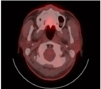
조직검사 확인 후 시행한 양전자 컴퓨터 단층촬영 검사 상 잔존 암조직 및 다른 전이소견은 관찰되지 않았다. 수술 부위에 200 cGy × 33회의 방사선치료 시행하였으며 수술 후 약 8년째 추적관찰 중 재발 및 전이소견 보이지 않았다 (Fig. 3).
C B
A
Fig. 1. Preoperative imaging. The transverse ultrasonography shows 2 × 2.6 cm, well-defined, homogenous hypoechoic lesion on right acessory parotid gland(A). Color Doppler scan shows incresed intralesional vascularity(B). The axial computed tomography scan shows a homogeneously enhancing tumor on the right masseter muscle. Note that tumor is more enhanced than both parotid and left accessory parotid gland(C).
C B
A
Fig. 2. Histopathologic finding. The section shows anastomosing vascular channels lined by atypical endothelial cells having epi- thelioid features. There are many branching vessels with staghorn appearance (H&E stain, x100)(A) and (H&E stain, x400)(B). The tu- mor is stained for CD34, a highly specific endothelial marker(x200)(C).
Fig. 3. Postoperative PET-CT. There is no evidence of recurrence
and metastasis in 5 years after surgery.
- 56 -
고 찰
부이하선은 비교적 흔히 관찰되는 해부학적 변이로 시 신연구에서 21 ~ 56%의 빈도로 보고되었다. 부이하선은 0.5 ~ 1.0 cm 크기로 이하선 앞쪽으로 약 6 mm 정도 떨어져 있으며 안면신경 협근지와 이하선관과 인접하여 위치한 다.
6)
조직학적 연구에서 일부 부이하선은 장액샘꽈리 (serous acini)와 점액샘꽈리(mucous acini)를 모두 포함하고 있어 장액샘꽈리로 구성된 이하선과의 차이점이 보고되었 다.7)
하지만 부이하선에서 발생하는 종양은 이하선에서 발생하는 종양과 큰 차이는 없으며, 부이하선의 악성종양 으로는 점액표피양 암종이 가장 흔히 보고되었고, 샘꽈리 세포암종(acinic cell carcinoma)과 B세포림프종 등이 흔히 보고되었다. 현재까지 혈관육종이 보고된 예는 없다.3,4)
혈관육종(angiosarcoma)은 혈관내피세포에서 기원하는 매우 공격적인 악성종양 중의 하나로, 방사선 노출의 기왕 력, 만성 임파선부종, 폴리염화비닐(polyvinyl chloride), 이 산화 토륨(thorium dioxide), 비소(arsenic), 라듐(radium) 등 의 화학물질에의 노출 등이 혈관육종을 증가시키는 것으 로 알려져 있다.
8)
혈관육종은 도플러 초음파 검사 상 혈관성을 가진 비교 적 균질한 에코 소견이 관찰되며, 전산화 단층 촬영에서는 조영 증강되는 경계가 분명하고 불균질한 종양 소견이 관 찰되는데, 본 증례에서도 전형적으로 이러한 소견들이 관 찰되었다.
9)
부이하선 종양의 세침흡인검사는 이하선 종양의 세침흡 인검사에 비하여 정확도와 민감도가 낮게 보고되었다. 따 라서 세침흡인검사에서 양성종양의 소견을 보이더라도 저 도 악성종양의 가능성을 완전히 배제할 수 없음을 고려해 야 한다.
10)
본 증례에서도 세침흡인검사상 위음성을 나타 내었고 수술 후 조직검사에서 악성으로 진단되었다.부이하선의 악성종양의 수술적 치료 시 이하선 전절제 수술의 필요성에 대하여 논란이 있다. 이하선 전절제수술 없이 광범위 절제수술로 재발 없이 완치된 결과가 보고되 었으며, 다른 연구에서는 이하선 전절제수술이 추가되어 도 광범위 절제수술과 치료 결과는 비슷하였다고 보고되
었다.
3,11)
부이하선종양의 수술적 접근을 위해서 전이개 절개, 종양 부위 피부절개, 또는 구강내 절개의 방법이 시행 될 수 있으며, 저작근 앞쪽 경계보다 뒤쪽에 위치한 부이하 선종양은 전이개 절개를 시행하고, 저작근 앞쪽 경계보다 앞쪽에 위치한 부이하선종양은 종양 부위 피부절개 또는 구강내 절개를 시행할 것이 주장되었다.
12)
얼굴 및 두피부의 혈관육종 증례들을 분석한 연구에서 두경부의 혈관육종은 절제 가능한 병변을 가진 젊은 환자 에게서는 수술적 치료와 방사선치료 및 항암치료의 복합
요법이 가장 추천되는 방법으로 제시하였다.
2)
다발성 전 이 등의 진행된 혈관육종에서는 항암요법이 시행될 수 있 으며, 특히 고령의 환자에게도 주저하지 말고 부작용에 유 의하며 항함요법을 시행할 것을 추천하고 있다.13)
두경부에 발생한 혈관육종은 국소재발 및 원격 전이가 많이 발생하여 예후가 매우 불량하며, 피부 및 연부조직의 혈관육종도 원격전이로 인하여 예후가 불량한 것으로 보 고되었다.
14)
하지만 구강 및 침샘에 생긴 혈관육종은 피부 나 연부조직 등의 다른 부위에 생긴 혈관 육종에 비해 양호 한 예후를 보였으며, 특히 구강의 혈관육종은 진단 및 치료 까지의 시간이 짧은 이유로 좋은 예후를 나타내는 것으로 보고되었다.15)
본 증례는 방사선치료의 과거력이 없는 중년 남성의 부 이하선에서 발생한 혈관육종으로, 이하선 절제수술 없이 종양 부위 피부절개를 통하여 종양 절제 및 방사선 치료를 시행하였으며, 8년간 재발 및 전이소견 없이 추적되었다.
비교적 젊은 나이에 발생하였고, 피부나 연부조직이 아닌 부이하선에서 발생하였으며, 종양의 크기가 크지 않았던 점이 본 증례의 좋은 예후와 관계될 것으로 판단된다.
중심 단어:혈관육종ㆍ침샘ㆍ부이하선 종양.