A Case of Pulmonary Artery Sarcoma
Presented as Cavitary Pulmonary Lesions
Daniel Min, M.D., Ji-Hyun Lee, M.D., Hye-Cheol Jeong, M.D., Jung-Hyun Kim, M.D., Suk-Pyo Shin, M.D., Hong-Min Kim, M.D., Kyu Hyun Han, M.D., Hye Yun Jeong, M.D. and Eun-Kyung Kim, M.D.
Department of Internal Medicine, Bundang CHA Medical Center, CHA University College of Medicine, Seongnam, Korea
Pulmonary artery sarcoma (PAS) is a rare, poorly differentiated malignancy arising from the intimal layer of the pulmonary artery. Contrast-enhanced chest computed tomography (CT) is a good diagnostic modality that shows a low- attenuation filling defect of the pulmonary artery in PAS patients. An 18-year-old man was referred to our hospital for the evaluation and management of cavitary pulmonary lesions that did not respond to treatment. A contrast-enhanced CT of the chest was performed, which showed a filling defect within the right interlobar pulmonary artery. The patient underwent a curative right pneumonectomy after confirmation of PAS. Although lung parenchymal lesions of PAS are generally nonspecific, it can be presented as cavities indicate pulmonary infarcts. Clinicians must consider the possibility of PAS as well as pulmonary thromboembolism in patients with pulmonary infarcts. So, we report the case with PAS that was diagnosed during the evaluation of cavitary pulmonary lesions and reviewed the literatures.
Keywords: Pulmonary Artery; Sarcoma; Pulmonary Infarction
chest computed tomography (CT) is a good initial diagnostic modality that shows a low-attenuation filling defect of the pulmonary artery. However, PAS is frequently misdiagnosed as pulmonary thromboembolism, and further evaluations are needed for differential diagnosis. Lung parenchymal lesions shown by chest radiography or non-enhanced chest CT are mostly nonspecific
4. Here, we report a case of an 18-year-old male patient with PAS who was diagnosed during evaluation of pulmonary cavitary lesions as seen on initial chest radiogra- phy and non-enhanced chest CT.
Case Report
An 18-year-old man was referred to our institution from a primary clinic for further evaluation and management of cavitary pulmonary lesions. He had no significant medical history and had visited the primary clinic 3 months before he was referred to our institution. He presented with complaints of cough, blood-tinged sputum, and right chest wall pain. The initial chest radiography at the primary clinic showed patchy consolidations in the right upper lobe (Figure 1), and non- enhanced chest CT was performed. A 5-cm cavitary consoli- dation and multifocal nodular consolidations were observed Copyright © 2014
The Korean Academy of Tuberculosis and Respiratory Diseases.
All rights reserved.
Introduction
Pulmonary artery sarcoma (PAS) is a rare, poorly differenti- ated malignancy arising from the intimal layer of the pulmo- nary artery. The first published case was reported from an autopsy by Mandelstamm in 1923
1. Since then, only approxi- mately 250 cases have been described, mostly as case reports
2. PAS is not common, with an incidence of 0.001−0.03%, and hence, initial diagnosis is challenging
3. Contrast-enhanced
Address for correspondence: Eun-Kyung Kim, M.D.
Department of Internal Medicine, Bundang CHA Medical Center, CHA University College of Medicine, 59 Yatap-ro, Bundang-gu, Seongnam 463-712, Korea
Phone: 82-31-780-5205, Fax: 82-31-780-6143 E-mail: [email protected]
Received: Oct. 14, 2013 Revised: Oct. 21, 2013 Accepted: Oct. 23, 2013
cc
It is identical to the Creative Commons Attribution Non-Commercial
License (http://creativecommons.org/licenses/by-nc/3.0/).
in the right lung, mainly in the right upper lobe posterior segment and the right lower lobe (Figure 2A). The patient was treated with medication, including empirical antibiotics, and follow-up non-enhanced chest CT was performed every month for the next 3 months. Despite medical treatment, his symptoms did not improve and the lung parenchymal lesions persisted. Follow-up CT showed changes in the pulmonary lesions, decrease in the size of the main cavitary consolidation and increase in the number and size of adjacent nodular con- solidations (Figure 2B−D). Eventually, the patient was referred to our institution for further diagnosis and treatment.
We suspected the possibility of various conditions that can cause cavitary lesions, including bacterial infection, fungal infection, tuberculosis, autoimmune disease, vasculitis, ma- lignancy, and other rare diseases. The findings of laboratory examinations were unremarkable. The patient’s leukocyte count was 7,870/mL with a C-reactive protein level of 0.44 mg/
dL. We could not detect any microorganisms on the microbio- logic studies including sputum culture, acid-fast bacilli stain, interferon-gamma release assay and aspergillous antigen test.
The tests of autoantibodies such as antinuclear antibody and antineutrophil cytoplasmic antibody were negative. Further, no endobronchial lesion was observed on bronchoscopy. Per- cutaneous needle aspiration was performed, targeting the cav- itary consolidation of the right upper lobe, but the pathologic results were unremarkable. Pathology examination showed parenchymal necrosis and fibrosis, interstitial lymphocytic infiltration, and type II pneumocyte hyperplasia. We did not detect granuloma, evidence of vasculitis, or any other possible causes of the pulmonary lesions.
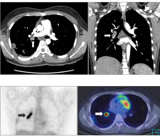
Next, the patient underwent contrast-enhanced chest CT, which showed a filling defect within the lumen of the right interlobar pulmonary artery, raising the possibility of either pulmonary thromboembolism or PAS (Figure 3). The patient received low molecular weight heparin, considering the possi- bility of pulmonary thromboembolism. Deep vein thrombosis was not detected on venous duplex examination of the lower extremities. Positron emission tomography (PET)-CT with
18F- fluorodeoxyglucose (FDG) showed increased FDG uptake, with a maximum standardized uptake value of 9.6 along the right interlobar pulmonary artery, suggesting the presence of a malignant lesion (Figure 4).
To confirm the diagnosis, open thoracotomy and excisional Figure 1. Initial chest radiography shows patchy consolidations in
the right upper lobe.
Figure 2. Non-enhanced chest computed
tomography (CT) images. (A) Initial non-
enhanced chest CT shows a 5-cm cavi-
tary consolidation in the right upper lobe
posterior segment. (B−D) Follow-up CT
show changes in the pulmonary lesions,
decrease in size of the main cavitary con-
solidation and increase in the number
and size of adjacent nodular consolida-
tions.
biopsy were performed. At surgery, frozen biopsy specimens obtained from the right interlobar pulmonary artery revealed a PAS. Accordingly, the patient underwent subsequent cura- tive right pneumonectomy.
Grossly, a 1.5×1 cm gray-tan soft tumor was detected in the lumen of the right interlobar pulmonary artery (Figure 5A).
The distal pulmonary artery was occluded with thrombus. A gray-tan-yellow infarct, measuring 4×2×2 cm, was observed in the right upper lobe, and multiple, small gray-tan nodules were scattered in the remaining lung.
Histopathologic examination of the intravascular tumor revealed abundant spindle cells with high cellularity, frequent mitoses, and nuclear pleomorphism (Figure 5B). Immuno- histochemical staining was positive for smooth-muscle actin and desmin (Figure 5C, D). Staining for CD34 and CD56 was negative. Pathologic finding of the distal parenchymal lesions were ischemic necrosis and fibrosis, indicated pulmonary infarct. Also, there were hematogenous spread of occlusive tumor emboli and multiple parenchymal involvements within the right lung.
The patient recovered uneventfully and received 3 cycles of adjuvant chemotherapy with ifosfamide and doxorubicin.
However, treatment was terminated because of side effects, which included severe gastrointestinal symptoms and neu- tropenia. There was no evidence of local recurrence or distant metastasis 6 months after surgery.
Discussion
PAS is a rare tumor of the cardiovascular system, and very few cases have been reported in the literature. In most cases, PAS occurs in middle-aged patients, but cases of PAS in young patients, including a case of a 13-year-old child
5, have also been reported. In our case, the patient was an adolescent, and therefore, it appears that PAS can occur in any age.
A variety of conditions is associated with pulmonary cavi- tary lesions. Infectious conditions include common bacterial infection; necrotizing pneumonia; lung abscess; septic emboli;
pulmonary tuberculosis; fungal infection; non-tuberculosis mycobacterial infection; and other rare infections such as mucormycosis, actinomycosis, and cryptococcosis. Examples of non-infectious conditions are neoplasm, vasculitis such as Behcet’s disease, Wegener’s granulomatosis and pulmonary infarct due to pulmonary embolism or trauma
6,7. In our case, the pulmonary cavitary lesion was eventually diagnosed as a pulmonary infarct due to PAS.
Because of its rarity and nonspecific clinical manifestations, PAS is often mistaken for pulmonary embolism, leading to inappropriate therapy such as prolonged anticoagulation or thrombolysis and thus delaying optimal therapy that might include surgery
8. The absence of predisposing factors for pulmonary embolism (e.g., no deep vein thrombosis and no finding suggestive of a hypercoagulable state) and the lack of response to anticoagulation therapy are factors suggestive of Figure 3. Contrast-enhanced chest com- puted tomography shows a filling defect within the lumen of the right interlobar pulmonary artery (arrows).
Figure 4. Positron emission tomography- computed tomography shows increased
18