207 Introduction
Differential diagnosis of an intracardiac mass in the tricus- pid valve after valvular surgery includes thrombosis, tumor, and vegetation, among others. Normal tissue such as fatty material and pectinate muscle surrounding the tricuspid an- nulus also may appear as a pseudo-tumor.1) In our case, be- cause the patient had undergone annuloplasty, thrombosis was suspected as most likely and therefore thrombolytic therapy was instituted.2) Because the size of the cardiac mass did not change, however, we finally chose a surgical method and the mass was found to be a myxoma. To our knowledge, this is first reported case of tricuspid valve myxoma in Korea.
Case
In November 2009, a 55-year-old man (who had smoking as a risk factor) was admitted to the hospital with progressive resting dyspnea. On physical examination, the patient was dyspneic and tachypneic. His jugular veins were distended.
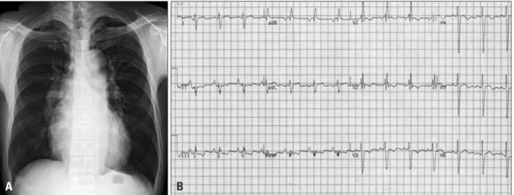
Initial laboratory work-up, including complete blood count, liver function tests, and cardiac enzyme level determination did not reveal any abnormality. He had an abnormal chest ra- diograph which showed cardiomegaly and a marked pulmo- nary trunk, and electrocardiogram reflected right ventricular strain pattern with normal sinus rhythm (Fig. 1). NT-pro BNP level was 5,592 pg/mL (normal: < 198 pg/mL), and D-dimer level was 2,296 ng/mL (normal: < 500 ng/mL). Transthoracic echocardiography (TTE) revealed right cardiac dysfunction with elevated right ventricular systolic pressure (RVSP). Pul- monary embolism was suspected and chest 3-dimensional computed tomography (3D CT) with contrast enhanced for pulmonary embolism showed a massive pulmonary thrombo- embolism (Fig. 2). No deep vein thrombosis in venous Dop- pler for the lower extremities or hypercoagulability in the laboratory test was apparent. Although the patient was he- modynamically stable, hypoxemia was noticed in arterial blood gas analysis (pH = 7.493, pCO2 = 27.9 mm Hg, pO2 =
pISSN 1975-4612/ eISSN 2005-9655 Copyright © 2011 Korean Society of Echocardiography www.kse-jcu.org http://dx.doi.org/10.4250/jcu.2011.19.4.207
CASE REPORT J Cardiovasc Ultrasound 2011;19(4):207-210
Tricuspid Valvular Myxoma: Unusual Case of Tricuspid Valve Myxoma Mimicking
Thrombus after Pulmonary Artery
Embolectomy and Tricuspid Annuloplasty in Pulmonary Thromboembolism Patient
Min Yong Park, MD1, Sung Uk Kwon, MD1, Sung Yun Lee, MD1, Boram Kang, MD1, Hyung Yoon Kim, MD1, Yu Jung Cho, MD1, Woo-Ik Chang, MD2 and Sun Hee Chang, MD3
1Departments of Cardiology, 2Thoracic Surgery, 3Pathology, Inje University Ilsan Paik Hospital, Goyang, Korea
A 55-year-old man with massive pulmonary thromboembolism underwent thrombolysis, pulmonary artery embolectomy and tricuspid annuloplasty. Nine months later, a mobile echogenic intra-cardiac mass was found in the tricuspid valve. Because the patient had undergone annuloplasty, thrombosis was suspected as the most likely diagnosis and thrombolytic therapy was instituted. However, the size of the cardiac mass did not change and after surgical excision the mass was found to be a myxoma.
Cardiac valvular tumors are uncommon and when they occur they are usually slow growing fibroelastomas. In this case, the rapid growing cardiac myxoma on the tricuspid valve was found after the occurrence of pulmonary thromboembolism. To our knowledge, this is first reported case of tricuspid valve myxoma in Korea.
KEY WORDS: Pulmonary thromboembolism · Thrombosis · Valvular myxoma.
• Received: July 6, 2011 • Revised: August 23, 2011 • Accepted: November 30, 2011
• Address for Correspondence: Sung Uk Kwon, Vision 21 Cardiac and Vascular Center, Inje University Ilsan Paik Hospital, 170 Juhwa-ro, Ilsanseo-gu, Goyang 411-706, Korea Tel: +82-31-910-7835, Fax: +82-31-910-7829, E-mail: [email protected]
• This is an Open Access article distributed under the terms of the Creative Commons Attribution Non-Commercial License (http://creativecommons.org/licenses/by-nc/3.0) which permits unrestricted non-commercial use, distribution, and reproduction in any medium, provided the original work is properly cited.
online © ML Comm
Journal of Cardiovascular Ultrasound 19 | December 2011
208
61.9 mm Hg). Therefore he was thrombolysed with recombi- nant tissue plasminogen activator and experienced a rapid im- provement in oxygenation.3) As follow-up, a second echocar- diography and chest 3D CT was performed 7 days later. TTE showed impairment in right ventricular function even though RVSP was reduced from 119 to 96 mm Hg and a second CT showed a partially resolved status. There was multifocal exten- sive pulmonary thromboembolism in both pulmonary arteries (Fig. 3). Because further treatment was required to reduce pul- monary hypertension, we performed open pulmonary embo-
lectomy with removal of thrombus from both pulmonary ar- teries, and De Vega’s tricuspid annuloplasty to correct severe tricuspid valve regurgitation. The patient experienced no postoperative complications and made a satisfactory recovery.
At 2 weeks after surgery, his TTE findings showed improve- ment in right heart function and RVSP (97 → 47 mm Hg).
The patient was discharged from the hospital on warfarin.
Nine months later, however, we found a mobile echogenic in- tra-cardiac mass (1.49 × 2.2 cm sized) at follow-up. The mass was attached to the septo-posterior leaflet portion of the tri-
Fig. 1. A: Chest X-ray showed cardiomegaly with right atrial enlargement and a prominent pulmonary trunk. B: Electrocardiogram showed characteristic features of RV strain with prominent S wave in lead I, Q wave in lead III, with T-wave inversion in lead III. RV: right ventricle.
A B
Fig. 2. A: Chest CT scanning detected a large thrombus in the right pulmonary artery. B: D-shaped LV was shown. C: Vmax was 4.96 m/s. Estimated right ventricular systolic pressure was 119 mm Hg (assumed right atrial pressure = 20 mm Hg, inferior vena cava dilatation and plethora was found).
LV: left ventricle.
Fig. 3. A: A second computed tomogram showed a partially resolved multifocal extensive pulmonary thrombus. B: D-shaped LV remained after thrombolysis. C: Vmax was 4.46 m/s. After thrombolysis, estimated right ventricular systolic pressure was slightly reduced. LV: left ventricle.
A
A
B
B
C
C
Tricuspid Valvular Myxoma | Min Yong Park, et al.
209 cuspid valve and protruded to the ventricular side. For differen-
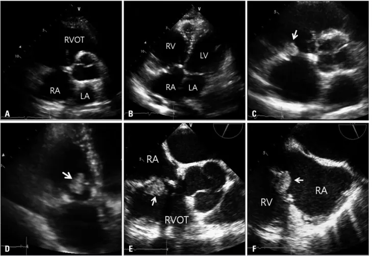
tial diagnosis, trans-esophageal echocardiography was carried out and revealed a mobile mass attached to the annuloplasty groove (Fig. 4). Also his prothrombin time was occasionally inappropriate fluctuating from 1.58 to 2.65 for 9 months.
Based on a suspicion of thrombus formation, we decided to do thrombolysis with tissue plasminogen activator (TPA).
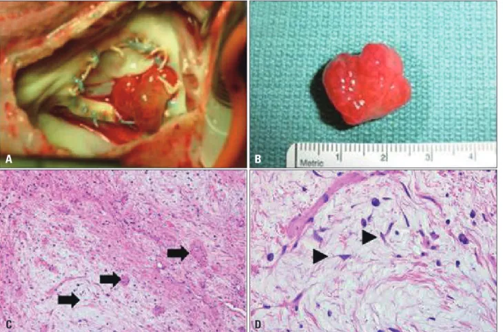
TPA (Alteplase®) was infused 100 mg, 10 mg as a bolus and 40 mg over 2 hours followed by 50 mg over 5 hours.2) How- ever, the mass did not respond to thrombolysis, and the pa- tient underwent surgery. Gross examination of the operative specimen revealed a polypoid mass with smooth surfaces; his- tological examination showed a myxoid matrix composed of an acid-mucopolysaccharide - rich stroma and embedded po- lygonal cells in hematoxylin and eosin stain, consistent with myxoma (Fig. 5).
Discussion
Thrombosis is first suspected when an intracardiac mass is observed in the tricuspid valve after valvular surgery. Patients with a right atrial thrombus have several therapeutic options.
For the patient at high risk for surgery, anticoagulation with heparin followed by an ongoing regimen of warfarin may be the treatment of choice.4) Thrombolytic therapy is also report- ed to be successful in some patients,5) although the possibility of pulmonary embolism caused by a fragmented thrombus due to thrombolytic therapy is a serious concern. In our case, given the patient’s previous history of valvuloplasty, the valvu- lar mass shown in TTE was suspected to be thrombus, so thrombolytic therapy was performed.2) After thrombolytic ther- apy, the size of the valvular mass did not change. We needed to exclude the possibility of other types of cardiac tumor and therefore decided to examine by surgical excision. The valvu- lar mass was found to be myxoma. The differential diagnoses of intraatrial masses include atrial myxoma, thrombus related to atrial fibrillation, vegetation, metastatic tumors, and pri- mary benign or malignant tumors.6) Although a mass usually shows up differently in an echocardiographic image, it is diffi- cult to discriminate between thrombus and tumor. However, in our case it was difficult to make an accurate preoperative di- agnosis. The patient underwent surgical excision of the tricus- pid valvular mass. Histopathology allowed diagnosis of the
Fig. 4. A and B: Parasternal short axis view and apical four-chamber view. There was no visible mass in the immediate postoperative image. C and D: Parasternal short axis view and apical four-chamber view. There was a visible mass (arrow) with stalk 9 months later. E and F: Mid-esophageal RV inflow-outflow view in 64 degrees and mid-esophageal RV inflow view in 116 degrees. An intracardiac mass (arrow) was obvious (size: 2.2 × 1.5 cm2).
After the 5th day of thrombolysis, the mass size remained unchanged. RA: right atrium, RV: right ventricle, RVOT: right ventricular outflow track, LA:
left atrium, LV: left ventricle.
D A
E B
F C
Journal of Cardiovascular Ultrasound 19 | December 2011
210
mass as a tricuspid valvular myxoma.
Myxomas are the most common type of primary benign cardiac tumor, accounting for 50% of all primary cardiac tu- mors. Myxomas may develop in any chamber of the heart: ap- proximately 75% of myxomas develop in the left atrium, 23%
in the right atrium, and the remaining 2% in the ventricles.
Primary tumors of the cardiac valves and chordae are uncom- mon. When they do occur, they are usually slow-growing fi- broelastomas which manifest after several years.7) The interest- ing aspect of our case was the rapid growing cardiac myxoma which became apparent on the tricuspid valve within 9 months after tricuspid annuoplasty. To our knowledge, this is first re- ported case of tricuspid valve myxoma in Korea.
References
1. Korean Society of Echocardiography. 2009 Training program for au-
thenticated echocardiographist [Internet]. 2009 [cited 2011 Feb 21].
Available from:http://www.ksecho.org/infomation/contents/notice126/11_1.
pdf.
2. Pierre-Justin G, Pierard LA. Management of mobile right heart thrombi:
a prospective series. Int J Cardiol 2005;99:381-8.
3. Piazza G, Goldhaber SZ. Fibrinolysis for acute pulmonary embolism.
Vasc Med 2010;15:419-28.
4. Porath A, Avnun L, Hirsch M, Ovsyshcher I. Right atrial thrombus and recurrent pulmonary emboli secondary to permanent cardiac pacing--a case report and short review of literature. Angiology 1987;38:627-30.
5. Mügge A, Gulba DC, Jost S, Daniel WG. Dissolution of a right atrial thrombus attached to pacemaker electrodes: usefulness of recombinant tissue- type plasminogen activator. Am Heart J 1990;119:1437-9.
6. Reynen K. Cardiac myxomas. N Engl J Med 1995;333:1610-7.
7. Graça A, Nunes R, Costeira A, Almeida J, Bastos P. Cardiac papil- lary fibroelastoma of a mitral valve chordae revealed by stroke. Rev Port Cardiol 1999;18:937-9.
Fig. 5. A: After right atrial opening, photograph showed a polypoid mass attached between annulus and tricuspid valve. B: After excision, mass size was similar to echocardiographically measured size (resected tumor size was approximately 2.5 × 1.5 cm2). C: Histological findings with Hematoxylin- Eosin staining. Histological examination of the mass revealed that it was composed of scattered small oval or stellate cells with abundant myxoid matrix, consistent with a diagnosis of nonmalignant cardiac myxoma. The tumor was hypocellular with prominent myxoid stroma and thick walled blood vessels (arrows, × 100). D: The tumor cells were elongated or stellate (arrowheads). They had round to elongated nuclei and eosinophilic cytoplasm. Lymphoplasma cells and histiocytes are present (× 400).
C A
D B