http://dx.doi.org/10.4046/trd.2012.72.2.218 ISSN: 1738-3536(Print)/2005-6184(Online) Tuberc Respir Dis 2012;72:218-222
CopyrightⒸ2012. The Korean Academy of Tuberculosis and Respiratory Diseases. All rights reserved.
폐색전증으로 오인된 폐동맥내막육종 1예
인제대학교 의과대학 일산백병원
1내과학교실,
2영상의학교실,
3병리학교실,
4흉부외과학교실,
5치료방사선과학교실
김진숙1, 박혜경1, 이혜란1, 강승대1, 배상철1, 김수영2, 장선희3, 장우익4, 강승희5, 이성순1A Case of Pulmonary Artery Intimal Sarcoma Masquerading as Pulmonary Embolism
Jin Suk Kim, M.D.
1, Hye Kyeong Park, M.D.
1, Hye Ran Lee, M.D.
1, Seung Dae Kang, M.D.
1, Sang Chul Bae, M.D.
1, Su Young Kim, M.D.
2, Sun Hee Chang, M.D.
3, Woo Ik Chang, M.D.
4, Seung Hee Kang, M.D.
5, Sung-Soon Lee, M.D.
1Departments of
1Internal Medicine,
2Radiology,
3Pathology,
4Cardiosurgery, and
5Radiation Oncology, Ilsan Paik Hospital, Inje University College of Medicine, Goyang, Korea
Pulmonary artery intimal sarcoma is a rare tumor with no characteristic symptoms. It is frequently misdiagnosed as pulmonary embolism. We report a case of pulmonary artery intimal sarcoma in a 48-year-old man with dyspnea, cough and blood-tinged sputum. He was initially suspected and treated as a pulmonary embolism. Computed tomography of the chest showed filling defects occupying the entire luminal diameter of the right and left pulmonary artery as well as extraluminal extension of the intraluminal mass. Surgical resection of the tumor confirmed pulmonary artery intimal sarcoma. After surgery, he received 8 cycles of combined chemotherapy consisting of doxorubicin and ifosfamide. After 8 cycles, Computed tomography of the chest showed interval regression of the residual tumor. Radiotherapy was done as total 6,000 cGy for 5 weeks, following the 8th chemotherapy. The patient's condition was successfully stabilized with chemotherapy and radiotherapy.
Key Words: Vascular Neoplasms; Sarcoma; Pulmonary Embolism; General Surgery; Chemotherapy, Adjuvant
Address for correspondence: Sung-Soon Lee, M.D.
Division of Pulmonary and Critical Care Medicine, Depart- ment of Internal Medicine, Ilsan Paik Hospital, Inje Universi- ty College of Medicine, 2240, Daehwa-dong, Ilsanseo-gu, Goyang 411-706, Korea
Phone: 82-31-910-9782, Fax: 82-31-910-7219 E-mail: [email protected]
Received: Dec. 31, 2011 Revised: Jan. 18, 2012 Accepted: Feb. 6, 2012
서 론
폐동맥내막육종은 매우 드문 악성 종양이며, 예후가 극 히 불량하다. 주로 호흡곤란, 기침, 객혈 혹은 흉통으로 내원하여 흔히 폐색전증으로 오인되어 많은 경우 초기에 항응고요법을 받기도 하며, 수술이나 부검을 통해 진단된 다1. 광범위한 수술적인 절제를 통해 임상증상 및 생존기 간을 연장할 수 있다고 하며2, 항암치료와 방사선치료에
대해 일부 연구에서 효과를 보고하고 있으나3,4, 확립된 치 료는 아니어서 현재까지는 폐동맥내막육종에 대한 표준 치료는 없다. 하지만 최근 광범위절제술을 포함한 다각적 인 치료법이 생존율을 증가시킬 수 있다고 하는 보고들이 있다3,5-7.
저자들은 노작성 호흡곤란으로 내원하였고, 광범위한 폐색전증 의증으로 초기 항응고치료를 시작했으나, 폐색 전증의 위험요인이 전혀 없고, 정상 심초음파 소견, 흉부 전산화 단층촬영 및 양전자 단층촬영에서 폐동맥내막육 종 및 폐침윤이 의심되어, 폐동맥절제술 및 조직절제술을 시행하여 폐동맥내막육종을 확인하였다. 이후 약 9개월간 항암치료 및 방사선치료 후 경과가 호전되어 추적관찰 중 인 1예를 경험하였기에 보고한다.
Case Report
Figure 1. Chest radiograph on admission showed poor- ly-defined multiple nodular and mass-like opacities in both lower lung fields. Dilatation of both pulmonary arterial shadows were suspected.
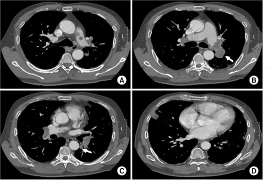
Figure 2. Axial enhanced CT scans obtained with pulmonary embolism pro- tocol showed multifocal low attenuating filling defects within the proximal left pul- monary artery (A), distal right and left pulmonary ar- teries, left lower lobar ar- tery and right middle and lower lobar arteries (B, C).
Main lesion was luminal ex- pansile (arrow). Subpleural nodular consolidations were seen in the right middle lobe and left lower lobe (D).
CT: computed tomography.
증 례
환 자: 조○○, 48세, 남자 주 소: 호흡곤란, 기침 및 객혈
현병력: 환자는 약 1개월 전부터 경사진 곳을 오르거나 평지에서 빨리 걸을 때 호흡곤란이 발생하였으며, 기침 및 소량의 객혈이 있어 인근병원을 방문하였고 흉부 전산 화 단층촬영에서 폐동맥의 충만결손이 발견되어 평가 및 치료를 위해 외래로 내원하였다.
과거력 및 가족력: 특이사항이 없었다.
직업력: 교포로 괌에서 슈퍼마켓을 운영하고 있었다.
진찰 소견: 내원 당시 환자의 혈압은 120/80 mm Hg, 맥박은 분당 102회, 호흡은 분당 20회, 체온은 36.5oC였 고, 키 166 cm, 체중 66 kg이었다. 급성 병색을 보였으며, 의식은 명료한 상태로 흉부진찰상 수포음이나 천명음은 들리지 않았으며, 심음은 빈맥을 보였으나 심잡음은 들리 지 않았다.
방사선 및 검사실 소견: 내원 후 시행한 단순 흉부촬영 에서 양측 폐하부에 여러 개의 경계가 불분명한 음영 증가 소견 및 좌측 폐문부에 둥근 종괴 음영이 보였다(Figure 1). 말초혈액 검사에서 혈색소 13.1 g/dL, 백혈구 9,760/μ L, 혈소판 245,000/μL였으며, 혈액화학 검사상 혈청요소 질소 16 mg/dL, 크레아티닌 0.9 mg/dL, 혈청 D-이합체 973.85 ng/mL, 대기 중 동맥혈가스분석에서 PH 7.46,
PaCO2 38.8 mm Hg, PaO2 82.1 mm Hg, HCO3 27.1 mmol/L, 96.8%였다. 심전도는 동성 빈맥 소견을 보였다.
흉부 전산화 단층촬영에서 양측 폐동맥 내경을 채우는 다 발성의 저음영 충만결손이 관찰되었다(Figure 2). 폐동맥 내 충만결손 소견이 동맥 내에 한정되지 않고 혈관밖으로 팽창하는(expansile) 양상이 관찰되었다. 또한, 양측 중,
Figure 3. (A∼C) Axial fusion images of FDG-PET/CT showed multifocal strong FDG uptake, correspond- ing to the filling defects on CT scan. (D) Maximum in- tensity projection image of FDG-PET scan showed in- creased uptake of FDG in the left pulmonary hilar lesion. FDG: fluorodeox- yglucose positron; PET:
emission tomography; CT:
computed tomography.
Figure 4. The anaplastic tumor cells appear storiform- patterned arrangement (H&E stain, ×200).
하엽에는 흉막하 결절들이 관찰되었다. 하지 도플러 검사 에서는 심부정맥 혈전 소견은 보이지 않았고, 심초음파상 심구출율은 79%로 심근수축기능은 정상이었으며, 폐동맥 고혈압 및 삼첨판 역류 소견은 보이지 않았다. 흉부 전산 화 단층촬영 소견이 전형적인 폐색전증의 양상이 아니었 으므로, 혈관 기원의 종양과의 감별을 위해 양전자방출 단층촬영을 시행하였다(Figure 3). 검사에서 좌측 폐문부 주변 종괴에서 Standard Uptake Value (SUV)가 14.7로 강한 대사증가 소견이 관찰되었고, 양측 폐동맥 내 충만결 손부위도 대사증가(SUV=4.9)를 보였다.
수술 소견: 입원 6일째 폐혈관 내 종양이 의심되는 소견 으로 수술을 시행하였다. 우측 폐동맥 내 종괴 및 좌측
폐동맥 내부를 완전히 막고 있으며, 제2분지 동맥까지 연 결된 1 cm 크기의 종괴가 관찰되었고, 혈관내벽 침범 소 견도 보였다.
병리조직학적 소견: 육안 검사에서 폐동맥으로부터 얻 어진 조직은 혈전덩어리와 유사하였다. 현미경 소견에서 방추형 세포들이 무질서하게 증식되어 있는 소견이 보였 다(Figure 4). 종양세포들은 핵의 과염색성과 다형성이 심 한 역형성을 보였고, 유사분열이 다수 동반되어 있었다.
면역조직화학 염색에서 종양세포는 Vimentin 양성 반응 을 나타내었고, CD34와 Desmin에는 반응하지 않았다. 조 직학적 소견과 면역조직화학 염색을 기반으로 폐동맥혈 관 내막에서 기원한 내막육종으로 진단하였다.
경과: 환자가 호흡곤란, 기침 및 객혈을 호소하고, 흉부 전산화 단층촬영에서 폐색전증 의심 소견이 있어 항응고 치료를 시작하였으나, 광범위 폐색전증 소견임에도 심장 초음파에서 폐동맥고혈압 소견 및 폐색전증에 합당한 소 견이 관찰되지 않고, 폐색전증이 생길 만한 위험요인이 전혀 없는 점, 흉부 전산화 단층촬영 및 양전자방출 단층 촬영에서 폐혈관 내 종괴 의심 소견이 보여 조직 확인을 위해 수술을 진행하였다. 수술 당시 좌측 폐혈관 내 종괴 가 혈관벽을 침범하여, 폐실질 내로 침윤을 동반하고 있었 으며 반대편 우측 폐혈관 내 종괴도 수술장 내 동결 생검 에서 악성 종양으로 밝혀져 양측 침범한 경우로 판단되었 기에 광범위한 수술적 절제는 시행하지 못했고, 증상 완화 를 위하여 혈관내 종괴 조직만 가능한 한도에서 제거하였
다. 진단 이후 항응고치료는 중단하였다. 수술 3주 후부터 doxorubicin과 ifosfamide로 항암치료를 시작하였고, 6개 월 동안의 항암치료 중에 호전경과를 보였다. 이후 8차 항암치료 후에 시행한 흉부 전산화 단층촬영에서 좌측 폐 혈관 내 종양 및 주변 폐침윤부 암 진행 소견이 관찰되어 5주간 6,000 cGy의 방사선치료를 시행했고, 치료 2개월 후 시행한 흉부 전산화 단층촬영에서 종양 및 폐침윤부 종양이 현저히 감소된 부분관해 소견으로 증상호소 없는 상태로 경과관찰 중이다.
고 찰
폐동맥내막육종은 폐동맥 내막에서 유래된 매우 드문 악성 종양으로, 1923년 Mandelstamm이 부검을 통해 처 음 발표하였는데8, 잠행성으로 진행되기 때문에 임상적으 로 증상이 나타날 때에는 국소적 침윤과 원격전이 등을 동반하는 경우가 많다. 약 50%에서 인접한 폐, 기관지벽, 임파선 또는 심근으로의 직접 침윤을 일으키며 약 16∼
25% 정도만 신장, 뇌, 부신으로의 원격전이가 일어난다9. 전형적으로 폐동맥간으로부터 기원하며 뒤쪽으로는 폐동 맥판과 우심방, 앞쪽으로는 폐동맥분지로 뻗어나간다. 폐 주변부에는 색전, 경색, 전이 등을 동반하게 되며 폐혈관 의 말초에 있는 색전들은 우심실의 후부하를 증가시켜 우 심부전을 초래하게 된다10,11.
임상증상은 폐혈관폐쇄에 의해 나타나며 비특이적인 데, 호흡곤란이 가장 많고, 흉통이나 배부통, 기침, 객혈, 체중감소, 전신쇠약감, 실신, 열 등이 동반될 수 있다. 이 런 증상과 흉부 전산화 단층촬영 소견으로 인해 흔히 폐색 전증으로 오인하게 된다6,10,12.
흉부 전산화 단층촬영에서 폐색전증에 비해 폐동맥육 종의 경우 주폐동맥 혹은 근위부폐동맥의 전체 내경을 차 지하는 저음영의 충만결손이 특징적이며, 편측성 분포를 보이는 경우가 많고, 혈관 내 결손부위가 침범된 동맥으로 연속해서 뻗어나가는 소견을 보이거나, 충만결손 혹은 혈 관 내 종괴가 혈관 밖으로 팽창되어 있는 모습이 관찰된 다. 종양의 혈관분포 상태, 출혈, 괴사로 인한 균일하지 않은 조영증강도 폐색전증과의 감별에 도움을 준다13,14. 자기공명 영상에서 가돌리늄 투여 후의 불균질한 조영 증강은 폐동맥 육종을 진단하는데 조금 더 특이적이며 조 영증강의 정도는 종양분화도의 정도와 연관이 있고 이런 소견은 폐색전증과의 감별에 도움을 준다. 종양은 혈전에 비해 대사적으로 활동성을 띄기 때문에 양전자 단층촬영
에서 섭취율이 증가한다15. 심초음파는 우심방 확장 및 출 구폐쇄를 보여주지만, 폐쇄부위가 주폐동맥에 있다면 색 전과 종양을 구분하는 것은 어렵다.
폐혈관내막육종의 예후는 매우 불량하여, 치료에 있어 서 근치적인 절제술이 증상을 개선시키고, 생존기간을 연 장시키지만 진단이 늦어지는 경우가 많아 문헌으로 보고 된 증례의 60%가 부검을 통해 확인되었다12. 표준적인 치 료방법이 확립되지는 않았지만, 적극적인 수술적 치료에 항암치료나 방사선치료 혹은 두 가지 방법을 병행하는 것 이 수술 단독보다 생존기간을 연장시킨다는 보고들이 있
다3,5-7. 수술이 불가능하거나 수술 후 재발된 경우에도 항
암치료 혹은 방사선치료가 시도될 수 있다4,7. 항암요법은 doxorubicin, epirubucin,carboplatin, ifosfamide, cyclo- phosphamide, cyclophosphamide, taxane 등이 사용된다3-7. 질병의 조기진단이 환자의 생존연장에 중요하므로 폐 동맥육종의 조기진단율을 높이기 위해서는 폐혈관색전증 혹은 심부정맥혈전증의 임상적인 양상과 맞지 않거나, 의 심할 만한 임상적인 병력 및 위험인자가 없는 경우, 항응 고치료에도 불구하고 증상이 지속되는 경우 혹은 폐혈관 근위부 내경을 채우는 충만결손 등이 보이는 경우 폐동맥 육종의 가능성이 있는지 의심하고 흉부 전산화 단층촬영 소견에서의 특징적인 양상이 있는지 확인해야 하며, 자기 공명 영상 혹은 양전자 단층촬영술이 도움이 될 수 있겠 다. 진단 당시 근치적인 절제술이 어려운 경우가 많으므 로 수술을 포함한 항암치료 및 방사선치료 등의 다각적인 치료방법을 통해 생존기간을 높이는 노력이 필요하다. 국 외의 증례에서는 폐동맥내막육종환자에서 수술적인 제거 후 항암치료 및 방사선치료를 시행하고 장기 추적관찰하 여 호전경과를 보인 경우들이 있었으나3-5,7, 국내에서는 수술적인 절제 후 항암치료 및 방사선치료를 시행하고 9 개월 경과관찰 중에 종양 소견의 현저한 호전경과를 보이 고 있는 경우에 대한 사례는 보고된 적이 없어서 보고하는 바이다.
참 고 문 헌
1. Delany SG, Doyle TC, Bunton RW, Hung NA, Joblin LU, Taylor DR. Pulmonary artery sarcoma mimicking pulmonary embolism. Chest 1993;103:1631-3.
2. Mattoo A, Fedullo PF, Kapelanski D, Ilowite JS. Pul- monary artery sarcoma: a case report of surgical cure and 5-year follow-up. Chest 2002;122:745-7.
3. Head HD, Flam MS, John MJ, Lipnik SS, Slater DL,
Stewart RD. Long-term palliation of pulmonary artery sarcoma by radical excision and adjuvant therapy. Ann Thorac Surg 1992;53:332-4.
4. Hirose T, Ishikawa N, Hamada K, Inagaki T, Kusumoto S, Shirai T, et al. A case of intimal sarcoma of the pul- monary artery treated with chemoradiotherapy. Intern Med 2009;48:245-9.
5. Genoni M, Biraima AM, Bode B, Shan AC, Wilkler MB, Turina MI. Combined resection and adjuvant therapy improves prognosis of sarcomas of the pulmonary trunk. J Cardiovasc Surg (Torino) 2001;42:829-33.
6. Blackmon SH, Rice DC, Correa AM, Mehran R, Putnam JB, Smythe WR, et al. Management of primary pulmo- nary artery sarcomas. Ann Thorac Surg 2009;87:977-84.
7. Xu Y, Wang K, Geng Y, Shao Y, Yin Y. A case of in- timal sarcoma of the pulmonary artery successfully treated with chemotherapy. Int J Clin Oncol 2011 Oct 27. [Epub]. DOI: 10.1007/s10147-011-0338-8.
8. Mandelstamm M. Über primäre neubildungen des herzens. Virchows Archiv 1923;245:43-54.
9. Furest I, Marn M, Escribano P, Gómez MA, Cortinac J, Blanquer R. Intimal sarcoma of the pulmonary ar- tery: a rare cause of pulmonary hypertension. Arch Bronconeumol 2006;42:148-50.
10. Anderson MB, Kriett JM, Kapelanski DP, Tarazi R, Jamieson SW. Primary pulmonary artery sarcoma: a re- port of six cases. Ann Thorac Surg 1995;59:1487-90.
11. Mayer E, Kriegsmann J, Gaumann A, Kauczor HU, Dahm M, Hake U, et al. Surgical treatment of pulmo- nary artery sarcoma. J Thorac Cardiovasc Surg 2001;
121:77-82.
12. Krüger I, Borowski A, Horst M, de Vivie ER, Theissen P, Gross-Fengels W. Symptoms, diagnosis, and therapy of primary sarcomas of the pulmonary artery. Thorac Cardiovasc Surg 1990;38:91-5.
13. Yi CA, Lee KS, Choe YH, Han D, Kwon OJ, Kim S.
Computed tomography in pulmonary artery sarcoma:
distinguishing features from pulmonary embolic disease. J Comput Assist Tomogr 2004;28:34-9.
14. Choi EY, Yoon YW, Kwon HM, Kim D, Park BE, Hong YS, et al. A case of pulmonary artery intimal sarcoma diagnosed with multislice CT scan with 3D recon- struction. Yonsei Med J 2004;45:547-51.
15. Tueller C, Fischer Biner R, Minder S, Gugger M, Stoupis C, Krause TM, et al. FDG-PET in diagnostic work-up of pulmonary artery sarcomas. Eur Respir J 2010;35:
444-6.