Clinical Characteristics and Treatment Outcomes of Primary Pulmonary Artery Sarcoma in Korea
Pulmonary artery sarcomas (PAS) are rare malignant neoplasms. Right heart failure due to tumour location is the main cause of death in PAS patients. The hemodynamic influence of PAS may effect prognosis, but this has not been proven. We aimed to identify the clinical characteristics and prognostic factors of PAS in Korea, their association with pulmonary hypertension (PH). PAS patients treated at the Asan Medical Center between 2000 and 2014 were reviewed. We examined demographic characteristics, diagnostic and treatment modalities. Potential prognostic factors were evaluated by univariate and multivariate analysis. Twenty patients were diagnosed with PAS. Ten patients were male, the median age was 54 years (range, 33-75 years). The most common symptom observed was dyspnea (65%). The most common histologic type was spindle cell sarcoma (30%). Ten patients had a presumptive diagnosis of pulmonary embolism (PE) and received
anticoagulation therapy. Seventeen patients underwent surgery, but only 5 patients had complete resection. Eleven patients received post-operative treatment (chemotherapy = 3, radiotherapy = 5, chemoradiotherapy = 3). PH was observed in 12 patients before treatment and in 6 patients after treatment. Overall median survival was 24 months. Post- treatment PH was associated with poor prognosis (HR 9.501, 95% CI 1.79-50.32;
P = 0.008) while chemotherapy was negatively associated with mortality (HR 0.102, 95%
CI 0.013-0.826; P = 0.032) in univariate analysis. Post-treatment PH was also associated with poor prognosis in multivariate analysis (HR 5.7, 95% CI 1.08-30.91; P = 0.041). PAS patients are frequently misdiagnosed with PE in Korea. Post-treatment PH is associated with a poor prognosis.
Keywords: Diagnosis; Pulmonary Artery Sarcoma; Pulmonary Hypertension; Survival;
Treatment Yunkyoung Lee,¹ Hyun Jung Kim,²
Heeyoung Yoon,¹ Chang-Min Choi,¹ Yeon-Mok Oh,¹ Sang-Do Lee,¹ Chae-Man Lim,¹ Woo-Sung Kim,¹ Younsuck Koh,¹ and Jae Seung Lee1
1Division of Pulmonary and Critical Care Medicine, Department of Internal Medicine, University of Ulsan College of Medicine, Asan Medical Center, Seoul, Korea; 2Department of Internal Medicine, Kyungpook National University Hospital, Kyungpook National University School of Medicine, Daegu, Korea
Received: 4 March 2016 Accepted: 20 July 2016 Address for Correspondence:
Jae Seung Lee, MD
Division of Pulmonary and Critical Care Medicine, Department of Internal Medicine, University of Ulsan College of Medicine, Asan Medical Center, 88 Olympic-ro 43-gil, Songpa-gu, Seoul 05505, Korea
E-mail: [email protected]
http://dx.doi.org/10.3346/jkms.2016.31.11.1755 • J Korean Med Sci 2016; 31: 1755-1760
INTRODUCTION
Pulmonary artery sarcoma (PAS) is an extremely rare, malig- nant neoplasm. The etiology is unknown, and the disease re- sults in significant morbidity and high mortality. Without surgi- cal intervention, the mean survival time after diagnosis is 1.5 months (1). There have been few reports of the long-term re- sults of PAS treatment, especially in Asia. Due to its nonspecific clinical manifestations, including dyspnea on exertion, chest pain and cough, PAS is easily misdiagnosed as a pulmonary vascular disease such as pulmonary thromboembolism (PE) (2,3). These types of misdiagnoses can delay proper treatment.
Therefore, the ability to distinguish PAS from PE is an important issue (4,5).
Radiation therapy and chemotherapy have been recommend- ed for some patients with PAS (6), although the efficacy of these treatments is largely unproven. Thus, surgical resection remains the mainstay for PAS treatment, as complete resection offers the only chance for a potential cure (7) and prolonged survival
(6,8-13). Although TNM staging reflects prognosis and is used as the standard for many cancers, there is no standard TNM staging system for PAS. Instead hemodynamic influence may affect to prognosis of PAS. The origination of PAS from the pul- monary trunk and/or main pulmonary artery leads to right heart failure as the main cause of death due to obstruction of the right ventricular outflow track (RVOT). There have been some case reports of PH due to PAS (4,11), but none of these studies reported the correlation between PH and prognosis.
Interestingly, the largest series of PH due to PAS were report- ed by pathologists and surgeons (14,15). In our study, from a physician’s point of view, we report our experience with 20 PAS patients over a period of 14 years at the Asan Medical Center in Seoul, Korea. We analyse demographics, other baseline charac- teristics, echocardiographic findings, diagnostic and treatment modalities, prognostic factors, postoperative outcomes, and long-term results. We also report on the influence of hemody- namics on PAS prognosis.
Respiratory Diseases
MATERIALS AND METHODS Study patients
We retrospectively reviewed the clinical records of all patients with a pathological diagnosis of PAS at the Asan Medical Center in Seoul, Korea between January 2000 and December 2014. Pa- tients with insufficient medical data were also excluded. Diag- nosis was confirmed by histological examination and immuno- histochemistry. Although some reviews have designated PAS as a primary cardiac sarcoma (16,17). Here we describe only pri- mary PAS and not cardiac-origin sarcoma. A total of 20 patients were included in this study.
Data collection
Baseline data, including age at diagnosis, sex, duration of symp- toms until diagnosis, symptoms at diagnosis, smoking history, diagnostic and treatment modalities, operation records, and pathologic findings were collected from medical charts. Echo- cardiographic studies were performed to identify PH. Trans- thoracic echocardiography (TTE) is the standard technique for diagnosing PH (18). European guidelines define the echocar- diographic diagnosis of PH as “likely” when the tricuspid regur- gitation velocity (TR Vmax) is > 3.4 m/s or the pulmonary ar- tery systolic pressure is > 50 mmHg with/without additional echocardiographic variables (19). We diagnosed PH when TR Vmax was > 3.4 m/s. Other echocardiographic measurements, such as the ejection fraction of the left ventricle, the presence of an inferior vena cava plethora or a pericardial effusion, were also checked. Survival status and date of death were determined based on medical charts and the expiration date of the Korean national health insurance service. Patients were only classified as deceased if they died during the follow-up period. All other cases, including cases lost to follow-up, were censored.
Statistical analysis
All values were expressed as the median (range) for continuous variables and as the number or percentage for categorical vari- ables. Survival analysis was performed using the Kaplan-Meier method, and the log-rank test and Cox proportional hazard model were used for univariate and multivariable analysis re- spectively. Variables used in multivariable analysis included age, sex and variables showing P value < 0.10 in univariate anal- ysis (e.g. Receipt of chemotherapy and post-treatment PH). A P value of < 0.05 was considered to be statistically significant. Anal- ysis was performed using PASW Statistics 18 (SPSS Inc., Chica- go, IL, USA).
Ethics statement
This study was approved by the institutional review board of the Asan Medical Center. We did not seek informed consent from the patients because it was a retrospective study (IRB No. 2015-0122).
RESULTS
Patient characteristics
Of the 20 patients included in the analyses, 10 (50%) were male, and the median age was 54 years (range, 33-75 years) at diag- nosis. Patient characteristics are summarized in Table 1. The median duration between symptom onset and diagnosis was 2 months (range, 0-28 months). Disease evaluation included com- puted tomography (CT; n = 20), TTE (n = 19), magnetic reso- nance imaging (MRI; n = 10), lung perfusion scan (n = 12), and 18F-fluorodeoxyglucose positron emission tomography (18FDG- PET; n = 17). Ten patients (50%) had a presumptive diagnosis of acute or chronic PE and received anticoagulation therapy preoperatively. Tumour tissues were directly obtained by sur- gery (n = 13), open biopsy (n = 2), endobronchial ultrasound transbronchial needle aspiration (EBUS-TBNA; n = 2), percuta- neous needle biopsy (PCNA; n = 2), or pulmonary angiography with endovascular biopsy (n = 1). There were no statistically significant differences in survival by histologic subtype. PH was observed in 12 patients before treatment (median TR Vmax 3.8 m/s, range 3.2-4.4 m/s) and in 6 patients after treatment (medi- an TR Vmax 3.75 m/s, range 2.9-4.0 m/s). Right ventricular dys- function was observed in 11 patients before treatment and 8 patients after treatment. The bilateral pulmonary arteries and/
or pulmonary trunk were affected in 60% of patients, while the unilateral pulmonary artery was involved in 30% of patients.
The tumour extended to the heart in 5 patients.
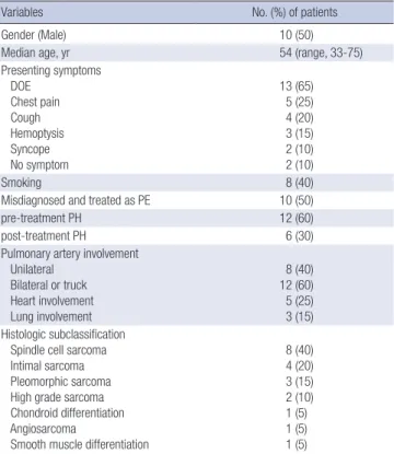
Table 1. Characteristics of the pulmonary artery sarcoma patients (n = 20)
Variables No. (%) of patients
Gender (Male) 10 (50)
Median age, yr 54 (range, 33-75)
Presenting symptoms DOE
Chest pain Cough Hemoptysis Syncope No symptom
13 (65) 5 (25) 4 (20) 3 (15) 2 (10) 2 (10)
Smoking 8 (40)
Misdiagnosed and treated as PE 10 (50)
pre-treatment PH 12 (60)
post-treatment PH 6 (30)
Pulmonary artery involvement Unilateral
Bilateral or truck Heart involvement Lung involvement
8 (40) 12 (60) 5 (25) 3 (15) Histologic subclassification
Spindle cell sarcoma Intimal sarcoma Pleomorphic sarcoma High grade sarcoma Chondroid differentiation Angiosarcoma
Smooth muscle differentiation
8 (40) 4 (20) 3 (15) 2 (10) 1 (5) 1 (5) 1 (5)
DOE = dyspnea on exertion, PE = pulmonary embolism, PH = pulmonary hypertension.
Treatment
Seventeen patients (85%) underwent surgery, although only five of these (30%) received complete resections. Surgery for PAS included pneumonectomy (n = 4), lobectomy (n = 4), pul- monary endarterectomy/tumour debulking with or without pulmonary artery reconstruction (n = 8), and heart/lung trans- plantation (n = 1). The choice of procedure was dependent on factors such as tumour location and distal extension. There were three peri-operative deaths, two of which had unresolved severe PH after surgical resection, and one of which had acute respiratory distress syndrome after surgery. Eleven patients (55%) received post-operative treatment, 3 (15%) received che- motherapy, 5 (25%) received radiotherapy, and 3 (15%) receiv- ed chemotherapy plus radiotherapy. The remaining 2 patients (10%) had inoperable disease involving both pulmonary arter- ies and received only chemotherapy, as complete resection was not possible. These patients were still alive at follow-up (22 and 37 months, respectively). One patient refused treatment and died due to disease progression.
Survival
The median overall survival (OS) for all 20 study patients was 24 months. We used Cox proportional hazard model analysis to
identify factors influencing mortality (Table 2). We found that post-treatment PH was significantly associated with increased mortality (HR 9.501, 95% CI = 1.794-50.32; P = 0.008) (Fig. 1), while chemotherapy was negatively associated with mortality (HR 0.102, 95% CI 0.013-0.826; P = 0.032) (Fig. 2). Post-treat- ment PH was significantly associated with increased mortality in multivariable analysis (HR 5.946, 95% CI 1.551-31.7; P = 0.037) (Table 3). There were no significant differences in survival be- tween patients who were misdiagnosed with PE and those who were not. There is no standard TNM staging system for primary PAS at present. Two previous studies have used their own stag- ing system, described in Table 4 (15,20). There were no statisti- cally significant differences in mortality between these two stag- ing systems in our current patients. The longest surviving pa- tient (188.7 months) had pulmonary artery reconstruction and pulmonary valve replacement along with postoperative che- motherapy and radiotherapy. He developed thyroid metastases 2 years later and adrenal gland metastasis 6.7 years later, both of which were treated with sequential metastasectomies (21).
Table 2. Univariate Cox proportional hazards analysis of variable factors for mortality in the pulmonary artery sarcoma patients
Variables Univariate analysis
HR (95% CI) P value Misdiagnosis and treatment as PE 0.40 (0.10-1.60) 0.370
Smoking history 1.60 (0.42-6.10) 0.490
Pre-treatment PH 0.34 (0.73-1.59) 0.153
Surgical treatment 2.24 (0.28-18.08) 0.403
Chemotherapy 0.10 (0.01-0.83) 0.032
Radiotherapy 0.28 (0.034-2.28) 0.234
Anticoagulation 0.28 (0.03-2.21) 0.225
Treatment modalities > 1 0.30 (0.06-1.46) 0.115
Post-treatment PH 9.50 (1.79-50.32) 0.008
Recurrence to distant site 1.59 (0.40-6.37) 0.512
Metastasis 1.41 (0.34-5.80) 0.632
Heart involvement 0.71 (0.15-3.48) 0.667
Both pulmonary artery or trunk involvement 1.39 (0.34-5.58) 0.641
Complete resection 0.99 (0.209-4.75) 0.988
HR = hazard ratio, CI = confidence interval, PE = pulmonary embolism, PH = pulmo- nary hypertension, RV = right ventricle.
No PAH after treatment (Median 805 days) PAH after treatment (Median 33 days)
Overall survival (%)
Time (day)
2,000 4,000 6,000 8,000 150
100
50
0
P = 0.008
Fig. 1. Kaplan-Meier survival curve of pulmonary artery sarcoma patients with or without pulmonary hypertension after treatment.
Chemotherapy No chemotherapy
Overall survival (%)
Time (day)
2,000 4,000 6,000 8,000 150
100
50
0
P = 0.032
Fig. 2. Kaplan-Meier survival curve of pulmonary artery sarcoma patients with or without chemotherapy.
Table 3. Multivariable Cox proportional hazard analysis of prognostic factors for mor- tality in the pulmonary artery sarcoma patients
Variables Multivariable analysis
HR (95% CI) P value
Age 0.96 (0.91-1.03) 0.249
Sex 1.31 (0.22-7.85) 0.765
Chemotherapy 0.15 (0.02-1.29) 0.083
Pulmonary hypertension (post-treatment) 5.95 (1.12-31.70) 0.037 HR = hazard ratio, CI = confidence interval.
Recurrence
Twelve (60%) patients had recurrence or disease progression (median recurrence-free survival, 15 months; range, 3-45 months).
Lung or locoregional (lymph nodes, chest wall) recurrence was observed in seven (60%) patients, while distant and/or multiple metastases (brain, thyroid, bone, adrenal gland, distant skin, muscles) were observed in five (40%) patients. Eleven patients (91.6%) received one or more treatment. Table 5 summarizes the treatments given after recurrence. Four patients died due to disease progression. Chemotherapy performance (HR 0.331, 95% CI 0.102-1.069; P = 0.053), and lung involvement (HR 5.394, 95% CI 1.423-20.454; P = 0.006) were correlated with recurrence in univariate analysis but not in multivariable analysis.
DISCUSSION
There have been few reports concerning the long-term results of PAS treatment. The 20 patients evaluated in this study consti- tute one of the largest single-centre studies in the published lit- erature to date in Asia. The long-term outcomes of all patients are presented. The reported median age at PAS diagnosis (48.4- 57.0 years) (6,10,13,15,20), and median OS (8-36 months) after treatment (10,15,20), were similar to our study.
Due to absence of a gold standard diagnostic test, multiple tests are used to diagnose PAS. Chest radiography findings are not specific to PAS (22). Although CT provides more informa- tion on the anatomy of the intravascular process, it cannot easi- ly differentiate between PE and PAS (2,22,23). CT findings that favour PAS diagnosis include a heterogeneous enhancement of the mass occupying the main or the proximal pulmonary ar- tery, and extravascular spread of the lesion (24). Echocardiog- raphy is a readily available tool for detecting PAS. The sensitivi- ties of TTE and transoesophageal echocardiography in detect- ing these primary masses are reported as 93% and 97%, respec- tively (25). PAS patients may show a bulging mass impacted in the main pulmonary artery (MPA) on echocardiographic ex- amination, which differs from the linear mass usually found in acute PE. The involvement of the pulmonary valves and attach- ment to the RVOT are other differential findings that suggest a malignant mass with infiltration (5,26,27). When thrombi are
visualized in the MPA of patients with acute PE, they are usually located in its distal part near the bifurcation and often extend to a pulmonary artery (28,29). Despite the findings mentioned above, echocardiography is non-diagnostic for PAS. But TTE has no harmful effect, short-term follow-up echocardiographic examinations represent an attractive method for differential di- agnosis (5). Gadolinium-enhanced MRI can be helpful in dis- tinguishing a soft tissue mass from a thrombus by lesion enhan- cement (26,30). Some experts have suggested that further inves- tigation with MRI is warranted in patients who do not respond to initial anticoagulation (15). Ventilation/perfusion scintigra- phy is used for assessing the likelihood of PE but it is not helpful for distinguishing PAS from thromboembolic disease (31,32).
18FDG-PET plays an important role in the evaluation of pleu- ropulmonary neoplasms (2,23) and is a non-invasive method for differential diagnosis of diseases showing a contrast-filling defect in the pulmonary artery by CT angiography, as blood thrombi do not take up 18F-FDG but malignant tumours such as PAS do (2,23,33,34). In our study, malignancy was preopera- tively suspected in 12/17 patients on the basis of PET (sensitivi- ty 70%) and 9/10 patients on MRI (sensitivity 90%). Suspicion is justified when CT indicates a filling defect in the pulmonary ar- tery that looks atypical for PE or when anticoagulant treatment fails to alleviate pulmonary perfusion abnormalities and system- ic symptoms. 18FDG-PET, MRI or short-term follow-up TTE can help with differential diagnosis in this situation.
In our centre, tumour tissues are mostly obtained intraopera- tively but can also be obtained by PCNA, EBUS-TBNA, or angi- ography. Some studies have reported the use of EBUS-TBNA to differentiate PAS from thromboembolism (35). However, proxi- mal obstruction of the pulmonary arteries by sarcoma or throm- boembolic material can cause hypertrophy of the systemic bron- chial arteries, increasing the risk of haemorrhage from EBUS- TBNA (36).
Reported histopathologic classifications of PAS vary widely.
Fibrous Histiocytoma-type pleomorphic fascicular sarcoma was the most common type of PAS (70%) in a French study (10), pleomorphic-fascicular sarcoma was frequently observed (65%) in an American pathology study (14), and approximately 30%
of PAS were undifferentiated sarcomas in another study (37). In primary cardiac sarcomas, histologic grade of the malignancy Table 4. The number of PAS patients for each stages. Pulmonary artery sarcoma
stages defined in two previous studies
Stages No. of patients
Han Hsi Wong’s stage (20) Operable and non-metastatic
Non-operable and/or metastatic 18
2 Blackmon et al. (15) stage
I (Tumor limited to the main pulmonary artery)
II (Tumor involving one lung plus a main pulmonary artery) III (Bilateral lung involvement)
IV (Extrathoracic spread)
12 2 1 5
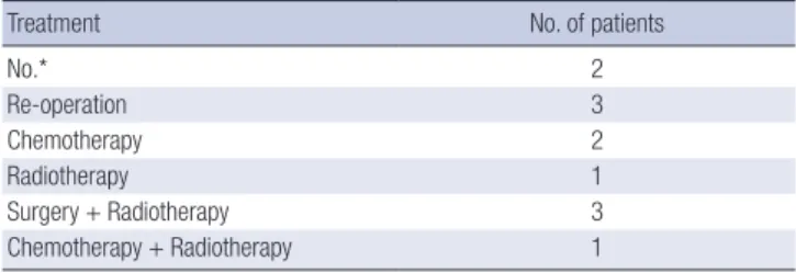
Table 5. Treatment modalities for pulmonary artery sarcoma after recurrence (n = 12)
Treatment No. of patients
No.* 2
Re-operation 3
Chemotherapy 2
Radiotherapy 1
Surgery + Radiotherapy 3
Chemotherapy + Radiotherapy 1
*One patient received angioplasty to relieve symptoms.
has the greatest correlation with survival (38). In PAS, however, there were no statistically significant differences in survival by histologic grade, mitotic count, and presence of necrosis except in low-grade myofibroblastic sarcomas (14). In our present study, spindle cell sarcoma was the most common classification (40%) and there were no differences in survival between different his- tologic classifications in our patient series. The fourth edition of the World Health Organization (WHO) Classification of Tumours of Soft Tissue and Bone was updated in 2013 with immunohis- tochemical and genetic/molecular data for established tumour types, which might necessitate histopathological reviews of our study data.
In this study, complete surgical resection was only performed in a small number of patients, as PAS usually presents late as a bilateral, advanced disease. The possibility of complete resec- tion did not significantly affect mortality, although definite con- clusions are difficult due to the small sample size. According to recent studies, patients treated with chemotherapy or multimo- dality have increased median survival compared with those treated with monotherapy (9,20). Our study showed that che- motherapy may confer a survival benefit, but further studies are required to assess the role and regimen of chemotherapy in PAS. The only targeted agent approved for use in soft tissue sar- coma is the tyrosine kinase inhibitor pazopanib (39), but the use of it for PAS has not been reported. In this study, one patient who had progression after chemotherapy (ifosfamide with mes- na, Doxorubicin, and Dacarbazine) was administered pazopanib as a second-line chemotherapy. However, we only had one month of follow-up and need more time to sufficiently evaluate its ef- fects.
In our study, long-lasting PH after treatment was associated with poor prognosis. Therefore, we suggest that further studies are required focused on post-operative hemodynamic influenc- es as the main treatment target and development of a staging system.
DISCLOSURE
The authors have no potential conflicts of interest to disclose.
AUTHOR CONTRIBUTION
Conception and design: Choi CM, Oh YM, Lee SD, Lim CM, Koh Y, Lee JS. Acquisition of clinical data: Lee Y, Kim HJ, Yoon H. Statistical analysis and interpreted the results: Lee Y, Lee JS.
Writing manuscript: Lee Y, Kim HJ, Lee JS. Revision and final approval of manuscript: all authors.
ORCID
Yunkyoung Lee http://orcid.org/0000-0001-7364-5609
Hyun Jung Kim http://orcid.org/0000-0002-1878-1111 Heeyoung Yoon http://orcid.org/0000-0001-7109-1374 Chang-Min Choi http://orcid.org/0000-0002-2881-4669 Yeon-Mok Oh http://orcid.org/0000-0003-0116-4683 Sang-Do Lee http://orcid.org/0000-0001-8189-4509 Chae-Man Lim http://orcid.org/0000-0001-5400-6588 Woo-Sung Kim http://orcid.org/0000-0002-1254-1264 Younsuck Koh http://orcid.org/0000-0001-5066-2027 Jae Seung Lee http://orcid.org/0000-0003-4130-1486 REFERENCES
1. Krüger I, Borowski A, Horst M, de Vivie ER, Theissen P, Gross-Fengels W.
Symptoms, diagnosis, and therapy of primary sarcomas of the pulmo- nary artery. Thorac Cardiovasc Surg 1990; 38: 91-5.
2. Coskun U, Sinan UY, Calpar I, Yildizeli B, Yanartas M, Filinte D, Kucukog- lu MS. Pulmonary artery sarcoma masquerading as chronic pulmonary thromboembolism. Tex Heart Inst J 2014; 41: 518-22.
3. Dornas AP, Campos FT, Rezende CJ, Ribeiro CA, Amaral NF, Corrêa Rde A. Intimal sarcoma of the pulmonary artery: a differential diagnosis of chronic pulmonary thromboembolism. J Bras Pneumol 2009; 35: 814-8.
4. Kerr KM. Pulmonary artery sarcoma masquerading as chronic thrombo- embolic pulmonary hypertension. Nat Clin Pract Cardiovasc Med 2005;
2: 108-12.
5. Kim MJ, Kim MS, Park JH, Park KI, Lee CS, Na MH, Lee JH, Choi SW, Jeong JO, Seong IW. Pulmonary artery angiosarcoma confused with acute pul- monary thromboembolism: focusing on clinical and echocardiographic features in the differentiation of two categories. J Cardiovasc Ultrasound 2015; 23: 44-7.
6. Mayer E, Kriegsmann J, Gaumann A, Kauczor HU, Dahm M, Hake U, Sch- mid FX, Oelert H. Surgical treatment of pulmonary artery sarcoma. J Tho- rac Cardiovasc Surg 2001; 121: 77-82.
7. Maruo A, Okita Y, Okada K, Yamashita T, Tobe S, Tanimura N. Surgical experience for the pulmonary artery sarcoma. Ann Thorac Surg 2006; 82:
2014-6.
8. Xue H, Wu QY. Surgical treatment of primary pulmonary artery sarcoma.
Chin Med J (Engl) 2011; 124: 461-3.
9. Gan H, Zhang J, Feng L, Zhang Z, Liang L, Zhu G, Chen D. Diagnosis and surgical treatment of pulmonary artery sarcoma. Zhonghua Yi Xue Za Zhi 2014; 94: 1252-4.
10. Mussot S, Ghigna MR, Mercier O, Fabre D, Fadel E, Le Cesne A, Simon- neau G, Dartevelle P. Retrospective institutional study of 31 patients treat- ed for pulmonary artery sarcoma. Eur J Cardiothorac Surg 2013; 43: 787- 93.
11. Rossi A, Zompatori M, Tchouante Tchouanhou P, Amadori M, Palazzini M, Conficoni E, Galiè N, Poletti V, Gavelli G. Rare causes of pulmonary hypertension: spectrum of radiological findings and review of the litera- ture. Radiol Med 2014; 119: 41-53.
12. Bacha EA, Wright CD, Grillo HC, Wain JC, Moncure A, Keel SB, Donahue DM, Mathisen DJ. Surgical treatment of primary pulmonary sarcomas.
Eur J Cardiothorac Surg 1999; 15: 456-60.
13. Huo L, Moran CA, Fuller GN, Gladish G, Suster S. Pulmonary artery sar- coma: a clinicopathologic and immunohistochemical study of 12 cases.
Am J Clin Pathol 2006; 125: 419-24.
14. Tavora F, Miettinen M, Fanburg-Smith J, Franks TJ, Burke A. Pulmonary artery sarcoma: a histologic and follow-up study with emphasis on a sub- set of low-grade myofibroblastic sarcomas with a good long-term follow- up. Am J Surg Pathol 2008; 32: 1751-61.
15. Blackmon SH, Rice DC, Correa AM, Mehran R, Putnam JB, Smythe WR, Walkes JC, Walsh GL, Moran C, Singh H, et al. Management of primary pulmonary artery sarcomas. Ann Thorac Surg 2009; 87: 977-84.
16. Reardon MJ, Walkes JC, Benjamin R. Therapy insight: malignant primary cardiac tumors. Nat Clin Pract Cardiovasc Med 2006; 3: 548-53.
17. Blackmon SH, Patel A, Reardon MJ. Management of primary cardiac sar- comas. Expert Rev Cardiovasc Ther 2008; 6: 1217-22.
18. Milan A, Magnino C, Veglio F. Echocardiographic indexes for the non-in- vasive evaluation of pulmonary hemodynamics. J Am Soc Echocardiogr 2010; 23: 225-39.
19. Galiè N, Hoeper MM, Humbert M, Torbicki A, Vachiery JL, Barbera JA, Beghetti M, Corris P, Gaine S, Gibbs JS, et al. Guidelines for the diagnosis and treatment of pulmonary hypertension: the Task Force for the Diag- nosis and Treatment of Pulmonary Hypertension of the European Soci- ety of Cardiology (ESC) and the European Respiratory Society (ERS), en- dorsed by the International Society of Heart and Lung Transplantation (ISHLT). Eur Heart J 2009; 30: 2493-537.
20. Wong HH, Gounaris I, McCormack A, Berman M, Davidson D, Horan G, Pepke-Zaba J, Jenkins D, Earl HM, Hatcher HM. Presentation and man- agement of pulmonary artery sarcoma. Clin Sarcoma Res 2015; 5: 3.
21. Choi YM, Jang EK, Ahn SH, Jeon MJ, Han JM, Kim SC, Han DJ, Gong G, Kim TY, Shong YK, et al. Long-term survival of a patient with pulmonary artery intimal sarcoma after sequential metastasectomies of the thyroid and adrenal glands. Endocrinol Metab (Seoul) 2013; 28: 46-9.
22. Khadir MM, Chaturvedi A, Nguyen MS, Wandtke JC, Hobbs S, Chaturve- di A. Looking beyond the thrombus: essentials of pulmonary artery im- aging on CT. Insights Imaging 2014; 5: 493-506.
23. Dias OM, Lombardi EM, Canzian M, Soares Júnior J, Vieira Lde O, Terra Filho M. 18F-fluorodeoxyglucose positron emission tomography as a non- invasive method for the diagnosis of primary pulmonary artery sarcoma.
J Bras Pneumol 2011; 37: 817-22.
24. Yi CA, Lee KS, Choe YH, Han D, Kwon OJ, Kim S. Computed tomography in pulmonary artery sarcoma: distinguishing features from pulmonary embolic disease. J Comput Assist Tomogr 2004; 28: 34-9.
25. Meng Q, Lai H, Lima J, Tong W, Qian Y, Lai S. Echocardiographic and patho- logic characteristics of primary cardiac tumors: a study of 149 cases. Int J Cardiol 2002; 84: 69-75.
26. Viana-Tejedor A, Mariño-Enríquez A, Sánchez-Recalde A, López-Sendón
JL. Intimal sarcoma of the pulmonary artery: diagnostic value of different imaging techniques. Rev Esp Cardiol 2008; 61: 1363-5.
27. Chaachoui N, Haik W, Tournoux F. Pulmonary artery sarcoma: a rare cause of dyspnoea. Eur J Echocardiogr 2011; 12: E20.
28. Wittlich N, Erbel R, Eichler A, Schuster S, Jakob H, Iversen S, Oelert H, Mey- er J. Detection of central pulmonary artery thromboemboli by transesoph- ageal echocardiography in patients with severe pulmonary embolism. J Am Soc Echocardiogr 1992; 5: 515-24.
29. Oser RF, Zuckerman DA, Gutierrez FR, Brink JA. Anatomic distribution of pulmonary emboli at pulmonary angiography: implications for cross-sec- tional imaging. Radiology 1996; 199: 31-5.
30. Hsing JM, Thakkar SG, Borden EC, Budd GT. Intimal pulmonary artery sarcoma presenting as dyspnea: case report. Int Semin Surg Oncol 2007;
4: 14.
31. Cox JE, Chiles C, Aquino SL, Savage P, Oaks T. Pulmonary artery sarco- mas: a review of clinical and radiologic features. J Comput Assist Tomogr 1997; 21: 750-5.
32. Palevsky HI, Cone L, Alavi A. A case of “false-positive” high probability ventilation-perfusion lung scan due to tuberculous mediastinal adenop- athy with a discussion of other causes of “false-positive” high probability ventilation-perfusion lung scans. J Nucl Med 1991; 32: 512-7.
33. Chong S, Kim TS, Kim BT, Cho EY, Kim J. Pulmonary artery sarcoma mim- icking pulmonary thromboembolism: integrated FDG PET/CT. AJR Am J Roentgenol 2007; 188: 1691-3.
34. Tueller C, Fischer Biner R, Minder S, Gugger M, Stoupis C, Krause TM, Carrel TP, Schmid RA, Vock P, Nicod LP. FDG-PET in diagnostic work-up of pulmonary artery sarcomas. Eur Respir J 2010; 35: 444-6.
35. Park JS, Chung JH, Jheon S, Choi DJ, Yoon HI, Lee JH, Lee CT, Lee SW.
EBUS-TBNA in the differential diagnosis of pulmonary artery sarcoma and thromboembolism. Eur Respir J 2011; 38: 1480-2.
36. Montani D, Jaïs X, Sitbon O, Dartevelle P, Simonneau G, Humbert M. EBUS- TBNA in the differential diagnosis of pulmonary artery sarcoma and throm- boembolism. Eur Respir J 2012; 39: 1549-50.
37. Croitoru AG, Klein MJ, Galla JD, Fallon JT. Primary pulmonary artery leio- myosarcoma. Cardiovasc Pathol 2003; 12: 166-9.
38. Basso C, Valente M, Poletti A, Casarotto D, Thiene G. Surgical pathology of primary cardiac and pericardial tumors. Eur J Cardiothorac Surg 1997;
12: 730-7.
39. van der Graaf WT, Blay JY, Chawla SP, Kim DW, Bui-Nguyen B, Casali PG, Schöffski P, Aglietta M, Staddon AP, Beppu Y, et al. Pazopanib for meta- static soft-tissue sarcoma (PALETTE): a randomised, double-blind, pla- cebo-controlled phase 3 trial. Lancet 2012; 379: 1879-86.