응급 폐동맥 색전 제거술로 진단 및 치유된 폐동맥내 융모막 암종
Choriocarcinoma in the Pulmonary Artery Diagnosed and Treated by Emergency Pulmonary Embolectomy
A 43-year-old woman who had had an invasive mole 5 years previously required emergent pulmonary embolectomy under cardiopulmonary bypass. Curative resection was impossible because the tumor invaded the right main pul- monary artery and left lower pulmonary artery. The pathologic diagnosis made by the tumor emboli specimens was choriocarcinoma. The patient received post-operative chemotherapy over a 6-month period and had complete remis- sion. Although rare, choriocarcinoma should be considered in the differential diagnosis of fertile women presented with pulmonary embolism.
(Korean J Thorac Cardiovasc Surg 2003;36:531-534) Key words
:1. Pulmonary embolism
2. Choriocarcinoma
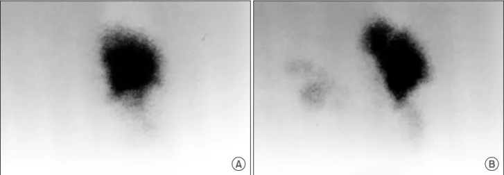
Fig. 1. Lung perfusion scan: preoperative (A) and 14 days after the operation (B).
Fig. 2. Preoperative computed tomograph shows a completely filling defect of right main pulmonary artery and a partialy one of left lower interlobar artery.
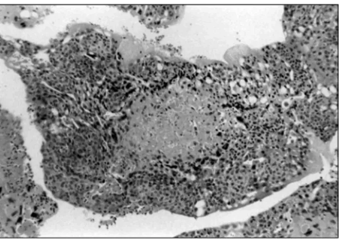
Fig. 3. Microscopic finding shows some tumor nests and marked necrosis (H & E, ×100).
Fig. 4. The tumor is characterized by biphasic pattern which consists of cytotrophoblasts and syncytiotrophoblasts (H & E,
×200).
=국문 초록=
중심 단어