Altered Cellular Kinetics in Growth Plate
according to Alterations in Weight Bearing
Hoon Park, Sun Young Kong, Hyun Woo Kim, and Ick Hwan Yang
Department of Orthopaedic Surgery, Yonsei University College of Medicine, Seoul, Korea.
Received: July 12, 2011 Revised: August 16, 2011 Accepted: August 18, 2011
Corresponding author: Dr. Ick Hwan Yang, Department of Orthopaedic Surgery, Yonsei University College of Medicine, 211 Eonju-ro, Gangnam-gu, Seoul 135-720, Korea.
Tel: 82-2-2019-3416, Fax: 82-2-573-5393 E-mail: [email protected]
∙ The authors have no financial conflicts of interest.
© Copyright:
Yonsei University College of Medicine 2012
This is an Open Access article distributed under the terms of the Creative Commons Attribution Non-Commercial License (http://creativecommons.org/ licenses/by-nc/3.0) which permits unrestricted non-commercial use, distribution, and reproduction in any medium, provided the original work is properly cited.
Purpose: To examine the effects of change in weight bearing on the growth plate metabolism, a simulated animal model of weightlessness was introduced and the chondrocytes’ cellular kinetics was evaluated. Materials and Methods: Unloading condition on the hind-limb of Sprague-Dawley rats was created by fixing a tail and lifting the hind-limb. Six rats aged 6 weeks old were assigned to each group of un-loading, reun-loading, and control groups of unloading or reloading. Unloading was maintained for three weeks, and then reloading was applied for another one week thereafter. Histomorphometry for the assessment of vertical length of the growth plate, 5-bromo-2’-deoxyuridin immunohistochemistry for cellular kinetics, and bio-tin nick end labeling transferase-mediated deoxyuridine triphosphate-biobio-tin nick end labeling (TUNEL) assay for chondrocytes apoptosis in the growth plate were performed. Results: The vertical length of the growth plate and the proliferative po-tential of chondrocytes were decreased in the unloading group compared to those of control groups. Inter-group differences were more significant in the proliferative and hypertrophic zones. Reloading increased the length of growth plate and prolif-erative potential of chondrocytes. However, apoptotic changes in the growth plate were not affected by the alterations of weight bearing. Conclusion: Alterations in the weight bearing induced changes in the chondrocytic proliferative potential of the growth plate, however, had no effects on the apoptosis. This may explain why non-weight bearing in various clinical situations hampers normal longitudinal bone growth. Further studies on the factors for reversibility of chondrocytic proliferation upon variable mechanical stresses are needed.
Key Words: Cellular kinetics, growth plate, changes in weight bearing
INTRODUCTION
Muscles and skeleton play a role in combination, and maintain the body balance and induce movement. For example, the anti-gravity muscles in the lower extremi-ties continuously perform contraction activity to maintain the posture, and skeleton plays a role of the lever that transmits muscle actions. On the other hand, in the cases when weight bearing is reduced or under insufficient condition, the body no longer receives normal weight bearing stress, and atrophy and weakness
phenome-hind limbs. The fore limbs were in contact with the plastic grid floor of the model, but some loss of weightbearing might have occurred. The suspended rats could eat food and drink water (Fig. 1).
Experimental animals and classification
For each group, 6 weeks old 6 Sprague-Dawley rats (weigh-ing around 100 g) were used. By obtain(weigh-ing unload(weigh-ing and reloading conditions by the use of the lower limb suspen-sion method, the change of weight bearing and redistribu-tion of body fluid were induced. Animals were divided to the experiment groups experiencing the lower limb suspen-sion for 3 weeks (unloading group, 9 weeks old) and re-loading for 1 week after the lower limb suspension (reload-ing group, 10 weeks old), and the correspond(reload-ing control group (age-matched 2 control groups; 9 weeks & 10 weeks old, respectively).
Animals were sacrificed under ether inhalation anesthe-sia, and the knee joint area containing the proximal tibia was extracted by performing osteotomy on the tibia and the femoral neck area. The extracted tissues were fixed with 4% paraformaldehyde, decalcified for 2 weeks with 10% ethylenediaminetetracetic acid, and embedded in paraffin by a conventional method. Subsequently, the samples were sectioned as 4 µm in thickness, fixed using silane coating slides, stained with hematoxylin and eosin, and examined na are developed by muscle contracted protein and bone
matrix loss. Furthermore, reduction of number of osteo-blasts, reduction of osteoid volume of cortical bone and tra-becular bone, and decrease of bone density by abnormal for-mation of minerals in the cortical bone, etc. are detected.1-5
Changes occurring in the skeletal system are a frequently developed pathological phenomenon in the cases which re-quire long bed rest due to trauma, or various internal or sur-gical diseases, in disabled individuals who depend on a wheel chair in person with autonomic imbalance, or in the elderly with reduced activity. If exercise were performed to overcome this, it is difficult to withstand the loading deliv-ered suddenly, and ensuring complications and impairment are very severe. Previous studies on the changes of the skel-etal system are biased on the observation of osteoporosis caused by the lack of weight bearing or the change of bone associated with bone loss.6,7 In the case of animal
experi-ments, since the periosteal bone formation is shown to be substantially faster than humans, it is extremely difficult to interpret the results and to apply clinically. In addition, the remodeling of bone is controlled not only by the intramem-branous bone formation but also by the endochondral bone formation. It is, therefore, essential to observe the pattern of the change of cell activity in the growth plate according to the change of weight bearing.
In our present study, the change of activity of chondro-cytes in the growth plate of rats by the change of weight bearing was examined by the analysis of histomorphomet-ric measurement and cellular kinetics.
MATERIALS AND METHODS
Lower-limb suspension method
As a lower-limb suspension method, the method of immo-bilizing the back and the tail of Sprague-Dawley rats was used by improving the model of Nyhan, et al.3 To create the
unloading state by lifting the lower extremities of rats, ty-gon tubing was fixed to the dorsal skin, and subsequently, the tail was immobilized using a tape. The tubing connect-ed to the rat was connectconnect-ed to the restraint equipment of the lower-limb suspension equipment, and the angle was al-ways maintained constant. The animals were thus free to move in a 360° arc. The rats were suspended with 30° head-down tilt in order to initiate a fluid shift similar to that experienced during space flight. This method of suspension
Examination of the apoptosis of chondrocyte within the growth plate (biotin nick end labeling TUNEL assay)
Each tissue slide was pretreated with Tris-HCl solution (pH 8.0) for 10 minutes, and subsequently treated with proteinase K for 20 minutes. After washing with phosphate buffered sa-line (PBS), endogenous tissue peroxidase was inactivated with 3% H2O2, and the samples were washed again with
PBS. The positive control of immunohistochemical exami-nation was pretreated with DNA buffer for 10 minutes, and subsequently reacted with DNase I for 20 minutes, and thus DNA was fragmented artificially. After the pretreatment with transferase-mediated deoxyuridin triphosphated buffer for 15 minutes, reacted with terminaldeoxytransferase and Biotin-16-2’-deoxy-uridine-5’-triphosphate (dUTP) at 37°C for 2 hours and 30 minutes, and thus dUTP was labeled. The nega-tive control group was not treated with terminaldeoxytrans-ferase. The reaction was terminated by immersing the sam-ples in TB buffer for 10 minutes, and subsequently blocked with 2% bovine serum albumin for 15 minutes, washing them with PBS, reacting them with streptavidin-peroxidase for 30 minutes, and then staining them with DAB. For coun-ter staining, nuclear fast red was used, and the samples were dehydrated again with 70, 90, and 100% ethyl alcohol, and mounted. To count the number of TUNEL positive-staining cells, three areas in each slide were selected, the number of entire cells and the number of stained cells in a rectangle of 150×250 µm in size were counted, and the rate of positive cells within the growth plate was calculated as the ratio of positive cells to the entire cells.
Statistical analysis
As a statistical method, Wilcoxon signed rank test was ap-plied, and a statistical significance was determined at p=0.05.
RESULTS
The length of the growth plate of each group
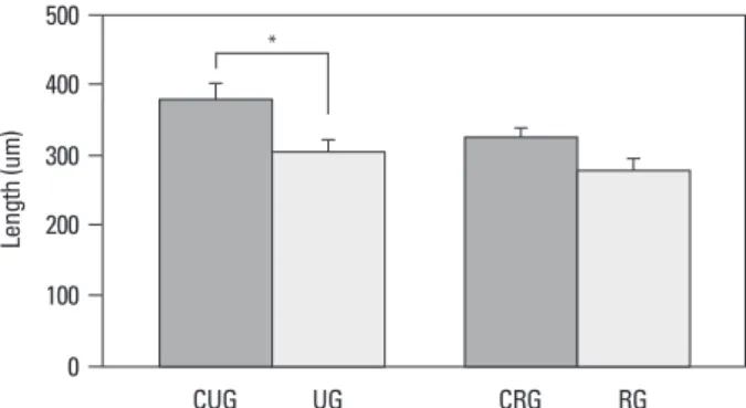
In the case of the change of the growth plate of each group during the experiment period, in the unloading group and the reloading group, the entire length of the growth plate was shorter than the control group (unloading control group; 382.9±11.7 µm, reloading control group; 324.5±13.8 µm), and it was 305.3±14.2 µm (79.7% of the control group) and 281.7±10.3 µm (86.8% of the control group), respectively, and the recovery of the reduction of the length after reload-under a light microscope.
Experimental methods
Histomorphological assessment within the growth plate
In the unloading and reloading group as well as their con-trol group, the lengths of the entire growth plate, the resting zone, the proliferation zone and the hypertrophic zone were measured using the ImagePro® program.8 Regarding all
his-tological examination, four areas were selected randomly, and the entire length as well as the length of each zone were measured vertically.
Measurement of the proliferative capacity of chondrocyte within the growth plate
5-bromo-2’-deoxyuridin (BrdU) injected peritoneally (100 mg/kg) twice, 25 hours and 1 hour prior to the sacrifice of experiment animals. The slides prepared by a conventional method were treated with xylen for 10 minutes 3 times, and for rehydration, the samples were washed with 100, 90, 70% ethanol and double distilled water sequentially. Endog-enous tissue peroxidase was inactivated with 3% hydrogen peroxide (H2O2), and the tissues were reacted with 0.4%
pepsin for 20 minutes. The samples were denatured with 2N HCl for 30 minutes, reacted with goat serum diluted to 20%, and reacted with BrdU primary antibody (SIGMA, St. Lou-is, MO, USA) at room temperature for 12 hours. By apply-ing the Avidin-biotin method, immunohistochemistry was performed. The samples were stained with 3,3’-diaminoben-zidine (DAB), counterstained with Mayer’s hematoxylin, and examined under a light microscope. To count the num-ber of BrdU positive-staining cells, three areas of each slide were selected randomly, and the stained cells within a rect-angle of 150×250 µm in size among the entire cells within the growth plate were counted, and in addition, the positive cell rate in each zone of the growth plate was calculated.
Fig. 2. Total length of the growth plate (*p<0.05). UG, unloading group; CUG,
control for unloading; RG, reloading group; CRG, control for reloading group. 0 100 200 300 400 500 CUG UG CRG RG * Le ng th (u m )
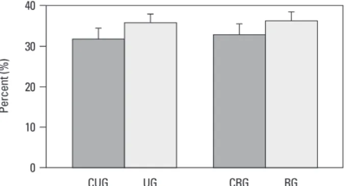
ed (Fig. 6), and in the unloading control group and the re-loading control group, 31.9±8.7% and 32.9±9.4% positive cell rate was shown respectively. However, the unloading group and the reloading group showed 35.8±7.2% and 36.3±6.9% apoptosis level, respectively, and a significant difference among the groups was not detected (Fig. 7), in the assessment of each zone, a significant difference among the groups was not detected (data not shown).
DISCUSSION
Rats are the animals most often used in experiments study-ing the effects of locomotor disuse. Hindlimb rat suspen-sion is considered to be the model of choice for simulating the effects of hypoactivity and weightlessness, with its use having been described in >800 articles.9 With respect to the
skeletal system, tail suspension is an appropriate model for bone disuse studies, as shown by Bloomfield, et al.,10 who
established a correlation between suspension and bone mor-ing was detected (Fig. 2).
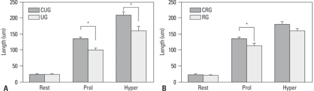
In addition, in the case of the comparison of the length of each zone within the growth plate, a large difference of the length according to the change of loading in the proliferation zone and the hypertrophic zone could be detected (Fig. 3). Cell proliferation capacity of the growth plate of each group
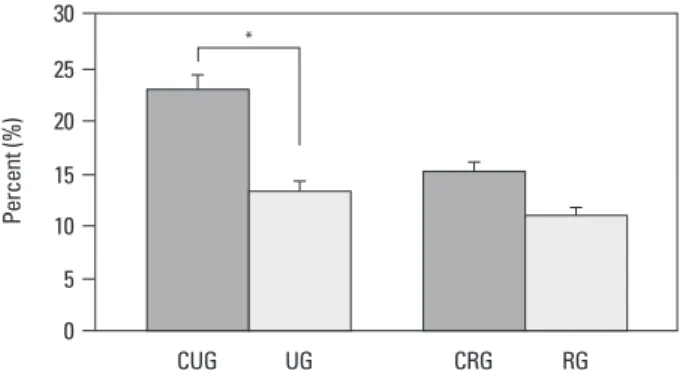
In all groups, BrdU positive cells representing proliferation were detected in the proliferation zone of the growth plate (Fig. 4), and in the unloading control group and reloading control group, 22.9±7.4% and 15.1±4.8% positive cell rate was shown. In the cases of the unloading group, the posi-tive cell rate was 13.3±4.1%, and it was measured to be 57.8% of the control group, on the other hand, in the loading cases, it was 11.0±3.8%, and the result of the re-covery to 86.8% of the control group was shown (Fig. 5). Apoptosis level in the growth plate of each group In all four groups, apoptosis in the growth plate was
detect-Fig. 3. Length of each zone of the growth plate (*p<0.05). (A) Comparison between unloaded and control group. (B) Comparison between
reloaded and control group. rest, resting zone; prol, proliferative zone; hyper, hypertrophic zone; CUG, control for unloading; RG, reloading group; CRG, control for reloading group; UG, unloading group.
Fig. 4. Findings of BrdU immunohistochemistry (×100). (A) Control group. (B) Unloading group. (C) Control for reloading group. (D) Reloading group. BrdU, 5-bromo-2’-deoxyuridin.
A B A B C D 0 0 50 50 100 100 150 150 200 200 250 250
Rest Prol Hyper Rest Prol Hyper
* * Le ng th (u m ) Le ng th (u m ) * CUG UG CRG RG
ing in the distance between cells becomes expanded and progresses to the hypertrophic zone. In the hypertrophic zone, the accumulation and release of calcium in chondro-cytes and the calcification of the stroma are achieved, and simultaneously, the neovasculature infiltrates from the me-taphysis, and cells required for bone formation, osteogenic precursor cells and chondroclasts are infiltrated. In addition, together with the infiltration of these cells, while undergo-ing the apoptosis process or the death process, the length grows transforming to the bone, and so called “cell kinet-ics” that undergoes the overall process such as the prolifera-tion, hypertrophy, apoptosis of chondrocytes, etc.11-16 On
the other hand, it has been well known that the activity of chondrocytes in the growth plate is controlled by biological factors, by various hormones, local growth factors, etc., nevertheless, actually in clinics, instead of the changes due to biological factors; the cases undergoing the reduction of dynamic weight bearing due to their condition are more prevalent.
In our study, the length of the growth plate of the sus-pended group was shorter than that control group because of the decrease in the length of the proliferating and hyper-trophic zones, while the resting zone were not changed. Af-ter reloading, we observed that the length of the hypertro-phic zone was recovered, while the proliferating zone didn’t. Our results are not consistent with previous studies made by examining of growth plates by electron and light
microsco-py.17,18 This studies reported that there was no significant
dif-ference in the width of growth plate between the spaceflight and weight-bearing control groups. However, these studies were performed in different experimental environment (spaceflight) and were conducted in short unloading and re-phometry. In our study, we took into consideration many of
the recommendations of Morey-Holton and Globus.9 We
used more than one control to correctly interpret the results. Two control groups were created (baseline controls) be-cause we used young adult animals that were still develop-ing and could present differences caused by maturation dur-ing the experimental period.10 Control for suspended group
was sacrificed at 9 weeks, control for reloading group was sacrificed at 10 weeks. The differing results of ultimate stress between the two control groups reinforce the correct-ness of our decision.
In the growth plate, histologically distinguishable 4 types of chondrocytes form a continuous columnar pattern, and it is classified to the resting zone, the proliferation zone, the hypertrophic zone and the mineralizing zone.4,11
Chondro-cytes in the resting zone are inactive flat cells that supply new cells for the proliferation to cartilage cells, cells in the proliferation zone repeat cell division and mature to a round shape, increase cell volumes and thus create a vertical space, and on the other hand, produce extracellular matrix
result-Fig. 6. Apoptosis in the growth plate (×100, TUNEL assay). (A) Control group. (B) Unloading group. (C) Control for reloading group. (D) Reloading group. TUNEL, transferase-mediated deoxyuridine triphosphate-biotin nick end labeling.
Fig. 5. Comparison of BrdU Immunohistochemistry between groups (*p<0.05). UG, unloading group; CUG, control for unloading; RG, reloading group; CRG, control for reloading group; BrdU, 5-bromo-2’-deoxyuridin.
A B C D 0 5 15 10 20 25 30 CUG UG CRG RG * Pe rc en t ( % )
drocytes in the proliferation zone rather than the defect in the process of the transformation to the bone in the lower hypertrophic zone. In addition, it is considered that to eluci-date whether such reduction of cell activity is caused by the change of the single process chondrocytes proliferation or the secondary due to the change of the expression of stress protein caused by the decrease of loading, additional stud-ies should be performed in future, and it is thought that studies characterizing kinetic factors involved in irrevers-ible cell proliferation within the growth plate are required.
In conclusion, due to the change within the growth plate in rats caused by the lack of weight bearing for 3 weeks, the reduction of the entire length, particularly, the reduction of the length in the proliferation zone and the hypertrophic zone was distinct, which was caused by the reduction of cell proliferation capacity. In addition, the change of load-ing did not influence the apoptosis occurrload-ing in the growth plate, and in the cases delivered reloading for 1 week after unloading, activation of chondrocytes was accelerated and thus the acceleration of the growth of the growth plate length and the recovery of the chondrocyte proliferation ca-pacity were induced.
ACKNOWLEDGEMENTS
This study was supported by Yonsei University College of Medicine (#6-2006-0069) and the Korea Healthcare tech-nology R&D Project, Ministry for Health, Welfare & Fami-ly Affairs, Republic of Korea (#A084120).
REFERENCES
1. Globus RK, Bikle DD, Morey-Holton E. Effects of simulated
loading periods, so it is not appropriate to compare to our study. Some studies19,20 experimented the simulated
weight-lessness using the method performed in our study. They re-ported that the skeleton of growing rats is capable of a rapid recovery from the adverse effects of simulated weightless-ness. However, this study has been biased toward the change of the bone matrix in the cortical bone or the sponge bone depending on the dynamic loading changes, and stud-ies on the change of the chondrocytes activity within the growth plate according to the change of dynamic loading changes and its reversibility have not been conducted.
Among a variety of methods to assess the cell kinetics of chondrocytes in the growth plate, in our study, BrdU immu-nohistochemistry measuring cell proliferation capacity was performed. This measures cell proliferation capacity by the application of the principle that BrdU substitutes thymidine during the S-phase of DNA replication, undergoes the cell division process, and labeled, proliferating cells were as-sessed.13,21,22 The cell proliferation rate in the growth plate
measured in our study of each control group was detected to be 22.9% and 15.1%, and it was found that with aging, the proliferation rate becomed lower. In addition, in the case of the experiment groups, the unloading group and the re-loading group, in comparison with the corresponding con-trol group, it showed 57.8% and 86.8%, and it was ob-served to be lowered noticeably in comparison with the normal group and the cell proliferation rate was on the re-covery by the reloading.
Apoptosis in the growth plate is a physiological cell change occurred during the process of the transformation to the bone through the calcification process in the lower hyper-trophic zone primarily,16,21,22 and the level substituted to the
bone could be detected, and apoptosis could be induced by the damage of the growth plate by the necrosis in the sec-ondary ossification center.16,23,24 In our study, all four groups
showed approximately 34% apoptosis rate, and a signifi-cant difference among the groups was not shown. Contrary to this result, Basso and Heersche25 reported that the
per-centage of apoptotic chondrocytes in the growth plate carti-lage was increased by unloading and that was associated with a 21% decrease in growth plate width. They suggested that apoptosis was likely a significant mechanism by which reduced load affected longitudinal growth. However, we thought that stress caused by weight bearing did not medi-ate a close effect on apoptosis, and it suggested that the re-duction of the length of the growth plate by unloading was caused by the defect in the proliferation process of
chon-Fig. 7. Comparison of apoptosis between the groups. UG, unloading group; CUG, control for unloading; RG, reloading group; CRG, control for reloading group. 0 10 20 30 40 CUG UG CRG RG Pe rc en t ( % )
562-72.
14. Gerber HP, Ferrara N. Angiogenesis and bone growth. Trends Cardiovasc Med 2000;10:223-8.
15. Karsenty G. Chondrogenesis just ain’t what it used to be. J Clin Invest 2001;107:405-7.
16. Matsuno T, Ishida O, Arihiro K, Sunagawa T, Mori N, Ikuta Y. Cell proliferation and death of growth plate chondrocyte caused by ischemia and reperfusion. Microsurgery 2001;21:30-6. 17. Duke PJ, Durnova G, Montufar-Solis D. Histomorphometric and
electron microscopic analyses of tibial epiphyseal plates from Cosmos 1887 rats. FASEB J 1990;4:41-6.
18. Sibonga JD, Zhang M, Evans GL, Westerlind KC, Cavolina JM, Morey-Holton E, et al. Effects of spaceflight and simulated weight-lessness on longitudinal bone growth. Bone 2000;27:535-40. 19. Shimano MM, Volpon JB. Biomechanics and structural
adapta-tions of the rat femur after hindlimb suspension and treadmill run-ning. Braz J Med Biol Res 2009;42:330-8.
20. Wronski TJ, Morey ER. Recovery of the rat skeleton from the ad-verse effects of simulated weightlessness. Metab Bone Dis Relat Res 1983;4:347-52.
21. Farnum CE, Wilsman NJ. Determination of proliferative charac-teristics of growth plate chondrocytes by labeling with bromode-oxyuridine. Calcif Tissue Int 1993;52:110-9.
22. Vanky P, Brockstedt U, Hjerpe A, Wikström B. Kinetic studies on epiphyseal growth cartilage in the normal mouse. Bone 1998; 22:331-9.
23. Roach HI, Erenpreisa J, Aigner T. Osteogenic differentiation of hypertrophic chondrocytes involves asymmetric cell divisions and apoptosis. J Cell Biol 1995;131:483-94.
24. Hatori M, Klatte KJ, Teixeira CC, Shapiro IM. End labeling stud-ies of fragmented DNA in the avian growth plate: evidence of apoptosis in terminally differentiated chondrocytes. J Bone Miner Res 1995;10:1960-8.
25. Basso N, Heersche JN. Effects of hind limb unloading and reload-ing on nitric oxide synthase expression and apoptosis of osteo-cytes and chondroosteo-cytes. Bone 2006;39:807-14.
weightlessness on bone mineral metabolism. Endocrinology 1984;114:2264-70.
2. Matsumoto T, Nakayama K, Kodama Y, Fuse H, Nakamura T, Fukumoto S. Effect of mechanical unloading and reloading on periosteal bone formation and gene expression in tail-suspended rapidly growing rats. Bone 1998;22(5 Suppl):89S-93S.
3. Nyhan D, Kim S, Dunbar S, Li D, Shoukas A, Berkowitz DE. Im-paired pulmonary artery contractile responses in a rat model of microgravity: role of nitric oxide. J Appl Physiol 2002;92:33-40. 4. Vico L, Lafage-Proust MH, Alexandre C. Effects of gravitational
changes on the bone system in vitro and in vivo. Bone 1998;22(5 Suppl):95S-100S.
5. Mayr W, Bijak M, Girsch W, Hofer C, Lanmüller H, Rafolt D, et al. MYOSTIM-FES to prevent muscle atrophy in microgravity and bed rest: preliminary report. Artif Organs 1999;23:428-31. 6. de Rooij PP, Siebrecht MA, Tägil M, Aspenberg P. The fate of
mechanically induced cartilage in an unloaded environment. J Biomech 2001;34:961-6.
7. Gardner TN, Mishra S. The biomechanical environment of a bone fracture and its influence upon the morphology of healing. Med Eng Phys 2003;25:455-64.
8. Kong SY, Kim HW, Park HW, Lee SY, Lee KS. Effects of multi-ple drilling on the ischemic capital femoral epiphysis of immature piglets. Yonsei Med J 2011;52:809-17.
9. Morey-Holton ER, Globus RK. Hindlimb unloading rodent mod-el: technical aspects. J Appl Physiol 2002;92:1367-77.
10. Bloomfield SA, Allen MR, Hogan HA, Delp MD. Site- and com-partment-specific changes in bone with hindlimb unloading in mature adult rats. Bone 2002;31:149-57.
11. Streeter GL. Developmental horizons in human embryos; a review of the histogenesis of cartilage and bone. Contrib Embryol 1949;33:149-68.
12. Chung UI. Essential role of hypertrophic chondrocytes in endo-chondral bone development. Endocr J 2004;51:19-24.
13. Wilsman NJ, Farnum CE, Green EM, Lieferman EM, Clayton MK. Cell cycle analysis of proliferative zone chondrocytes in growth plates elongating at different rates. J Orthop Res 1996;14: