J Korean Soc Radiol 2017;77(1):19-26 https://doi.org/10.3348/jksr.2017.77.1.19
INTRODUCTION
The demand for breast imaging is increasing as a result of na-tionwide breast cancer screening. Although mammography is a very useful imaging modality for detecting and diagnosing breast cancer, its sensitivity for detecting breast cancers is reported to
be as much as 50% lower in dense breasts (1-3). In recent years, breast ultrasonography (US) has undergone enormous growth and change, which are expected to continue. Therefore, supple-mental US for the detection of breast cancer is being widely rec-ommended in women with dense breasts (4-7). As a result, the issue of qualifications required for breast imaging and adequate
Radiology Residents’ Comprehension of the Breast Imaging
Reporting and Data System: The Ultrasound Lexicon and Final
Assessment Category
Breast Imaging Reporting and Data System에 대한 영상의학과
전공의들의 이해도: 초음파 검사 표준용어 및 최종평가분류
Sun Hye Jeong, MD
1, Yun Ho Roh
2, Jung Hyun Yoon, MD
3, Eun Hye Lee, MD
1,
Sung Hun Kim, MD
4, Ji Hyun Youk, MD
5, You Me Kim, MD
6, Min Jung Kim, MD
3*
1Department of Radiology, Soonchunhyang University College of Medicine, Bucheon Hospital, Bucheon, Korea 2Biostatistical Collaboration Unit, Yonsei University College of Medicine, Seoul, Korea
3Department of Radiology, Severance Hospital, Research Institute of Radiological Science, Yonsei University College of Medicine, Seoul, Korea 4Department of Radiology, Seoul St. Mary’s Hospital, College of Medicine, The Catholic University of Korea, Seoul, Korea
5Department of Radiology, Gangnam Severance Hospital, Yonsei University College of Medicine, Seoul, Korea 6Department of Radiology, Dankook University Hospital, Dankook University College of Medicine, Cheonan, Korea
Purpose: To evaluate radiology residents’ performance in interpretation and compre-hension of breast ultrasonographic descriptors in the Breast Imaging Reporting and Data System (BI-RADS) to suggest the adequate duration of training in breast ultraso-nography.
Materials and Methods: A total of 102 radiology residents working in the Depart-ment of Radiology were included in this study. They were asked to answer 16 questions about the ultrasonographic lexicon and 11 questions about the BI-RADS category. We analyzed the proportion of correct answers according to the radiology residents’ year of training and duration of breast imaging training.
Results: With respect to the duration of breast imaging training, the proportion of correct answers for lexicon descriptors ranged from 77.2% to 81.3% (p = 0.368) and
the proportion of correct answers for the BI-RADS category was highest after three-four months of training compared with after one month of training (p = 0.033). The
proportion of correct answers for lexicon descriptors and BI-RADS category did not differ significantly according to the year of residency training.
Conclusion: Radiology residents’ comprehension of the BI-RADS category on breast ultrasonography was not associated with their year of residency training. Based on our findings, radiology residents’ assessment of the BI-RADS category was significantly im-proved with three-four months of training compared with one month of training.
Index terms Breast Ultrasonography Comprehension Education Received June 18, 2016 Revised November 1, 2016 Accepted February 12, 2017
*Corresponding author: Min Jung Kim, MD
Department of Radiology, Severance Hospital, Research Institute of Radiological Science, Yonsei University College of Medicine, 50-1 Yonsei-ro, Seodaemun-gu,
Seoul 03722, Korea.
Tel. 82-2-2228-7413 Fax. 82-2-393-3035 E-mail: [email protected]
This is an Open Access article distributed under the terms of the Creative Commons Attribution Non-Commercial License (http://creativecommons.org/licenses/by-nc/4.0) which permits unrestricted non-commercial use, distri-bution, and reproduction in any medium, provided the original work is properly cited.
curriculum for breast US training has been raised. Nowadays, some countries have established their own training requirements for physicians interpreting mammography (8-10). However, there is a lack of established guidelines for breast US training in radiology residency programs.
The development of resident training guidelines for breast US should be based on actual radiology residents’ performance in determining the lesion description and final assessment, for example, their year of residency and the duration of breast im-aging training. However, there are few published reports about the actual radiology residents’ performance in breast US.
The Breast Imaging Reporting and Data System (BI-RADS) lexicon for US is a quality assurance tool designed to standardize reporting, interpretation and management recommendations (11, 12). Therefore, the BI-RADS is likely to be the most objective tool for assessing the training performance. The purpose of our study was to evaluate radiology residents’ performance in inter-pretation and comprehension of breast US descriptors in order to suggest the adequate duration of training in breast US.
MATERIALS AND METHODS
The Institutional Review Board approved this retrospective study. The study was based on a survey performed at an annual breast imaging conference for educating the radiology residents, which is hosted by the Korean Society of Radiology and the Korean Society of Breast Imaging. This conference is one of the largest meetings on breast imaging–many radiology residents from around the country gather for the breast imaging review. A total of 113 radiology residents who attended the conference in 2014 were enrolled in this study. The main theme of the con-ference was the fifth edition of BI-RADS, which was released in 2013 (11). The radiology residents were asked to independently complete a survey using a wireless remote control answering machine with a numerical keyboard. Each surveyee’s answers were gathered into the mainboard. Prior to conducting the survey, we explained to the radiology residents that the purpose of the survey was to assess their degree of understanding of breast US and to inform conference lecture planning. They were assured that their individual responses would be kept confidential. Among the 113 radiology residents, eleven radiology residents who answered less than three questions were excluded from our
study population. The remaining 102 radiology residents were included in the data analysis.
Questionnaire
We collected US images of 27 breast masses in 27 women from five academic institutions that were obtained between November 2013 and April 2014. Six dedicated breast imaging radiologists specializing in breast imaging with 3–13 years of experience from five institutions performed the ultrasound examinations.
Informed consent was obtained from all of the patients for establishing the image database for radiology resident educa-tion and evaluaeduca-tion. Breast masses were examined with one of the institution’s US systems (iU22, Philips Medical Systems, Both-ell, WA, USA; GE LOGIQ E9, GE Medical Systems, Milwaukee, WI, USA; SuperSonic Imagine, Aix-en-Provence, France; EUB-8500, Hitachi Medical, Tokyo, Japan); each US system being equipped with high-frequency linear array transducers. During scanning, spatial compounding was set on all units and harmon-ic imaging was used on two units (SuperSonharmon-ic Imagine and EUB-8500). Representative transverse and longitudinal gray-scale images of each mass were acquired and then consensus was reached for each imaging descriptor, and final assessment among the six radiologists was based on the fifth edition of BI-RADS (11). Questions were generated for US lexicon and final assessment categories, and grayscale US images were chosen from the database under the consensus of two radiologists. BI-RADS categories 4C and 5 were grouped into one category (4C and 5) because the clinical management of these two categories is the same and the number of answer choices which could be programmed into the answering machine system was limited to five. The survey consisted of 11 questions about the final as-sessment categories of BI-RADS (3 questions for category 2, 1 question for category 3, 1 question for category 4A, 2 questions for category 4B, and 4 questions for category 4C and 5) and 16 questions on the US lexicon (3 questions for shape, 2 questions for orientation, 5 questions for margin, 4 questions for echo pattern, and 2 questions for posterior feature). The descriptors of calcifications and associated features including vascularity and elasticity assessment were not included in this survey.
The histopathologic diagnoses of masses for the 11 questions about the BI-RADS category were collected from each institu-tion. We obtained the final histopathology results for each case
by US-guided core needle biopsy (n = 10) and US-guided vacu-um-assisted biopsy (n = 1).
Survey
Before starting the survey, general information, such as their year of residency and duration of breast imaging training they had received during training, was collected. Breast US images were displayed on a large screen in the front of an auditorium for review. For each question, ten seconds were given to watch the images and an additional five seconds to choose the answer from a multiple choice format. Radiology residents answered by using a wireless answering machine in which all answers were collected in a single database for analysis.
Statistical Analysis
The reference standard was based on the consensus among six breast imaging radiologists. The proportion of correct answers ac-cording to the BI-RADS US lexicon and the final assessment cate-gory was defined as the number of correct answers divided by the total number of responses. No answer was regarded as missing data and it was excluded from the statistical analysis. Among US lexicons, the proportion of correct answers was compared using the chi-square test. The proportion of correct or incorrect answers among radiology residents was also compared according to the year of training or the duration of breast imaging training using the chi-square test. Trends in each of the training duration catego-ries were tested with the use of linear by linear association test (Mantel-Haenszel trend test). We performed a post-hoc analysis for evaluation of significant differences according to the duration of breast imaging training. The results were expressed as
percent-ages and raw numbers. p-values less than 0.05 were deemed to in-dicate statistical significance. Statistical analysis was performed using statistical software (SAS version 9.2; SAS Institute, Cary, NC, USA).
RESULTS
After the survey, a total of 2343 answers were obtained, which were 79.2% of the 2958 expected answers. On average, 21 ques-tions were answered (median, 24; range, 7–27) by each partici-pant. Of the 77 participants who gave information regarding their year of residency training, 13 (16.9%) were in their first year of residency training, 16 (20.8%) were in their second year of residency training, 16 (20.8%) were in their third year of residen-cy training, and 32 (41.6%) were in their fourth year of residenresiden-cy training. Twenty-six radiology residents provided no response. Among the 68 participants who gave information regarding the duration of breast imaging training they had received, the dura-tion of breast imaging training was one month in 25 radiology residents (36.8%), two months in 24 radiology residents (35.3%), Table 1. The Proportion of Correct Answers According to Ultrasono-graphic BI-RADS Lexicon
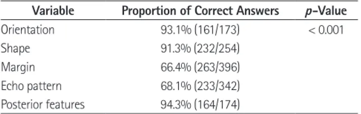
Variable Proportion of Correct Answers p-Value
Orientation 93.1% (161/173) < 0.001
Shape 91.3% (232/254)
Margin 66.4% (263/396)
Echo pattern 68.1% (233/342)
Posterior features 94.3% (164/174)
Data in parentheses are the number of correct answers/total number of answers.
BI-RADS = Breast Imaging Reporting and Data System
Fig. 1. Transverse (A) and longitudinal (B) ultrasonographic images showing an irregular hypoechoic mass with spiculated margins. The
propor-tion of correct answers for margin was 74.1% (60/81). The mass was confirmed as a complex sclerosing lesion. Complex sclerosing adenosis can be seen as a spiculated margin.
and three-four months in 19 radiology residents (27.9%). Thir-ty-five radiology residents provided no response.
With respect to the US BI-RADS lexicon descriptors, the pro-portion of correct answers for shape, orientation, and posterior feature was significantly higher than that for margin and echo pat-tern (p < 0.001) (Table 1). Among the margin descriptors, the proportion of correct answers for circumscribed (88.0%), indis-tinct (82.4%) and spiculated (74.1%) (Fig. 1) margins was higher than that for microlobulated (51.8%) and angular (30.0%) mar-gins. However, after dichotomizing the mass margin into ‘cir-cumscribed’ or ‘not cir‘cir-cumscribed’, the proportion of correct an-swers for ‘not circumscribed’ increased up to 97.4%. The pro-portion of correct answers for lexicon descriptors ranged from 69.2% to 81.9% according to the year of residency (p = 0.167) and from 77.2% to 81.3% according to the duration of breast imaging training (p = 0.368) (Table 2).
Table 2. The Proportion of Correct Answers for Ultrasonographic BI-RADS Lexicon According to Year in Resident Training and the Duration of Training for Breast Imaging
Number of Residents Proportion of Correct Answers p-Value
Year in resident training 0.167
1st 16 69.2% (119/172)
2nd 16 76.0% (171/225)
3rd 16 81.9% (186/227)
4th 33 79.9% (373/467)
Duration of training (months) for breast imaging 0.368
1 28 81.3% (273/336)
2 24 77.4% (271/350)
3–4 19 77.2% (203/263)
Data in parentheses are the number of correct answers/total number of answers. BI-RADS = Breast Imaging Reporting and Data System
Fig. 2. Transverse (A) and longitudinal (B) ultrasonographic images of an oval circumscribed hypoechoic mass showing BI-RADS category 3. The
proportion of correct answers for this BI-RADS category was 38.2% (26/68). The mass was confirmed to be a fibroadenoma. BI-RADS = Breast Imaging Reporting and Data System
A B
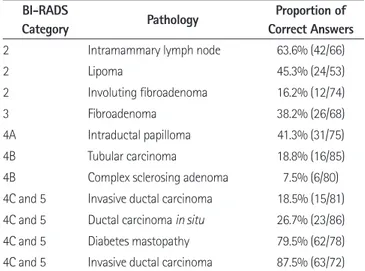
Table 3. The Proportion of Correct Answers According to BI-RADS Category
BI-RADS
Category Pathology
Proportion of Correct Answers
2 Intramammary lymph node 63.6% (42/66)
2 Lipoma 45.3% (24/53)
2 Involuting fibroadenoma 16.2% (12/74)
3 Fibroadenoma 38.2% (26/68)
4A Intraductal papilloma 41.3% (31/75)
4B Tubular carcinoma 18.8% (16/85)
4B Complex sclerosing adenoma 7.5% (6/80)
4C and 5 Invasive ductal carcinoma 18.5% (15/81)
4C and 5 Ductal carcinoma in situ 26.7% (23/86)
4C and 5 Diabetes mastopathy 79.5% (62/78)
4C and 5 Invasive ductal carcinoma 87.5% (63/72)
Data in parentheses are the number of correct answers/total number of answers.
With respect to the BI-RADS category, the proportion of cor-rect answers was 16.2–63.6% for category 2, 38.2% for category 3 (Fig. 2), 41.3% for category 4A, 7.5–18.8% for category 4B, and 18.5–87.5% for category 4C and 5 (Table 3). There was no sig-nificant difference in the proportion of correct answers accord-ing to the pathology (41.1% for benign vs. 36.3% for malignan-cy, p = 0.176) or the dichotomized BI-RADS category (39.9% for category 2 or 3 vs. 38.8% for category 4 or 5, p = 0.813). The proportion of correct answers for the BI-RADS category was 30.4% (n = 28 of 92) among the first-year radiology residents, 37.8% (n = 59 of 156) among the second-year radiology resi-dents, 39.6% (n = 55 of 139) among the third-year radiology residents, and 42.9% (n = 130 of 303) among the fourth-year ra-diology residents, but there were no significant differences (p = 0.186) (Table 4). The proportion of correct answers for the BI-RADS category was 35.4% (n = 76 of 215) after one month of breast imaging training, 37.0% (n = 87 of 235) after two months of breast imaging training, and 47.8% (n = 77 of 161) after three-four months of breast imaging training (p = 0.033) (Table 5). The trend in each of the 3 categories was statistically
signifi-cant (p = 0.018) (Table 5). Therefore, radiology residents’ com-prehension of the BI-RADS category on breast US had a ten-dency to improve according to the duration of training. Post-hoc analysis of these results revealed a significant improvement in the proportion of correct answers when comparing the re-spondents with three-four months of training to those with one month of training (Bonferroni corrected p-value = 0.044) (Table 5). In comparison with radiology residents’ performance after 1 month of training, radiology residents’ performance after two months of training was improved although it was not statistical-ly significant (Bonferroni corrected p-value = 0.096) (Table 5).
DISCUSSION
Although mammography is a very useful imaging modality for detecting and diagnosing breast cancer, its sensitivity for detect-ing breast cancers is reported to be as much as 50% lower in dense breasts (1-3). Therefore, supplemental US for the detection of breast cancer is being widely recommended in women with dense breasts (4-7). At this point, adequate training and experi-ence in breast US are very important, particularly in areas where dense breasts are common, such as Asia. However, there is a lack of guidelines that indicate the adequate amount of breast imaging training radiology residents should receive.
To assess the training performance in breast US, we used BI-RADS descriptors or final assessment whose reliability for the prediction of malignancy has been validated and the interob-server agreement has been found to be good in previous studies (13-15). In this report, the proportion of correct answers given by radiology residents for margin and echo pattern on breast US was lower than that for the other descriptors (Table 1). We as-Table 4. The Proportion of Correct Answers for BI-RADS Category
According to Year in Resident Training Number of Residents
Proportion of
Correct Answers p-Value
Year in resident training 0.186
1st 16 30.4% (28/92)
2nd 16 37.8% (59/156)
3rd 16 39.6% (55/139)
4th 33 42.9% (130/303)
Data in parentheses are the number of correct answers/total number of answers.
BI-RADS = Breast Imaging Reporting and Data System
Table 5. The Proportion of Correct Answers for BI-RADS Category According to the Duration of Training for Breast Imaging Duration of Training (Months) Number of Residents Proportion of Correct Answers p-Value
Among 3 Group Test for Trend vs. 1 vs. 2 vs. 3-4
1 28 35.4% (76/215) 0.033 0.018 0.032 0.096* 0.015 0.044* 2 24 37.0% (87/235) 0.032 0.096* 0.712 > 0.999* 3–4 19 47.8% (77/161) 0.015 0.044* 0.712 > 0.999* Data in parentheses are the number of correct answers/total number of answers.
*Bonferroni corrected p-value.
sumed that this is because margin and echo pattern are com-monly mixed with one or more descriptors and it might also result in a lower value of interobserver agreement. In previous reports that evaluated interobserver variability between breast imaging radiologists, the interobserver agreement was fair for mass margin (kappa value: 0.32–0.4) and echo pattern (kappa value: 0.29–0.37), while the interobserver agreement for shape, orientation and posterior acoustic features was substantial or moderate (14, 16, 17). However, in one previous study, the in-terobserver agreement for margin when simplified to ‘circum-scribed’ or ‘not circum‘circum-scribed’ was improved to moderate (17), which was also consistent with our results since the proportion of correct answers when it was dichotomized into these two categories was greater than 90%. Because one of the four de-scriptors of ‘not circumscribed’ margin should be considered as suspicious findings, variability in the selection among ‘not cir-cumscribed’ descriptors may not have a serious clinical impact. Biopsy is the optimal management for category 4 or higher breast lesions, and this may lessen the importance of variability among observers regarding the margin descriptors. Our results showed that the proportion of correct answers for indistinct or spiculated margins (82.4% and 74.1%, respectively) was higher than that for other margin descriptors (30–51.8%), which is consistent with previous results (17).
According to the year of resident training, the proportion of correct answers for lexicon descriptors and BI-RADS category increased with every year of training, but there was no significant difference among the years of training (p = 0.167 and 0.186, re-spectively) (Tables 2, 4). This finding is consistent with a previous domestic report using mammography which indicated that the year of training alone does not affect residents’ performance (18). We suppose that this is because the training schedule at each in-stitution is very different for each year of residency and breast imaging training is mostly performed in dedicated breast imag-ing sections, and it does not overlap with the other subspecialties of the radiology department.
In the present study, the proportion of correct answers for the BI-RADS category increased with an increasing duration of train-ing from one month to three-four months with a significant im-provement in the proportion of correct answers from one month to three-four months of training despite the lack of a significant difference in terms of lexicon descriptors. These findings are
consistent with a previous report on mammographic training, which showed that radiology residents’ performance in mam-mographic interpretation was correlated with the training period (p < 0.001) (18). The three-four month duration of training is identical to the period of formal training required by a physician to interpret mammograms independently (19). According to our results, three-four months of training will also improve radi-ology residents’ performance in interpretation of breast US. But, there was no significant improvement between two and three-four months of training. We assumed that these results might be associated with each institution’s quality of education. How-ever, we did not investigate each institution’s quality of education. Although post-hoc analysis did not show significant improve-ment between two and three-four months of training, radiology residents’ comprehension of the BI-RADS category on breast US had a tendency to improve according to the duration of training. In our opinion, three-four months of training is ideal for improv-ing radiology residents’ performance in interpretation of breast US, but 2 months of training would be acceptable as the minimal duration of breast US training. In our study, the duration of breast imaging training was one or two months in 72.1% of the par-ticipants who answered the question on the duration of breast imaging training.
This study has some limitations. The first limitation is the large amount of missing data. Second, mammographic information was not available during the survey; therefore, descriptors of calci-fications were inevitably excluded. Mammography is quite helpful for evaluating breast lesions, especially in cases of calcification. However, this study aimed to evaluate the comprehension of radi-ology residents in breast US. Third, we did not include questions about elasticity or color Doppler image which has been added to the fifth edition of BI-RADS because the likelihood of malignancy based on these descriptors is controversial. Finally, we did not consider the differences in training quality, the number of breast US examinations performed per person during training, whether other subspecialties overlapped during breast imaging training and the time interval between the last training and the survey.
Radiology residents’ performances in breast US interpretation and comprehension of BI-RADS descriptors and final assess-ments were variable. Comprehension of US BI-RADS categories and descriptors were not associated with the year of resident training, but they were improved with the duration of breast
im-aging training. Based on our findings, radiology residents’ assess-ment of the BI-RADS category was significantly improved with three-four months of training compared with one month of training and these results might be helpful in deciding the opti-mal duration of radiology residents’ training.
Acknowledgments
This work was supported by the Korean Society of Breast Im-aging & Korean Society for Breast Screening (KSBI & KSFBS- 2013-No. 001) and Soonchunhyang University Research Fund.
REFERENCES
1. Mandelson MT, Oestreicher N, Porter PL, White D, Finder CA, Taplin SH, et al. Breast density as a predictor of mammo-graphic detection: comparison of interval- and screen-de-tected cancers. J Natl Cancer Inst 2000;92:1081-1087 2. Kolb TM, Lichy J, Newhouse JH. Comparison of the
perfor-mance of screening mammography, physical examination, and breast US and evaluation of factors that influence them: an analysis of 27,825 patient evaluations. Radiology 2002;225:165-175
3. Berg WA, Gutierrez L, NessAiver MS, Carter WB, Bhargavan M, Lewis RS, et al. Diagnostic accuracy of mammography, clinical examination, US, and MR imaging in preoperative assessment of breast cancer. Radiology 2004;233:830-849 4. Leong LC, Gogna A, Pant R, Ng FC, Sim LS. Supplementary
breast ultrasound screening in Asian women with nega-tive but dense mammograms-a pilot study. Ann Acad Med
Singapore 2012;41:432-439
5. Kelly KM, Dean J, Comulada WS, Lee SJ. Breast cancer de-tection using automated whole breast ultrasound and mammography in radiographically dense breasts. Eur Radiol 2010;20:734-742
6. Parris T, Wakefield D, Frimmer H. Real world performance of screening breast ultrasound following enactment of Con-necticut Bill 458. Breast J 2013;19:64-70
7. Shen SJ, Sun Q, Xu YL, Zhou YD, Guan JH, Mao F, et al. [Comparative analysis of early diagnostic tools for breast cancer]. Zhonghua Zhong Liu Za Zhi 2012;34:877-880 8. Feig SA, Hall FM, Ikeda DM, Mendelson EB, Rubin EC, Segel
MC, et al. Society of Breast Imaging residency and fellow-ship training curriculum. Radiol Clin North Am 2000;38: 915-920, xi
9. Bassett LW, Monsees BS, Smith RA, Wang L, Hooshi P, Far-ria DM, et al. Survey of radiology residents: breast imaging training and attitudes. Radiology 2003;227:862-869 10. Burhenne LJ, Smith RA, Tabar L, Dean PB, Perry N, Sickles
EA. Mammographic screening: international perspective.
Semin Roentgenol 2001;36:187-194
11. American College of Radiology. ACR BI-RADS Atlas, Breast
Imaging Reporting and Data System. 5th ed. Reston, VA:
American College of Radiology, 2013
12. American College of Radiology. ACR BI-RADS Breast
Im-aging and Reporting Data System: Breast ImIm-aging Atlas,
4th ed. Reston, VA: American College of Radiology, 2003 13. Kim EK, Ko KH, Oh KK, Kwak JY, You JK, Kim MJ, et al.
Clini-cal application of the BI-RADS final assessment to breast sonography in conjunction with mammography. AJR Am J
Roentgenol 2008;190:1209-1215
14. Lazarus E, Mainiero MB, Schepps B, Koelliker SL, Livingston LS. BI-RADS lexicon for US and mammography: interob-server variability and positive predictive value. Radiology 2006;239:385-391
15. Hamy AS, Giacchetti S, Albiter M, de Bazelaire C, Cuvier C, Perret F, et al. BI-RADS categorisation of 2,708 consecu-tive nonpalpable breast lesions in patients referred to a dedicated breast care unit. Eur Radiol 2012;22:9-17 16. Park CS, Lee JH, Yim HW, Kang BJ, Kim HS, Jung JI, et al.
Observer agreement using the ACR Breast Imaging Re-porting and Data System (BI-RADS)-ultrasound, first edi-tion (2003). Korean J Radiol 2007;8:397-402
17. Lee HJ, Kim EK, Kim MJ, Youk JH, Lee JY, Kang DR, et al. Ob-server variability of Breast Imaging Reporting and Data Sys-tem (BI-RADS) for breast ultrasound. Eur J Radiol 2008;65: 293-298
18. Lee EH, Lyou CY. Radiology residents’ performance in screen-ing mammography interpretation. J Korean Soc Radiol 2013; 68:333-341
19. U.S. Food and Drug Administration. Mammography facilities. Web site. http://www.accessdata.fda.gov/scripts/cdrh/cfdocs/ cfMQSA/mqsa.cfm. Accessed Sep 2, 2015
Breast Imaging Reporting and Data System에 대한 영상의학과
전공의들의 이해도: 초음파 검사 표준용어 및 최종평가분류
정선혜
1· 노윤호
2· 윤정현
3· 이은혜
1· 김성헌
4· 육지현
5· 김유미
6· 김민정
3*
목적: 영상의학과 전공의들의 Breast Imaging Reporting and Data System (이하 BI-RADS) 초음파 검사 표준용어 및 최 종평가분류에 대한 이해도를 분석하고 적절한 유방초음파 수련기간을 제시하고자 하였다. 대상과 방법: 102명의 영상의학과 전공의들을 대상으로 하였다. 초음파 검사 표준용어에 관련한 질문 16개, 최종평가분 류에 관련한 질문 11개가 주어졌다. 이들의 답을 연차 및 수련기간에 따라 분석하였다. 결과: 수련기간에 따른 분석에서 표준용어에 대한 정답률은 77.2%에서 81.3%로 기간에 따른 차이가 없었으나(p = 0.368), 최종평가분류에 대한 정답률은 3~4개월간 수련받은 그룹에서 1개월간 수련받은 그룹보다 의미 있게 높았다(p = 0.033). 연차에 따른 분석에서는 표준용어와 최종평가분류에서 모두 의미 있는 차이를 보이지 않았다. 결론: 전공의들의 BI-RADS 초음파 검사 표준용어 및 최종평가분류에 대한 이해도는 연차에 따라 의미 있는 차이가 없었 다. 최종평가분류에 대한 이해도는 1개월과 비교하여, 3~4개월간 수련을 받은 경우 의미 있게 향상되었다. 1순천향대학교 의과대학 부천병원 영상의학과, 2연세대학교 의과대학 연구부 통계지원실, 3연세대학교 의과대학 세브란스병원 방사선의과학연구소 영상의학과, 4가톨릭대학교 의과대학 서울성모병원 영상의학과, 5연세대학교 의과대학 강남세브란스병원 영상의학과, 6단국대학교 의과대학 단국대학교병원 영상의학과