http://dx.doi.org/10.14405/kjvr.2014.54.1.1
1
<원례보저>
6-Hydroxydopamine 유발 SH-SY5Y 세포주 손상에 대한 resveratrol의 신경보호 효과
장건천1·김형춘2·위명복1,
*
1강원대학교 수의과대학 및 동물의학연구소, 2강원대학교 약학대학 (접수: 2013년 8월 29일, 수정: 2013년 12월 3일, 게재승인: 2013년 12월 24일)
Neuroprotective effects of resveratrol on 6-hydroxydopamine-induced damage of SH-SY5Y cell line
Geon-Cheon Chang
1, Hyoung-Chun Kim
2, Myung-Bok Wie
1,*
1College of Veterinary Medicine and Institute of Veterinary Science, and 2Neuropsychopharmacology and Toxicology Program, College of Pharmacy, Kangwon National University, Chuncheon 200-701, Korea
(Received: August 29, 2013; Revised: December 3, 2013; Accepted: December 24, 2013)
Abstract : Parkinson’s disease is known to exhibit progressive degeneration of the dopaminergic neurons in the substantia nigra via inhibition of glutathione metabolism. It is well known that 6-Hydroxydopamine (6-OHDA) induces Parkinson’s disease-like symptoms, while resveratrol (3,5,4´-trihydroxystilbene) has been shown to have anti-inflammatory and antioxidant effects. In the present study, we investigated the neuroprotective effects of resveratrol, a phytoalexin found in grapes and various plants, on 6-OHDA-induced cell damage to the SH-SY5Y human neuroblastoma cell line. Resveratrol (5 and 10 µM) inhibited 6-OHDA (60 µM)-induced cytotoxicity in SH-SY5Y cells and induced a reduction of the number of apoptotic nuclei caused by 6-OHDA treatment. Additionally, the total apoptotic rate of cells treated with both resveratrol (10 µM) and 6-OHDA (60 µM) was less than that of 6-OHDA treated cells.
Resveratrol also dose-dependently (1, 5 and 10 µM) scavenged reactive oxygen species (ROS) induced by 6-OHDA in SH-SY5Y cells and prevented depletion of glutathione in response to the 6-OHDA-induced cytotoxicity in the glutathione assay. Overall, these results indicate that resveratrol exerts a neuroprotective effect against 6-OHDA-induced cytotoxicity of SH-SY5Y cells by scavenging ROS and preserving glutathione.
Keywords : 6-hydroxydopamine, apoptosis, Parkinson’s disease, resveratrol, SH-SY5Y cell line
서 론
파킨슨병은 중뇌의 흑색질(substantia nigra)에 분포하는 도 파민성 뉴런의 선택적 퇴행변화를 일으키는 대표적 신경계 질환 가운데 하나로 강직(rigidity), 서동(bradykinesia), 안정 시 진전(resting tremor) 및 자세 불안정(postural instability) 을 특징으로 하며 아직 이 질환의 발병원인은 확실하게 밝 혀지지 않았지만, 노화(aging), 미토콘드리아의 기능 이상, 체 내 대표적 항산화 물질 가운데 하나인 글루타티온 양의 뇌 내 감소 및 고갈과 더불어 산화적 스트레스 증가의 결과로 인한 apoptosis가 주로 병인과 관련이 있는 것으로 알려져
왔다 [12, 14].
최근 들어 환경오염의 증가로 인해 현대인에게 다양한 형 태의 신경퇴행성 질환이 급증하는 추세와 더불어 식이보충 형태로 섭취하는 각종 항산화제가 예방의학적으로 매우 중 요하게 인식되고 있다 [1]. 레스베라트롤(resveratrol, 3,5,4´- trihydroxystilbene)은 포도, 포도주, 땅콩 등에 주로 존재하는 phytoestrogen 혹은 phytoalexin의 일종으로 곰팡이 공격이나 손상에 반응해서 포도나무 같은 종자식물에 의해 합성되는 비플라보노이드 계열의 페놀화합물의 일종으로 인간건강에 유익한 것으로 알려져 왔으며, 레스베라트롤의 효능에 대하 여는 항산화, 지질 및 지단백 대사 조절, 혈소판 응집억제,
*Corresponding author
Tel: +82-33-250-8676, Fax: +82-33-259-5625 E-mail: [email protected]
혈관 이완, 항암효과 등이 보고되고 있는데, 특히 암 예방에 유용할 뿐만 아니라 백혈병세포, 유방암세포 등 다양하게 암 세포의 성장을 억제함이 보고되었다 [2, 3, 7, 9]. 또한, 레 스베라트롤은 중국과 일본에서 오랫동안 심장병 및 뇌졸중 을 치유하는 효능이 알려진 전통 천연약물 Itadori tea의 구 성성분으로도 사용돼 왔으며 [3], 최근 심혈관계질환과 암뿐 만 아니라 헌팅턴병, 파킨슨병, 알츠하이머 치매 같은 퇴행 성 신경질환의 치료와 예방에도 레스베라트롤 같은 stilbenoid 화합물이 도움을 줄 수 있다는 사실이 점차 알려지고 있다 [15]. 그동안 파킨슨병 실험모델과 관련한 레스베라트롤의 세 포보호 차원의 약리학적 작용기전에 관한 연구로는 인간 신 경암세포종(human neuroblastoma)인 SH-SY5Y 세포에서 과량의 도파민 유발 신경 독성으로 인한 apoptosis 신호 단 백질인 caspase-3 억제 및 anti-apoptotic 단백질인 Bcl-2의 발현 증가 [10]와 실험적 파킨슨병 유발물질로 알려진 6- hydroxydopamine(6-OHDA)을 rat 선조체 투여하여 흑질부위 에서 염증과 관련한 cyclooxygenase-2(COX-2) 및 TNF- alpha mRNA의 변화 감소 관찰 [8]이 있었고, 또 다른 실험 적 파킨슨병 유발물질로 알려진 1-methyl-4-phenyl-1,2,3,6- tetrahydropyridine(MPTP) 투여로 인한 마우스 흑질 신경세 포 손상에 대해 레스베라트롤이 항산화 작용을 통해 보호작 용을 나타냄을 알 수 있다 [11].
실험적으로 중뇌 흑색질 혹은 선조체에 6-OHDA를 투여하 면 도파민성 뉴런의 손상을 볼 수 있는데, 이는 초기 단계의 파킨슨병과 유사한 증상을 나타낸다 [6, 10]. 따라서 본 실험 에서는 레스베라트롤이 갖는 신경세포 보호효능을 세포 수준 에서 알아보기 위해 파킨슨병 모델 유발 독성물질로 알려진 6-OHDA를 이용하여, 인간 신경암 세포종인 SH-SY5Y 신경 세포에서 6-OHDA 처리 시 신경세포의 apoptosis 및 reactive oxygen species(ROS) 증가 및 글루타티온 감소에 대해 레스 베라트롤이 방어 효과를 나타내는지 알아보고자 하였다.
재료 및 방법
세포 배양
Human neuroblastoma인 SH-SY5Y 세포는 Dulbecco’s Modified Eagle’s Medium(DMEM) 배지에 10% heat- inactivated fetal bovine serum(FBS)과 100 U/mL penicillin/
100µg/mL streptomycin을 첨가하여 배양하였다. 세포는 37oC 조건에서 5% CO2 humidified incubator에서 배양 유 지되었으며, 배지는 최종 실험 전까지 2일 간격으로 갈아주 었다.
6-OHDA에 의한 세포 손상
6-OHDA(CAS no. 28094-15-7; Sigma, USA)은 dimethyl- sulfoxide(DMSO)로 30 mM stock solution으로 만들어 준비 하였으며, 각각의 실험 시 DMSO에 희석하여 사용하였다.
배지 당 DMSO의 농도는 0.1%를 초과하지 않았다. 세포는 레스베라트롤을 2시간 전처리한 다음 6-OHDA를 24시간 동
안 처리하였다. 세포 손상은 3-(4,5-dimethylthiazol-2-yl)-2,5- diphenyltetrazolium bromide(MTT) assay, lactate dehydro- genase(LDH) assay로 측정하였으며, 세포의 형태학적 변화는 위상차현미경 혹은 DAPI(4´,6-diamidino-2-phenylindole) 염 색을 하여 형광현미경(Carl Zeiss, Germany)으로 확인하였다.
레스베라트롤 처리
레스베라트롤(CAS no. 501-36-0; Sigma)은 DMSO에 용 해하여 제조하였으며, DMEM 배지로 희석 사용하였다. 세포 배양에서 DMSO는 0.1%를 초과하지 않도록 하였다.
MTT assay
MTT assay는 cell viability를 측정하기 위해 사용하였다.
96-well 플레이트에 well 당 2.0 × 10⁴ 농도로 SH-SY5Y cells를 seeding하였다. 세포에 6-OHDA를 60 µM 농도로 첨 가하기 2시간 전에 레스베라트롤을 각각 5, 10 µM로 전처리 하였다. 6-OHDA (60 µM) 처리 후 24시간째 20 µL MTT (5 mg/mL in PBS)에 4시간 동안 처리한 다음 상등액을 제 거한 후 200 µL DMSO를 첨가하여 가볍게 흔들고 570 nm 흡광도에서 microplate spectrophotometer를 통해 reading하 였다.
LDH assay
세포 손상 정도를 알아보기 위한 또 다른 방법으로 LDH 분비 정도를 측정하였다. 세포는 24-well plate에 1.0 × 10⁵/ well 농도로 seeding하였다. 레스베라트롤을 2시간 전처리한 후 배양세포를 6-OHDA에 24시간 동안 노출했다. 노출 종 료 후 각각의 well로부터 25 µL씩 배양액을 96-well에 옮겨 담은 다음 NADH buffer(NADH 3 mg/0.1 M PBS 10 mL) 와 pyruvate buffer를 각각 100 µL, 25 µL씩 넣어 340 nm 흡광도에서 microplate spectrophotometer(Molecular Device, USA)로 kinetic program을 이용하여 측정하였다.
DAPI 염색
DAPI 염색을 위해 poly-D-lysine과 laminin으로 코팅된 coverglass-bottom dish에 2 × 10⁵ 농도로 seeding하여 키운 세포를 PBS로 씻은 후 70% ethanol로 30분 고정하였다.
PBS로 씻은 다음 1 : 1,000 비율로 DAPI(final 1 µg/mL) 염 색을 10분간 시행하고 다시 PBS로 씻은 후 fluorescence inverted microscope(Carl Zeiss, Germany)로 DNA 변화 여부를 관찰하였다.
Annexin V and Dead Cell assay
배양한 세포에 각각의 약물을 처리한 다음 24시간 후 1%
trypsin EDTA를 처리하여 세포를 배양하고 15 mL cornical tube에 모은 다음 600 × g에서 5분간 원심 분리하였다. 상등 액을 버린 다음 1% FBS를 함유한 DMEM으로 pipet을 이 용하여 세포를 단일세포로 만들고 이것을 2 × 10⁵/mL 농도가 되게 하였다. 100 µL를 microtube에 옮기고 Annexin V &
Dead Cell Kit(EMD Millipore, USA)를 이용하여 동량의 Annexin V & Dead Cell reagent를 첨가해 20분 동안 암 실온 조건에 방치한 후 세포분석기(Muse Cell Analyzer;
Millipore, USA)를 이용하여 분석하였다.
Thiobarbituric acid reactive substances (TBARS) assay
24-well의 세포를 2% SDS를 사용하여 용해시킨 다음 cell scrapper를 이용하여 배양하고 microtube에 세포를 모은 후 ice-cold 상태하에서 30초간 초음파로 분쇄하여 100 µL씩 sample을 채취하였다. TBARS assay kit(ZeptoMetrix, USA) 를 이용하여 각각 100 µL의 SDS 용액을 넣어 섞은 다음 2.5 mL TBA/Buffer reagent를 첨가하여 95oC에서 60분간 가열 후 ice bath에서 식히고 1,400 × g에서 30분간 원심 분 리한 후 상등액을 채취하여 532 nm에서 흡광도를 측정 비교 하였다. 또한, 동시에 malondialdehyde(MDA) standard(0~
100 nmol/mL) 용액을 이용하여 MDA 농도별로 표준검량선 을 만들어 값을 구하였다.
Glutathione assay
각각 약물 처리한 배양세포를 trypsin으로 처리한 후 4oC 에서 300 × g로 10분간 원심 분리하고 cold PBS(pH 7.4)로 한 번 세척하고 suspension 하였다. 이후 세포를 다시 동일 방법으로 원심 분리 및 세척해서 상등액을 버린 다음 5%(w/
v) metaphosphoric acid를 넣어 현탁액을 만들고 초음파로 분 쇄한 다음 5분간 얼음 위에 방치하였다가 4oC에서 12,000 × g로 5분간 원심 분리하여 상등액을 분리하고 글루타티온 검 출 kit(Enzo Life Science, USA)의 검출 방법에 따라 총 글루타티온과 산화 글루타티온을 405 nm에서 각각 측정하여 GSH/GSSG(pmole) 비율로 표시하였다.
통계분석
모든 실험 결과는 mean values ± standard deviation(SD)
으로 나타내었다. Statistical analysis는 Dunnet test를 따르 는 one-way ANOVA를 사용하였으며 통계적 유의성은 p <
0.05일 경우 유의한 차이가 있는 것으로 판정하였다.
결 과
6-OHDA 용량-독성반응
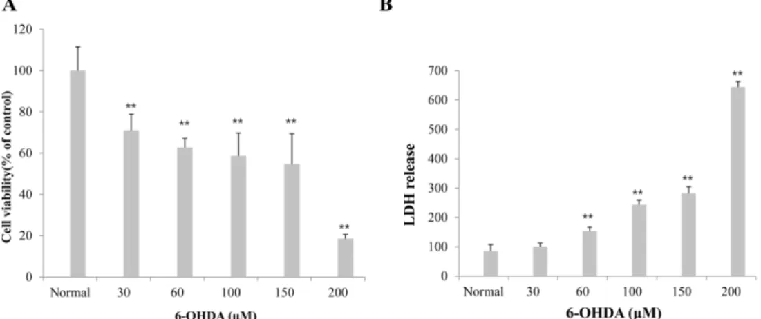
MTT 분석방법을 이용하여 6-OHDA을 SH-SY5Y세포에 처리한 세포독성검사에서는 normal과 비교하면 30, 60, 100, 150, 200µM의 6-OHDA를 투여한 군에서 각각 69%, 62%, 58%, 53%, 18%의 세포 생존율을 나타내어 처리용량 이 증가함에 따라 cell viability가 감소함을 나타내었다(Fig.
1A). 세포 독성평가의 또 다른 방법의 하나로 LDH 분비를 이용한 평가에서는 6-OHDA(30, 60, 100, 150, 200 µM)을 투여한 군에서도 6-OHDA 처리에 따른 세포 손상 증가에 따라 LDH 분비 증가가 관찰되었다(Fig. 1B). 따라서 6- OHDA의 신경세포 독성에 대한 레스베라트롤의 보호 효과 를 관찰하기 위해서 MTT 및 LDH 분석에서 나타난 세포 손상 정도를 동시에 고려하여 60 µM로 설정하였다.
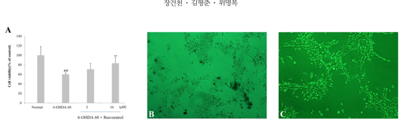
Resveratrol에 의한 신경보호 효과 및 형태학적인 증거 MTT 분석결과 6-OHDA(60 µM)를 투여한 군의 cell viability는 normal군보다 40% 가량 감소하였다. 하지만 레스 베라트롤 5 µM 및 10 µM을 전처리한 군에서는 normal군보 다 각각 31%, 17% 감소하여 cell viability가 6-OHDA 단 독 투여 군보다 증가함을 보여주었다(Fig. 2A). 위상차현미 경을 이용한 형태학적인 세포 관찰에서 6-OHDA 투여 시 많은 세포의 손상과 소실이 관찰되었으며(Fig. 2B) 레스베라 트롤을 전처리한 군에서는 세포 손상이 억제됨을 알 수 있 었다(Fig. 2C).
DAPI 염색을 이용한 resveratrol의 DNA 보호 효과 DNA 손상에 대한 형태학적인 관찰에서는 normal군(Fig.
Fig. 1. Dose-toxicity of 6-OHDA in SH-SY5Y cells. Cytotoxicity of SH-SY5Y cells was quantified by MTT assay (A) and LDH release measurement. Cultures were exposed with 30-200µM concentrations of 6-OHDA for 20~24 h. Values are presented as mean± SD. (n = 8, **p < 0.01 vs. normal).
3A)보다 6-OHDA(60 µM) 투여 군에서 cell loss가 많이 생 겨 염색된 핵이 감소한 것을 볼 수 있고, 분절된 핵과 핵 염색질 응축을 관찰할 수 있다(Fig. 3B). 이에 비해 레스베 라트롤(10 µM)을 전처리한 군에서는 세포의 분절화 및 핵응 축의 감소를 나타내었다(Fig. 3C).
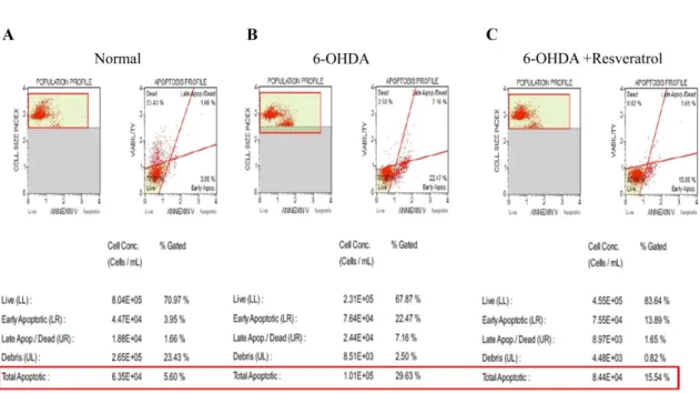
Annexin V/PI staining
normal군은 apoptotic cell rate가 5.6%(Fig. 4A)인데 반해 6-OHDA(60µM) 단독 투여 군에서는 29.6% 증가세를 보였 다(Fig. 4B). 레스베라트롤(10 µM)을 투여한 군에서는 15.5%
로 6-OHDA(60 µM) 단독 투여 군에 비해 뚜렷한 apoptosis 감소를 나타내었다(Fig. 4C).
TBARS and glutathione assay
지질과산화 정도를 나타내는 TBARS level이 6-OHDA(60 µM) 단독 처리 군에서 normal군보다 5배 가량 증가하였으 며, 레스베라트롤(1, 5, 10 µM)을 전처리한 군에서는 농도에 비례해서 TBARS level의 유의한 감소를 나타내었고(p <
0.01), 10µM 투여 군에서는 normal군에 가까운 수치를 보
여주었다(Fig. 5A). 글루타티온 양의 변화를 나타내는 GSH/
GSSG 비율의 변화에서는 normal군보다 6-OHDA(60 µM) 단독 투여 군에서 GSH/GSSG 비율이 감소하였으며, 레스베 라트롤(5, 10 µM) 전처리군에서 농도에 비례하여 GSH/
GSSG 비율의 감소가 유의하게 억제되었다(Fig. 5B).
고 찰
대표적 신경 퇴행성 질환의 하나인 파킨슨병의 원인은 아 직 정확히 알려진 바는 없으나 그동안 주로 free radical에 의해 생기는 산화적 스트레스와 mitochondria의 기능 이상, 염증 및 apoptosis 등이 관여하는 것으로 잘 알려져 왔다 [14]. 현재까지 알려진 치료방법으로는 L-dopa가 사용되나 증 상을 일시적으로 개선해 주거나 이 질환의 진행을 지연시켜 주는 정도이다. 특히 신경계 질환은 한번 발병하면 진행 경 과에 따라 부분적으로 진행속도를 늦출 수는 있으나 본래 상 태로의 완전한 회복이 어려운 치료적 한계성을 지니고 있어 점차 예방적 차원에서 신경계 질환의 발생을 낮추는 노력이 필요한 실정이다 [5]. 그동안 레스베라트롤의 신경보호 효능 Fig. 2. Protective effects of resveratrol on 6-OHDA-induced neurotoxicity in SH-SY5Y cells. Cultures were preincubated with 5 or 10µM resveratrol 2 h prior to the exposure to 6-OHDA for 20-24 h (A). Morphological evidence of protection by resveratrol on 6-OHDA-induced neurotoxicity (B and C). Phase contrast photomicrographs of cultured SH-SY5Y cells with 6-OHDA alone (B) or in the presence of both 10µM resveratrol and 6-OHDA (C).
Fig. 3. Detection of apoptosis in SH-SY5Y cells by DAPI (4´,6-diamidino-2-phenylindole) stain. Arrow indicates fragmented DNA nuclei. (A) Vehicle. (B) 60µM 6-OHDA. (C) 60 µM 6-OHDA + 10 µM resveratrol.
에 관한 몇몇 연구에서도 알 수 있듯이 여러 연구자는 효과 적으로 ROS를 제거하는 항산화 물질 혹은 파킨슨병의 특징 으로 나타나는 도파민 신경세포의 글루타티온 고갈 및 이에 따른 apoptosis를 억제할 수 있는 화합물의 발굴에 그 초점 이 맞추어져 왔다 [12]. 이번 실험을 통해 레스베라트롤은 비교적 낮은 농도(5~10 µM)에서 인간 신경암 세포종인 SH- SY5Y 세포에서 6-OHDA에 의한 신경 독성에 대해 보호 효과를 나타내었다. 이는 본 실험에서 사용한 세포주와 같은 인간 신경암 세포주를 이용한 다른 실험에서도 고농도의 도 파민으로 유발된 신경 독성에 대해 레스베라트롤이 5 µM에 서 유의한 보호 효과를 얻은 결과와 본 실험에 사용한 농도 보다 낮은 농도의 6-OHDA(25 µM)를 처리 시 레스베라트롤 이 1 µM 혹은 10 µM 농도에서 유의성 있는 신경보호 효과 를 나타낸 점과 비교해 볼 때 동일한 in vitro 신경계에서
본 실험결과와 비슷한 효과를 보인 것으로 보이며 [4, 10], 또 다른 도파민 신경독소로 알려진 1-methyl-4-phenylpyridi- nium(MPP+) 처리 시 나타나는 ROS 축적 및 글루타티온 고갈을 레스베라트롤이 예방하는 효과를 보여준 점은 본 실 험결과와 잘 일치하는 결과로 생각된다 [13]. 이러한 레스베 라트롤의 신경보호작용은 이미 예견하였듯이 이 결과에서 나 타난 강력한 지질 과산화 현상의 억제를 통해 나타나며, 6- OHDA에 의해 일어나는 ROS 축적 증가와 동시에 GSH/
GSSG 비율의 감소를 레스베라트롤이 잘 억제할 수 있다는 것을 보여준다.
마지막으로 DAPI 염색 및 Annexin V/PI 염색을 이용한 세포분석 검사에서도 apoptosis를 잘 억제함을 알 수 있었다.
최근 들어서는 레스베라트롤보다 신경보호 효능이 더 우수 한 다양한 레스베라트롤 유도체들에 대한 개발연구도 병행 Fig. 4. Resveratrol inhibits the apoptosis of SH-SY5Y cells induced by 6-OHDA by cell analyzer. The SH-SY5Y cells pretreated with for 2 h were exposed to 60µM 6-OHDA for 20-24 h. The SH-SY5Y cells were labeled with Annexin V/PI staining and quantified by cell analyzer. (A) Vehicle. (B) 60µM 6-OHDA. (C) 60 µM 6-OHDA + 10 µM resveratrol.
Fig. 5. Effect of resveratrol on TBARS formation (A) and GSH/GSSG ratio in SH-SY5Y cells obtained from neurotoxicity induced by 6-OHDA (60µM) exposure for 20~24 h. Values are expressed in mean ± SD. **p < 0.01 vs. 6-OHDA-treated cells, ##p < 0.01 vs. normal.
되고 있으므로 앞으로 더 자세한 기전 연구를 통해 파킨슨 병의 효과적 치료 및 예방 가능성을 추구해 볼 필요가 있을 것으로 생각한다.
References
1. Almeida IMC, Barreira JCM, Oliveira MBPP, Ferreira ICFR. Dietary antioxidant supplements: benefits of their combined use. Food Chem Toxicol 2011, 49, 3232-3237.
2. Blanchet J, Longpré F, Bureau G, Morissette M, DiPaolo T, Bronchti G, Martinoli MG. Resveratrol, a red wine polyphenol, protects dopaminergic neurons in MPTP-treated mice. Prog Neuropsychopharmacol Biol Psychiatry 2008, 32, 1243-1250.
3. Burns J, Yokota T, Ashihara H, Lean MEJ, Crozier A.
Plant foods and herbal sources of resveratrol. J Agric Food Chem 2002, 50, 3337-3340.
4. Chao J, Li H, Cheng KW, Yu MS, Chang RCC, Wang M.
Protective effects of pinostilbene, a resveratrol methylated derivative, against 6-hydroxydopamine-induced neurotoxicity in SH-SY5Y cells. J Nutr Biochem 2010, 21, 482-489.
5. Chao J, Yu MS, Ho YS, Wang M, Chang RCC. Dietary oxyresveratrol prevents parkinsonian mimetic 6-hydroxy- dopamine neurotoxicity. Free Radic Biol Med 2008, 45, 1019- 1026.
6. Deumens R, Blokland A, Prickaerts J. Modeling Parkinson’s disease in rats: an evaluation of 6-OHDA lesions of the nigrostriatal pathway. Exp Neurol 2002, 175, 303-317.
7. Frémont L. Biological effects of resveratrol. Life Sci 2000, 66, 663-673
8. Jin F, Wu Q, Lu YF, Gong QH, Shi JS. Neuroprotective effect of resveratrol on 6-OHDA-induced Parkinson’s disease in rats. Eur J Pharmacol 2008, 600, 78-82.
9. Johnson WD, Morrissey RL, Usborne AL, Kapetanovic I, Crowell JA, Muzzio M, McCormick DL. Subchronic oral toxicity and cardiovascular safety pharmacology studies of resveratrol, a naturally occurring polyphenol with cancer preventive activity. Food Chem Toxicol 2011, 49, 3319-3327.
10. Lee MK, Kang SJ, Poncz M, Song KJ, Park KS.
Resveratrol protects SH-SY5Y neuroblastoma cells from apoptosis induced by dopamine. Exp Mol Med 2007, 39, 376- 384.
11. Lu KT, Ko MC, Chen BY, Huang JC, Hsieh CW, Lee MC, Chiou RYY, Wung BS, Peng CH, Yang YL. Neuroprotective effects of resveratrol on MPTP-induced neuron loss mediated by free radical scavenging. J Agric Food Chem 2008, 56, 6910-6913.
12. Martin HL, Teismann P. Glutathione-a review on its role and significance in Parkinson's disease. FASEB J 2009, 23, 3263-3272.
13. Okawara M, Katsuki H, Kurimoto E, Shibata H, Kume T, Akaike A. Resveratrol protects dopaminergic neurons in midbrain slice culture from multiple insults. Biochem Pharmacol 2007, 73, 550-560
14. Orr CF, Rowe DB, Halliday GM. An inflammatory review of Parkinson’s disease. Prog Neurobiol 2002, 68, 325-340.
15. Richard T, Pawlus AD, Iglésias ML, Pedrot E, Waffo- Teguo P, Mérillon JM, Monti JP. Neuroprotective properties of resveratrol and derivatives. Ann N Y Acad Sci 2011, 1215, 103-108.