DOI: https://doi.org/10.3339/jkspn.2016.20.2.74 ISSN 2384-0250 (online)
Prognostic Factors of Renal Scarring on Follow-up DMSA Scan in Children with Acute Pyelonephritis
Purpose: Early diagnosis and treatment of urinary tract infection have been em- phasized to prevent renal scarring. If untreated, acute pyelonephritis could cause renal injury, which leads to renal scarring, hypertension, proteinuria, and chronic renal failure. The purpose of this study was to assess risk factors of renal scarring after treatment of acute pyelonephritis (APN).
Methods: The medical records of 59 patients admitted at Daegu Fatima Hospital because of APN between March 2008 and April 2015 whose renal cortical defects were confirmed by using initial technetium-99m dimercaptosuccinic acid (DMSA) scans were reviewed retrospectively. We divided 59 patients into 2 groups accor- ding to the presence of renal scar and assessed risk factors of renal scar, including sex, age at diagnosis, feeding method, hydronephrosis, bacterial species, vesi- coureteral reflux, and vesicoureteral reflux grade.
Results: Of 59 patients (41%), 24 showed renal scar on follow-up DMSA scan. No significant differences in sex, hydronephrosis, bacterial species, and fever duration were found between the renal-scarred and non-scarred groups. As for age at diag- nosis, age of >12 months had 5.8 times higher incidence rate of renal scarring.
Vesicoureteral reflux (VUR) affected renal scar formation. VUR grade III or IV had 14.7 times greater influence on renal scar formation than VUR grade I or II.
Conclusion: Our data suggest that the presence of VUR and its grade and age at diagnosis are risk factors of renal scar on follow-up DMSA scan after APN.
Key words: Acute pyelonephritis, Renal scar
Juyeen Lee, M.D.
Byung Woo Woo, M.D.
Hae Sook Kim, M.D.
Department of Pediatrics, Daegu Fatima Hospital, Daegu, Korea
*Announced on 23 October, 2015 fall conference of the Korean Pediatric Society Corresponding author: Hae Sook Kim, M.D.
Department of Pediatrics, Daegu Fatima Hospital, 99, Ayang-ro, Dong-gu, 41199, Daegu, , Korea
Tel: +82-53-940-7243 Fax: +82-53-940-7076 E-mail: [email protected] Received: 23 September 2016 Revised: 15 October 2016 Accepted: 19 October 2016
This is an open-access article distributed under the terms of the Creative Commons Attribu tion Non-Commercial License (http://
crea tivecom mons.org/licenses/by-nc/4.0/) which permits unrestricted non-commercial use, distribution, and reproduction in any medium, provided the original work is properly cited.
Copyright © 2016 The Korean Society of Pediatric Nephrology
Introduction
Five percent of febrile infants have a urinary tract infection
1). Most febrile patients with a urinary tract infection have acute pyelonephritis, and they can develop renal scarring. Patients with renal scars have a higher risk of hyper
tension and chronic renal failure
2). Early diagnosis and treatment of urinary
tract infection in children under 2 years of age are necessary because of the
higher incidence rate of renal scarring
3). The ultimate goal of treatment is to
prevent permanent renal injury. Although there is much controversy about
the real etiology of renal scarring, recent studies show that acute pyelonephritis
is one of the risk factors of renal scarring, and its severity is increased when it
is accompanied with vesicoureteral reflux (VUR) (and how this is correlated
with risk factors such as sex, age, hydronephrosis, and fee ding method), delay
of treatment, and recurrent urinary tract infections
4). Technetium99m dimercaptosuccinate (Tc99m DMSA) is the most sensitive diagnostic tool to identify acute pyelo
nephritis during the acute phase of a febrile urinary tract infection and furthermore pinpoint the area that will sub
sequently develop into a renal scar 3 to 6 months after the infection
5). About 4260% of patients with acute pyelon
ephritis develop renal scars
6). Therefore, we evaluated studies in which acute pyelonephritis was diagnosed and followed up with DMSA scans. The aim of this retrospective clinical study was to determine the incidence of renal scarring after acute pyelonephritis, duration of fever, level of Creactive protein (CRP), species of bacteria, and VUR and its grade.
Materials and methods
The medical records of 59 patients whose renal cortical defects were confirmed by DMSA scan when admitted to Daegu Fatima Hospital for their first febrile urinary tract infection from March 2008 to April 2015 were reviewed retrospectively. A followup DMSA scan was performed within 6 months after the last urinary tract infection with renal cortical defects. The DMSA scan was considered abnormal if one or more areas of decreased cortical uptake were noted with or without preservation of the cortical outline. Other urogenital tract anomalies were excluded.
We divided our clinical series of patients into the follo
wing 2 groups: the renalscarred group and the nonrenal
scarred group on the followup DMSA scan. Data on the following items were analyzed: age at diagnosis, sex, dura
tion of fever before treatment, total duration of fever, white blood cell count, CRP level, erythrocyte sedimentation rate (ESR), hydronephrosis, feeding method, and voiding cys
tourethrogram (VCUG) results.
Febrile urinary tract infection was defined as fever ≥38
℃, pyuria, or growth of at least 100,000 colonyforming units per milliliter of a single bacterial species from mid
stream or catheter specimens. Hydronephrosis was defined as ≥5 mm of renal pelvic anteroposterior (AP) diameter on sonograms. VCUG was used for detection and grading of VUR. VUR was graded as follows: Grade I reflux was de
fined as reflux limited to the ureter; grade II was reflux up to the renal pelvis; grade III was reflux into a mildly dilated
ureter and pelvicaliceal system; grade IV was a moderately dilated ureter and blunting of the fornix; and grade V was a tortuous ureter with severe dilatation of the ureter and pelvicaliceal system
7). Laboratory data that are markers of inflammation, including CRP and ESR, during acute py
elonephritis were collected. As for age at diagnosis, we divided our clinical series of patients into two groups: <12 months and ≥12 months. To investigate the protective ef
fect of breastfeeding against urinary tract infection, we grouped the patients into 3 groups by feeding method:
breastfed, formulafed, and breast and formulafed.
Results are presented as mean ± standard deviation. Sta
tistical analyses were conducted using the Statistical Pro
gram for the Social Sciences (SPSS) version 18.0, and P
values under 0.05 were reported as statistically significant.
The comparison of data and analysis of categorical variables were performed using either the Student ttest or the chi
square test. Logistic regression analysis was also performed to estimate the magnitude of association between renal scarring and risk factors.
Results
Of the 59 patients who had cortical defects on initial febrile urinary tract infection, renal scarring was found on followup DMSA scan in 24 (41%) patients. Table 1 shows the clinical characteristics and laboratory findings of all patients.
1. Renal Scarring and Age at Diagnosis
The 24 patients who had renal scarring on followup DMSA scan included 10 (42%) boys and 14 (58%) girls.
Table 1. Comparison of Clinical Characteristics and Laboratory Findings between Scarred and Non-scarred Group
No scar Scar P-
value Age at diagnosis (month) 10±15.7 23±24.5 0.02
Sex (male:female) 3:4 5:7 0.93
Duration of fever before treatment 2.5±1.0 2.2±1.1 0.31 Total duration of fever(day) 4.5±1.4 4.5±1.7 0.84
WBC (/m
3) 17,032±4,518 17,072±5,244 0.14
CRP (mg/dL) 7.45±5.4 8.59±8.2 0.56
ESR (mm/hr) 32.7±24.6 26.1±21.9 0.33
There was no significant difference between boys and girls in terms of developing renal scars ( P=0.93).
The mean age at diagnosis was 15.2 months. Patients with scars on followup DMSA scan were significantly older (mean age: 23±24.5 months) than those who did not have scars (mean age: 10±15.7 months).
We also divided patients into two groups according to age: under 12 months old and over 12 months old. Children over 12 months old were 5.83 times more likely to develop renal scars than those under 12 months) ( P=0.01, Table 2).
2. Feeding Method
As ongoing exclusive breastfeeding and an extended du
ration of breastfeeding are associated with a significantly lower risk of infection
8), we grouped patients by feeding method into the breastfed group, formulafed group, and breast and formulafed group. There was no statistically significant difference in feeding method (P=0.69, Table 3).
3. Hydronephrosis
We investigated correlations between hydronephrosis and renal scars. Twentyone (36%) of 59 patients had hydro
nephrosis on ultrasound. There was no statistically signifi
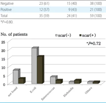
cant difference between patients with hydronephrosis and renal scars ( P=0.80, Table 4).
4. Distribution of Causative Organisms
A total of 48 patients (81%) out of 59 had positive urine cultures. There was no growth on urine culture in 11 out of 59 patients (Fig. 1). The most common pathogen was Escherichia coli (E. coli). We found no correlations between
the pathogens and the incidence of renal scars ( P=0.72).
5. CRP, ESR, and WBC
We found no difference between the groups with or with
out scars in regard to the level of CRP, ESR , or WBC count at the time of infection (Table 1).
6. VUR and VUR Grade
VUR was found in 23 (39%) patients after their first fe
brile urinary infection. There was a correlation between VUR and renal scarring on DMSA scan within 6 months after the first febrile urinary tract infection ( P=0.00). Renal scarring was significantly more common in patients with grade ≥3 reflux than in patients without reflux. VUR III groups had a 1.57 times greater incidence rate of scarring than the group with no reflux. Furthermore, VUR IIIIV groups had a 14.7 times greater incidence rate of scarring than a comparable group with no reflux (Table 5).
Table 2. The Difference of Renal Scars according to Age at Diag- nosis
Age No. (%) of patients
Total
No scar Scar
<12 months 30 (70) 13(30) 43(100)
≧ 12months 5 (31) 11 (69) 16 (100)
Total 35 (59) 24 (41) 59 (100)
*P=0.01. *Odds ratio=5.83.
Table 3. The Difference of Renal Scars according to Feeding Method
Feeding method No. (%) of patients
Total
No scar Scar
Breastmilk 17 (55) 14 (45) 31 (100)
Formula 11 (61) 7 (39) 18 (100)
Mixed 7 (70) 3 (30) 10 (100)
Total 35 (59) 24 (41) 59 (100)
*P=0.69.
Fig. 1. The Difference of Renal Scars according to Causative Organisms
0 5 10 15 20 25
scar(-) scar(+)
No. of patients
*
P=0.72
-21-