DOI: https://doi.org/10.3339/jkspn.2019.23.1.36 ISSN 2384-0250 (online)
A Renal Size Discrepancy among the Findings of Renal Sonogram in Children with Their First Episode of Pyelonephritis is One of the Useful Parameters to Predict the Presence of Cortical Defects on the Acute DMSA Renal Scan
Purpose: We investigated whether a renal size discrepancy on a renal sonogram (US) in children with febrile urinary tract infection (UTI) was correlated with the presence of cortical defects on their dimercaptosuccinic acid (DMSA) renal scan.
Methods: We examined 911 children who were admitted consecutively to our hospital with their first episode of febrile UTI from March 2001 to September 2014.
All enrolled children underwent a US and DMSA scan during admission. According to the US findings, including the renal size discrepancy, data were compared between children with positive and negative DMSA scan results. A positive DMSA scan result was defined as reduced or absent tracer localization and indistinct margins that did not deform the renal contour.
Results: Mean renal lengths of the right and left kidneys were larger in children with positive DMSA scan results than in children with negative DMSA scan results (63.2±11.3 mm vs. 58.4±7.8 mm, P<0.001; 64.9±11.2 mm vs. 59.9±7.9 mm, P<
0.001; respectively). A significant difference was observed in both renal lengths between children with positive and negative DMSA scan results (4.6±3.8 mm vs. 3.3±2.6 mm, P<0.001). A multiple logistic regression analysis, revealed that a small kidney, cortical thinning, and a renal length discrepancy on US findings were significant factors for predicting the presence of cortical defects on an acute DMSA scan [P=0.028, 95% confidence interval (CI) 1.054–2.547; P= 0.004, 95% CI 1.354– 4.810; P<0.001, 95% CI 1.077–1.190, respectively].
Conclusion: In conclusion, a renal size discrepancy on US findings in children with their first episode of febrile UTI was a helpful tool for predicting the presence of cortical defects on an acute DMSA scan.
Key words: Cortical defect, DMSA renal scan, Pyelonephritis, Renal sonogram, Urinary tract infection
Yoowon Kwon, M.D.
Bo kyeong Jin, M.D.
Seonkyeong Rhie, M.D., Ph.D.
Jun Ho Lee, M.D., Ph.D.
Department of Pediatrics, CHA Bundang Medical Center, CHA University School of Medicine, Seongnam-si, Korea
Corresponding author:
Jun Ho Lee, M.D, Ph.D.
Department of Pediatrics, CHA Bundang Medical Center, CHA University School of Medicine, 351 Yatap-dong, Bundang-gu, Seongnam-si, Gyeonggi-do, 13496, Korea Tel: +82-31-780-5011
Fax: +82-31-780-5011 E-mail: [email protected] Co-corresponding author:
Seonkyeong Rhie, M.D., Ph.D.
Department of Pediatrics, CHA Bundang Medical Center, CHA University School of Medicine, 351 Yatap-dong, Bundang-gu, Seongnam-si, Gyeonggi-do, 13496, Korea Tel: +82-31-780-1864
Fax: +82-31-780-5011 E-mail: [email protected] Received: 27 November 2018 Revised: 19 March 2019 Accepted: 26 March 2019
This is an open-access article distributed under the terms of the Creative Commons Attribu tion Non-Commercial License (http://
crea tivecom mons.org/licenses/by-nc/4.0/) which permits unrestricted non-commercial use, distribution, and reproduction in any medium, provided the original work is properly cited.
Copyright © 2019 The Korean Society of Pediatric Nephrology
Introduction
A renal ultrasonogram (US) is necessary in children with their first episode of pyelonephritis because of several congenital anomalies of the kidney and urinary tract, such as obstructive uropathy (hydronephrosis or hydroureter), duplex kidney, ectopic kidney, horseshoe kidney, hypoplastic kidney, cystic
kidney disease, ureterocele, bladder diverticulum, patent urachus, and stones, that have the risk of recurrent urinary tract infection (UTI) and can be easily detected. Renal size can be measured. A renal abscess, a complication of acute pyelonephritis (APN), can also be detected. US has a high negative predictive value (NPV; 98.2%) for detecting high grade vesicoureteral reflux (VUR)1).
However, US has low sensitivity (SS) and specificity (SP) for predicting positive dimercaptosuccinic acid (DMSA) scan results and detecting high grade VUR1,2). Therefore, a secondary imaging study is necessary in children with a recurrent febrile UTI or specific condition. Only pyelo
nephritis with cortical defects on a DMSA renal scan pro
duces acquired renal scarring, and only such recurrent pye
lonephritis can lead to chronic renal insufficiency. Patients with pyelitis, which has the same clinical manifestations as pyelonephritis with a negative DMSA scan result, do not develop acquired renal scarring. Therefore, discriminating pyelonephritis is very important among children with the first episode of febrile UTI.
Various reports have indicated a correlation or a lack of a correlation between abnormal US findings and positive acute DMSA scan results. However, no previous study has been investigated the correlation between a renal size dis
crepancy on US and positive acute DMSA scan results in acute phase.
In the present study, we investigated whether a renal size discrepancy on US in children with a febrile UTI was cor
related with the presence of cortical defects on a DMSA renal scans.
Materials and methods
Among 2,614 children consecutively admitted to our hospital with the diagnosis of UTI (N39.0) from March 2001 to September 2014, 911 with complete data were included in this retrospective study. In total, 320 children who had a UTI episode in the past or who did not satisfy the UTI cri
teria (defined as follow below) were excluded. A total of 1,138 children who did not undergo US or a DMSA scan during their admission and 245 children who did not have measured renal lengths medical records, or renal sonogram films were also excluded.
UTI was defined as follows: high fever ≥ 38℃, abnormal urinalysis (pyuria [>5 White blood cells (WBCs) /high
power field] and positive leukocyte esterase results), positive Creactive protein (CRP) results (>0.3 mg/dL), lack of an
other fever focus, and significant bacteriuria. A total of 118 children with a negative urine culture result, but with po
sitive DMSA scan results, were also included in the UTI group. A positive DMSA scan result (cortical defect) was defined as reduced or absent tracer localization and indi
stinct margins that did not deform the renal contour.
Urine was sampled by collecting a midstream urine spe
cimen in toddlers or in older children and using the clean catch bag method in nontoilet trained children. Signifi
cant bacteriuria was defined as the presence of ≥105 colony
forming units/highpower field of a singlestrain isolate.
All enrolled children with a UTI underwent US and a DMSA renal scan upon admission.
US was performed by a pediatric radiologist. Abnormal US findings included small kidneys (≤95% of the standard deviation of mean agematched renal size), hydroureter (≥
4 mm), cortical thinning, increased renal parenchymal echogenicity, hydronephrosis (anteroposterior diameter of renal pelvis ≥5 mm), swelling, bladder wall thickening and no difference between the corticomedullary junctions.
Renal lengths were measured as the longest diameter bet
ween the upper and lower pole of each kidney.
DMSA scans were performed using the planar technique with a dualheaded Symbia E gamma camera (Siemens, Erlangen, Germany) equipped with a lowenergy highre
solution collimator (140 KeV, 20% symmetric window) and were interpreted by two nuclear medicine consultants. A positive DMSA scan result was defined as reduced or absent tracer localization and indistinct margins that did not de
form the renal contour.
Written informed consent was obtained from the parents of all children with a UTI children prior to the DMSA scan.
The CHA University Institutional Review Board approved this study, including the consent procedure (CHA IRB No.
201807063).
All data were collected retrospectively and were com
pared between the children with positive and negative DMSA scan results according to their US findings.
1. Statistical analysis
All variables are presented as mean±standard deviation.
Continuous variables were analyzed using the Student’s ttest. Qualitative variables and correlations were analyzed using Pearson’s chisquare test and Pearson’s correlation coefficient analysis (twotailed probability), respectively.
Nonparametric variables were analyzed using Spearman’s rank correlation coefficient analysis. A multiple logistic re
gression analysis (MLRA) was conducted to test the ability of a renal size discrepancy on US to predict the presence of cortical defects on a DMSA scan. The areas under the re
ceiver operating characteristic (ROC) curves of relevant factors and the associated levels of significance were eva
luated when the value of the state variable was 1. The stati
stical analysis was performed using SPSS statistics 22 soft
ware (SPSS Inc, Chicago, IL, USA). A Pvalue ≤0.05 was considered significant.
Results
1. Comparison of the imaging data between children with positive and negative DMSA scan results A total of 911 children (616 males and 295 females) were included; 118 children (27.2%) with a positive DMSA scan result were diagnosed with culturenegative pyelonephritis.
Congenital hypoplastic kidney [relative renal uptake (RRU)
≤35% on DMSA scan] was detected in 17 children (1.9%).
A total of 434 children had positive DMSA scan results (47.6%), whereas 477 children had negative DMSA scan re
sults. A significant difference in age (12.8±24.5 vs. 7.5±15.3 months) was observed between the groups, but no diffe
rence was observed in the sex ratio. When the older child
ren were diagnosed with their first episode of UTI, more cortical defects were detected on the DMSA scan.
In this study population, mean renal length was larger in left kidneys than in right kidneys (62.3±10 vs. 60.7±9.9 mm, respectively; P<0.001). The left kidney was larger than the right kidney in 567 children. The mean difference in renal length was 3.9±3.3 mm.
Mean renal lengths of the right and left kidneys were larger in children with positive DMSA scan results than in children with negative DMSA scan results (63.2±11.3 vs.
58.4±7.8 mm, P<0.001; 64.9±11.2 vs. 59.9±7.9 mm, P<0.001;
respectively). No difference in the ratio of children with larger left kidneys than right kidney was observed between children with positive and negative DMSA scan results (1.56 vs. 1.72, P=0.07). Significant differences in both renal lengths were observed between children with positive and negative DMSA scan results (4.6±3.8 vs. 3.3±2.6 mm, P<
0.001).
Even after adjusting for age between DMSA scan positive and negative groups, the mean renal lengths of the right and left kidneys were larger in children with positive DMSA scan results than in children with negative DMSA scan re
sults (61.6±8.7 vs. 58.4±7.8 mm, P<0.001; 63.5±9.3. vs. 59.9±
7.9 mm, P<0.001; respectively). A significant difference was also found in both renal lengths between the ageadjusted groups (4.5±3.8 vs. 3.3±2.6 mm, P<0.001). This study in
cluded 807 infants of less than 12 monthold, 28 children of 13–24 monthold and 76 children of more than 25 month
old. There was no difference of these results between total study population and 76 children of more than 25 month
old, or between total study population and 835 children of less than 24 monthold (Table 1).
2. Significant correlations between parameters in this study population
The differences in both renal lengths on US correlated positively with age, cortical thinning, increased parenchy
mal echogenicity, hydronephrosis (anteroposterior dia
meter >15 mm), swelling, and positive DMSA scan results (r=0.109, P<0.001; r=0.064, P=0.021; r=0.070, P=0.012; r=
0.071, P=0.011; r=0.134, P<0.001; r=0.145, P<0.001, respec
tively).
The presence of cortical defects on the acute DMSA scan correlated positively with age, hydroureter, increased pa
renchymal echogenicity, swelling, and the difference in both renal lengths (r=0.075, P=0.009; r=0.083, P=0.012; r=
0.146, P<0.001; r=0.182, P<0.001; r=0.145, P<0.001, respec
tively).
3. Incidence and MLRA of abnormal US findings to predict the presence of cortical defects on an acute DMSA scan
The multiple logistic regression analysis revealed that a small kidney, cortical thinning, and a differences in both renal lengths on US findings were significant factors for
predicting the presence of cortical defects on the acute DMSA scan [P=0.028, 95% confidence interval (CI) 1.054–
2.547; P=0.004, 95% CI 1.354–4.810; P<0.001, 95% CI 1.077–1.190, respectively] (Table 2).
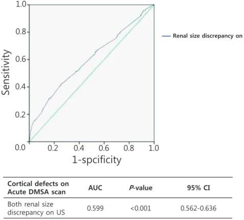
4. ROC curves and 95% CI of renal size discrepancy on US to predict the presence of cortical defects on an acute DMSA scan
The areas of the ROC curves and 95% CIs of both renal size discrepancies on US significantly predicted the pre
sence of cortical defects on an acute DMSA scan (0.599, 95
% CI 0.562–1.636, P<0.001) (Fig. 1).
5. Comparison of the imaging data between children with RRU >10% and RRU ≤10% on a DMSA scan The renal size discrepancies on US were 6.0±5.1 mm and 3.7±3.0 mm, when RRU >10% and RRU ≤10% on DMSA scan respectively. A significant positive correlation (r=
0.21813, P<0.001) was detected. Cortical thinning and in
creased parenchymal echogenicity on US were also corre
lated positively with the difference of RRU on the DMSA scan (P<0.001; P<0.001, respectively) (Table 3).
Discussion
There is no clear agreement about which imaging guide
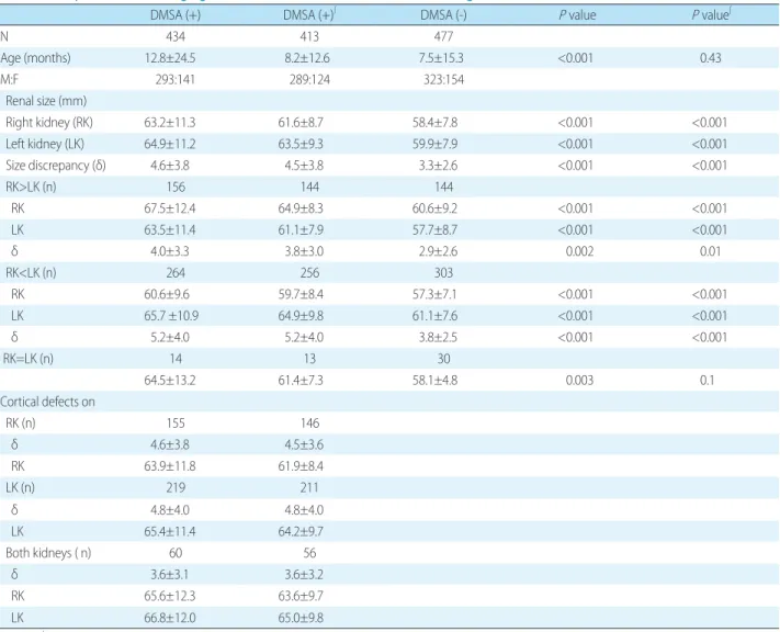
Table 1. Comparison of the Imaging Data between Children with Positive and Negative DMSA Scan Results
DMSA (+) DMSA (+)∫ DMSA (-) P value P value∫
N 434 413 477
Age (months) 12.8±24.5 8.2±12.6 7.5±15.3 <0.001 0.43
M:F 293:141 289:124 323:154
Renal size (mm)
Right kidney (RK) 63.2±11.3 61.6±8.7 58.4±7.8 <0.001 <0.001
Left kidney (LK) 64.9±11.2 63.5±9.3 59.9±7.9 <0.001 <0.001
Size discrepancy (δ) 4.6±3.8 4.5±3.8 3.3±2.6 <0.001 <0.001
RK>LK (n) 156 144 144
RK 67.5±12.4 64.9±8.3 60.6±9.2 <0.001 <0.001
LK 63.5±11.4 61.1±7.9 57.7±8.7 <0.001 <0.001
δ 4.0±3.3 3.8±3.0 2.9±2.6 0.002 0.01
RK<LK (n) 264 256 303
RK 60.6±9.6 59.7±8.4 57.3±7.1 <0.001 <0.001
LK 65.7 ±10.9 64.9±9.8 61.1±7.6 <0.001 <0.001
δ 5.2±4.0 5.2±4.0 3.8±2.5 <0.001 <0.001
RK=LK (n) 14 13 30
64.5±13.2 61.4±7.3 58.1±4.8 0.003 0.1
Cortical defects on
RK (n) 155 146
δ 4.6±3.8 4.5±3.6
RK 63.9±11.8 61.9±8.4
LK (n) 219 211
δ 4.8±4.0 4.8±4.0
LK 65.4±11.4 64.2±9.7
Both kidneys ( n) 60 56
δ 3.6±3.1 3.6±3.2
RK 65.6±12.3 63.6±9.7
LK 66.8±12.0 65.0±9.8
P<0.05, ∫age corrected sample. The laboratory examination results are expressed as mean±standard deviation.
DMSA (+), the existence of cortical defects on DMSA scan.
Abbreviations: DMSA, dimercaptosuccinic acid; N, number of patients.
Cortical defects on
Acute DMSA scan AUC P-value 95% CI Both renal size
discrepancy on US 0.599 <0.001 0.562-0.636
AUC, areas under the ROC curve; CI, 95% confidence interval; P<0.05.
Se nsit ivity
1-spcificity
Renal size discrepancy on us
0.0 0.2 0.4 0.6 0.8 1.0
0.2 0.4 0.6 0.8 1.0
Fig. 1. ROC curves and 95% CI of renal size discrepancy on US to predict the presence of cortical defects on acute DMSA scan.
Table 2. Incidence and MLRA of Abnormal US Findings to Predict the Presence of Cortical Defects on Acute DMSA Scan
US findings N Sensitivity Specificity PPV NPV OR CI P value
Small kidney 2 0.46%
(0.06–1.65) 99.79%
(98.84–99.99) 66.67%
(15.4–95.65) 52.42%
(52.23–52.61) 1.638 1.054–2.547 0.028
Hydroureter 89 20.51%
(16.81–24.62) 84.07%
(80.47–87.24) 53.94%
(47.02–60.71) 53.75%
(52.22–55.28) 0.144 0.012–1.765 0.129
Cortical thinning 1 0.23%
(0.01–1.28) 99.37%
(98.17–99.87) 25.00%
(3.36–76.15) 52.26%
(52.05–52.47) 2.552 1.354–4.810 0.004 Increased parenchymal
echogenicity 51 11.75%
(8.88–15.16) 96.44%
(94.35–97.91) 75.00%
(63.77–83.64) 54.57%
(53.61–55.52) 1.095 0.710–1.687 0.682 Hydronephrosis
APD≤15mm 106 24.42%
(20.45–28.75) 76.31%
(72.23–80.06) 48.40%
(42.68–54.17) 52.60%
(50.77–54.42) 0.512 0.070–3.739 0.509
APD>15mm 1 0.23%
(0.01–1.28) 98.95%
(97.57–99.66) 16.67%
(2.29–63.03) 52.15%
(51.90–52.41) – – 0.995
Swelling 26 5.99%
(3.95–8.65) 99.79%
(98.84–99.99) 96.30%
(77.99–99.48) 53.85%
(53.25–54.44) 1.884 0.150–23.738 0.624 Bladder wall thickening 2 0.46%
(0.06–1.65) 99.79%
(98.84–99.99) 66.67%
(15.40–95.65) 52.42%
(52.23–52.61) – – –
No differentiation
between C-M junction 1 0.23%
(0.01–1.28) 100.00%
(99.23–100.00) 100.00% 52.42%
(52.30–52.53) 1.260 0.801–1.984 0.318
Normal 230 53.00%
(48.18–57.77) 38.78%
(34.39–43.32) 44.06%
(41.28–46.88) 47.56%
(43.82–51.32) The laboratory examination results are expressed as mean±standard deviation.
Abbreviations: MLRA, multiple logistic regression analysis; DMSA, dimercaptosuccinic acid; N, number of patients; APD, anterioposterior diameter; C-M, corticomedullary; P<0.05.
lines in children with their first episode of febrile UTI are desirable considering all aspects of cost, radiation exposure, stress, or the probability of missing high grade VUR or renal scarring. Other authors have maintained our opinion that detecting the presence of congenital or acquired renal scar
ring in children with a febrile UTI is more important than detecting the presence of high grade VUR for the following reasons. In general, a VUR operation does not have to be performed in very young infants for the first episode of febrile UTI. The VUR operation was not performed on children without renal scarring on their DMSA scan. High grade VUR has a relatively high risk of recurrent UTI and accompanying congenital renal scarring, but all children experiencing recurrent UTI do not have high grade VUR and, moreover, severe congenital renal scarring is not com
monly detected1). Missing the renal scarring is worse for renal outcome than missing high grade VUR. Furthermore,
among children with a febrile UTI, acquired renal scarring developes only in some children with cortical defects on their DMSA scan, regardless of the presence of VUR.
A DMSA renal scan or computed tomography should be performed to prevent missing renal scarring, but to perform a DMSA scan on all patients with their first febrile UTI is not acceptable due to the low benefits compared with the risks, such as radiation exposure.
Trying to predict cortical defects on an acute DMSA scan in children with a febrile UTI is challenging. Procalcitonin and neutrophil gelatinaseassociated lipocalin are the best known parameters to predict the presence of cortical de
fects in children with a febrile UTI3,4), but the tests are ex
pensive and are not used to diagnose acute pyelonephritis in the Korean health care system because these parameters do not better predict the presence of cortical defects than CRP, WBC, or erythrocyte sedimentation rate in conside
ration of the cost to benefit ratio. Therefore, additional parameters to predict cortical defects in children with a febrile UTI are needed to more accurately predict pyelo
nephritis with cortical defects.
US is usually recommended in children with their first episode of febrile UTI. In our previous study1), US had low SS to predict positive DMSA scan results. However, Moh
kam et al.5) reported that US is a sensitive method to detect renal cortical defects in patients with pyelonephritic [SS 69.2% (95% CI 62.1–72.6), SP 89.3% (95% CI 80.2–94.9), positive predictive value (PPV) 65.5% (95% CI 60.5–70.9) and NPV 81.5% (95% CI 75.9–84.7)]. A few studies have suggested results similar with this study6,7). However, these studies did not specify what US findings predicted cortical defects on a DMSA scan. Furthermore, other reports have indicated that US is not sensitive to detect renal cortical defects in children with a febrile UTI810). This study suggests that only some US findings could predict renal cortical defects (Table 2).
If the difference in both renal lengths on US was ≥10 mm, the SS, SP, PPV, and NPV to predict cortical defects on the DMSA scan were 10.6% (95% CI 7.9–13.8), 98.5%
(95% CI 96.9–99.4), 87% (95% CI 75.4–93.6), and 53.5% (95
% CI 52.7–54.4), respectively. In cases with a renal length discrepancy ≤10 mm, mean renal length of both kidneys in children with a positive DMSA scan result was larger than in children with a negative DMSA scan result (normal), Table 3. Comparison of the Imaging Data between Children
with RRU >10% and RRU ≤10% on a DMSA Scan
ΔRRU>10% ΔRRU≤10% P value
N 90 821
Age (months) 15.2±25.1 9.4±19.7 0.010
M:F 53:37 563:258
Renal size (mm)
Right kidney (RK) 64.4±12.9 60.2±9.5 <0.001 Left kidney (LK) 65.2±12.7 61.9±9.6 0.003 Size discrepancy 6.0±5.1 3.7±3.0 <0.001
DMSA (+) (n) 73 361
DMSA (-) (n) 17 460
Small kidney 1 (1.1%) 2 (0.2%)
Hydroureter 19 (21.1%) 145 (17.7%) 0.532
Cortical thinning 3 (3.3%) 1 (0.1%) <0.001 Increased parenchymal
echogenicity 21 (23.3%) 47 (5.7%) <0.001 Hydronephrosis
APD≤15mm
APD>15mm 33 (36.7%) 186 (22.7%) 0.019
Swelling 2 (2.2%) 4 (0.5%) 0.050
Bladder wall thickening 9 (10.0%) 18 (2.2%) 0.002 No differentiation
between C-M junction 0 3 (0.4%)
Normal 0 1 (0.1%)
33 (36.7%) 489 (59.1%) <0.001 The laboratory examination results are expressed as mean±standard deviation.
DMSA (+), the existence of cortical defects on DMSA scan.
Abbreviations: DMSA, dimercaptosuccinic acid; N, number of patients;
APD, anterioposterior diameter; C-M, corticomedullary, P<0.05.
even after adjusting for age between the two groups, and when there was no difference between renal sizes (Table 1).
In this study, a significant positive correlation was de
tected between the renal size discrepancy on US and the difference in RRU on the DMSA scans (r=0.21813, P<0.001) (Table 3). However, if renal scarring was defined when the difference in RRU on the DMSA scans were ≥20%, there were no significant US findings for detecting renal scar
ring. Therefore, to prevent missing the children with con
genital renal scarring, a DMSA scan should be performed in children with a recurrent febrile UTI.
There has been known that there is no clear tool to discri
minate the differences between congenital scar and ac
quired scar. However, there exist differences between con
genital scar and cortical defects due to acute inflammation on DMSA scan. Congenital scar includes absent tracer lo
calization that deforms the renal contour and generalized decreased tracer. Hypoplastic kidney belongs to congenital scar. This study population included only children with the first episode of acute pyelonephritis, and 805 children of them were less than 12 monthold. Therefore, chronic defects due to repeated infection except congenital scarring could be excluded when DMSA scans were interpreted.
In conclusion, both renal size discrepancy on US in children with their first episode of febrile UTI was a useful tool to predict cortical detects on a DMSA scan. However, no US findings could detect all children with congenital renal scarring.
Disclosure
All the authors declared no competing interests.
ORCID
Jun Ho Lee [email protected]
Yoowon Kwon https://orcid.org/0000000292497493 Bo kyeong Jin–https://orcid.org/0000000290802001
Seonkyeong Rhie–https://orcid.org/000000033371
8310
Patient consent
This study was approved by the institutional review board (IRB), and the consent was waived due to the nature of the retrospective study [IRB number 201807063].
References
1. Lee JH, Kim MK, Park SE. Is a routine voiding cystourethrogram necessary in children after the first febrile urinary tract infection.
Acta Paediatr 2012;101:e105-9.
2. Shaikh N, Spingarn RB, Hum SW. Dimercaptosuccinic acid scan or ultrasound in screening for vesicoureteral reflux among child- ren with urinary tract infections. Cochrane Database Syst Rev.
2016 Jul 5;7:CD010657. Doi:10.1002/14651858.CD010657.pub2.
3. Leroy S, Gervaix A. Procalcitonin: a key marker in children with urinary tract infection. Adv Urol 2011;2011:397618.doi:10.1155/
2011/397618. Epub 2011 Jan 17.
4. Kim BK, Yim HE, Yoo KH. Plasma neutrophil gelatinase-associated lipocalin: a marker of acute pyelonephritis in children. Pediatr Nephrol 2017;32:477-84.
5. Mohkam M, Jahdavi C, Arad B, Moein HR, Tabar RV, Olyaie M, et al. The sensitivity of ultrasonography in detecting renal cortical defects in pyelonephritic patients with or without vesicoureteral reflux. J Ped Nephrol 2013;1:28-31.
6. Barrt BP, Hall N, Cornford E, Broderick NJ, Somers JM, Rose DH.
Improved ultrasound detection of renal scarring in children fol- lowing urinary tract infection. Clin Radiol 1998;53:747-51.
7. Bjorgvinsson E, Majd M, Egglil KD. Diagnosis of acute pyelone- phritis in children: Comparison of sonography and 99mTc-DMSA scintigraphy. AJR 1991;157:539-43.
8. Levocat MP, Granjon D, Allard D, Gay C. Imaging of pyelonephritis.
Pediatr Radiol 1997;27:159-65.
9. Moorthy I, Wheat D, Gordon I. Ultrasonography in the evaluation of renal scarring using DMSA scan as the gold standard. Pediatr Nephrol 2004;19:153-6.
10. Scherz HC, Downs TM, Caesar R. The selective use of dimercap- tosuccinic acid renal scans in children with vesicoureteral reflux.
J Urol 1994;152:628-31.