DOI: https://doi.org/10.3339/jkspn.2018.22.2.47
ISSN 2384-0250 (online)Comparison of 99mTc-DMSA Renal Scan and Power Doppler Ultrasonography for the Detection of Acute Pyelonephritis and Vesicoureteral Reflux
Purpose: Urinary tract infection (UTI) is one of the common infectious diseases in children. Several imaging modalities can be used to confirm the presence of acute pyelonephritis (APN). Among them the 99mTcdimercaptosuccinic acid renal scan (DMSA scan) is used as a gold standard for diagnosis. Ultrasonography technology is evolving. Therefore, in this study, we investigated the sensitivity and specificity of Power Doppler ultrasonography (PDU) compared to the results from the pre- vious study.
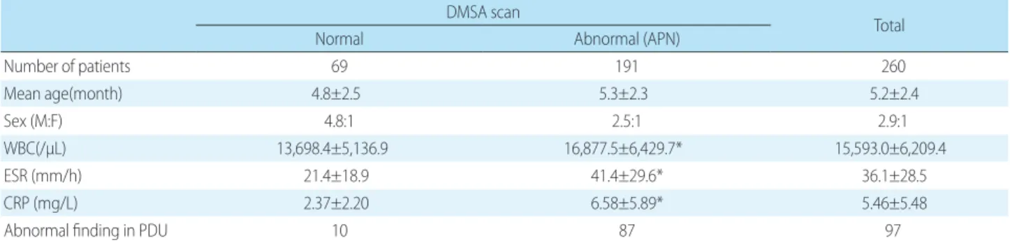
Methods: There were 260 patients included in this study, aged between 1 and 12 months old. The patients were admitted to the Yeungnam University Medical Center between January 2008 and December 2015. All patients underwent both DMSA scan and PDU within 5days of admission. Voiding cystourethrography (VCUG) was performed in 195 patients with abnormal DMSA scan or PDU.
Results: The diagnostic sensitivity of APN using PDU was 45.5% and specificity was 85.5% in 260 patients following detection of a defect on DMSA scan that was defined as APN. The diagnostic sensitivity and specificity of PDU for VUR were 65.5
% and 60.1%, respectively. The diagnostic sensitivity and specificity of DMSA scan for VUR were 95.7% and 14.1%, respectively.
Conclusion: PDU has a high specificity but low sensitivity, so there are limitations in using it to replace a DMSA scan for the diagnosis of APN in children. DMSA scan and PDU have different sensitivity and specificity in diagnosis of VUR, respectively.
Therefore, we suggest that the sensitivity and specificity of each test can be helpful in diagnosing APN and VUR when used in conjunction.
Key words: 99mTc-DMSA, Power Doppler Ultrasonography, Pyelonephritis, Ve- sicoureteral Reflux
Hee Jung Bae, M.D.
1Yong-Hoon Park, MD., Ph.D.
1Jae Ho Cho, M.D.
2Kyung Mi Jang, M.D.
1Department of Pediatrics
1, College of Medicine, Yeungnam University, Daegu, Korea, Department of Radiology
2, College of Medicine, Yeungnam University, Daegu, Korea
Corresponding author:
Kyung Mi Jang, M.D.
Department of Pediatrics, College of Medicine, Yeungnam University, 170 Hyunchungno, Nam-gu, Daegu 42415, Korea
Tel: +82-53-620-3533 Fax: +82-53-629-2252
E-mail: [email protected] Received: 14 September 2018 Revised: 8 October 2018 Accepted: 10 October 2018
This is an open-access article distributed under the terms of the Creative Commons Attribu tion Non-Commercial License (http://
crea tivecom mons.org/licenses/by-nc/4.0/) which permits unrestricted non-commercial use, distribution, and reproduction in any medium, provided the original work is properly cited.
Copyright © 2018 The Korean Society of Pediatric Nephrology
Introduction
Urinary tract infection (UTI) is one of the common bacterial infectious diseases in childhood. There are inherent difficulties in diagnosing infants because they frequently have nonspecific symptoms
1). Upper UTI in some infants can cause hypertension and chronic renal damage
2). Therefore, rapid diagnosis and treatment are critical.
Using a 99mTc-dimercaptosuccinic acid renal scan (DMSA scan), acute
pyelonephritis (APN) can be diagnosed at the acute phase, and renal scarring
can be diagnosed at 3-6 months following infection
3,4). However, DMSA scans
involve exposure to radiation as well as requiring intrave- nous drug admini stration and sedation. The use of com- puted tomography (CT) has a high sensitivity for APN diagnosis; however, it involves a high dose of radiation and requires administra tion of a contrast agent
5). Therefore, a DMSA scan is pre ferred in clinical practice. Magnetic re- sonance imaging (MRI) can also be used to diagnose APN, but it is not sui table for practical clinical applications be- cause of time con suming and high cost
6,7). Power Doppler ultrasonography (PDU) is a noninvasive technique that can be used to mo nitor the size and shape of the kidney
8-10). The presently used technique of color doppler ultrasonography can pro vide an image in color only when the speed of the blood flow is higher than a specific speed and does not ex- pose a vessel adequately if the angle between the insonation beam and vessel is sharp or if the vessel is in a region at a distance from the transducer. PDU was developed to over- come these disadvantages. This technique displays color on the basis of the total strength of the Doppler signal, thus, making it easy to examine weak blood flow. In addition, interference from the angle is relatively small; therefore, PDU is more sensitive than color doppler ultrasonography
11). About a decade ago, the author's hospital reported the sensitivity and specificity of PDU in the diagnosis of APN
12)