INTRODUCTION
Acute necrotizing encephalopathy (ANE) is a unique acute encephalopathy that predominantly affects infants and young children (1). It is characterized by the features of acute ence- phalopthy such as seizures and rapid alteration of conscious- ness after a nonspecific viral illness. Since Mizuguchi et al.
proposed this condition as a new entity in 1995 (2), it has occasionally been reported in both Asian and Western coun- tries. An increased level of serum aminotransferase activiy and cerebrospinal fluid protein, are the most common abnor- malities (1-4). Diagnosis is made mainly by the characteris- tic findings of computed tomography or magnetic resonance imaging (MRI), which typically show symmetric lesions in the thalami, with variable involvement of the white matter, basal ganglia, brainstem, and cerebellum (1, 4, 5). Regard- less of the treatment, the prognosis of ANE varies widely from complete recovery to death (1-6). With the widespread use of MRI, this unique condition is becoming more familiar.
However, the etiology, pathogenesis, guidelines of treatments, or prognostic factors still remain unclear. In this report, we described 6 children with ANE to elucidate the genetic pro- pensity, the clinical/neuroradiological characteristics, the res- ponse to medical treatment and prognostic factors.
MATERIALS AND METHODS
A total of 6 children with ANE (aged 1 yr to 7 yr at onset, 3 males and 3 females) was evaluated. The patients were ad- mitted to Kyungpook National University Hospital, Daegu, Korea, from 1999 through 2007. Diagnosis was made main- ly by clinical and radiological characteristics. Serological tests or polymerase chain reaction for certain viruses such as Her- pes simplex virus, cytomegalovirus, Epstein-Barr virus, influen- za virus, and mycoplasma were done in 5 out of them. Brain MRIs were taken in all patients at the time of initial presen- tation. In a search of other clinically plausible causes, mito-
449
Hye-Eun Seo1, Su-Kyeong Hwang1, Byung Ho Choe1, Min-Hyun Cho1, Sung-Pa Park2, and Soonhak Kwon1
Departments of Pediatrics1and Neurology2, School of Medicine, Kyungpook National University, Daegu, Korea
Address for Correspondence Soonhak Kwon, M.D.
Department of Pediatrics, Kyungpook National University Hospital and School of Medicine, 200 Dongdeok-ro, Jung-gu, Daegu 700-721, Korea Tel : +82.53-420-5704, Fax : +82.53-425-6683 E-mail : [email protected]
Clinical Spectrum and Prognostic Factors of Acute Necrotizing Encephalopathy in Children
This study was conducted to investigate the etiology, the clinical characteristics and prognosis of acute necrotizing encephalopathy (ANE) in Korean children. Six chil- dren (1 yr to 7 yr) patients with ANE were enrolled. They were diagnosed by clini- cal and radiological characteristics and their clinical data were retrospectively ana- lyzed. In a search of clinically plausible causes, brain MRI in all patients, mitochon- drial DNA studies for mitochondrial encephalomyopathy, lactic acidosis, and stroke- like episodes (MELAS) and myoclonus epilepsy and ragged red fibers (MERRF) in four patients, and genomic typing on HLA DRB/HLA DQB genes in three patients were performed. All had precedent illnesses and the main initial symptoms includ- ed mental change (83%), seizures (50%), and focal deficits (50%). MRI revealed increased T2 signal density in the bilateral thalami and/or the brainstem in all pati- ents. Mitochodrial DNA studies for MELAS and MERRF were negative in those chil- dren and HLA-DRB1*1401, HLA-DRB3*0202, and HLA-DQB1*0502 seemed to be significant. A high dose steroid was given to all patients, which seemed to be partly effective except for 2 patients. In conclusion, ANE is relatively rare, but can result in serious neurological complication in children. Early detection and appropriate treat- ment may lead to a better neurological outcome.
Key Words : Acute Necrotizing Encephalopathy; Mitochodrial; MELAS Syndrome; MERRF Syndrome; HLA- DR Antigens; HLA-DQ Antigens
Received : 13 January 2009 Accepted : 18 May 2009
ⓒ 2010 The Korean Academy of Medical Sciences.
This is an Open Access article distributed under the terms of the Creative Commons Attribution Non-Commercial License (http://creativecommons.org/licenses/by-nc/3.0) which permits unrestricted non-commercial use, distribution, and reproduction in any medium, provided the original work is properly cited.
RESULTS
A total of six children was enrolled in the study. Past medi- cal histories were uneventful in all patients and they had nor- mal developmental milestones. The family histories were also unremarkable. No patients were exposed to any drugs or chemi- cal substances known to cause toxic encephalopathies.
Clinical features of the subjects
Clinical characteristics of the subjects are summarized in Table 1. All had precedent illnesses and five out of them (83%) had fever. The initial neurological symptoms include men- tal change in five patients (83%), seizures (50%) and focal neurological signs (50%).
simplex virus, cytomegalovirus, Epstein-Barr virus, influenza virus, and mycoplasma were unremarkable. Mitochodrial DNA studies for MELAS and MERRF were done in 4 patients, and none of them showed any mutations. Genomic typing of HLA DRB/HLA DQB genes in three patients (patient 3, 4, and 5) using PCR-SSOP/SSP techniques with gel immunoelectro- phoresis showed HLA-DRB1*1401, HLA-DRB3*0202, and HLA-DQB1*0502 which were possibly significant (Table 3).
Cerebrospinal fluid study revealed normal protein levels in all patients and mild pleocytosis in one patient (patient 4).
Herpes IgM antibody, oligoclonal band and myelin basic pro- tein were unremarkable in all subjects.
Radiological findings
As shown in Fig. 1, brain MRI revealed increased signal
*Status epilepticus.
ANE, acute necrotizing encephalopathy; NSFI, non specific febrile illness; URI, upper respiratory tract infection; BG, Basal ganglia; T, Thalamus; TT, Thalamotegmantum; P, Pons; C, Cerebellum.
Patient No. Age (month) Sex Precedent illnesses Initial presentation Sites of involvement
1 93 F NSFI Diplopia BG, T, P, C
2 19 M URI & diarrhea Stupor/Rigidity TT
3 40 F NSFI Stupor/Seizure* BG, T
4 68 M NSFI Drowsiness/Gait disturbance TT
5 73 F NSFI Stupor/Seizure T, P, C
6 88 M URI Stupor/Seizure TT
Table 1. Clinical features of the subjects with ANE
ANE, acute necrotizing encephalopathy; AST, aspartate aminotransferase; ALT, alanine aminotransferase; ND, not done; MBP, myelin basic protein;
mtDNA, mitochondrial DNA.
Patient No.
AST/ALT (U/L)
Ammonia (mg/dL) Glucose
(mg/dL)
Cerebrospinal fluid Glucose (mg/dL) Protein
(mg/dL) Cell
count (mL) Oligoclonal MBP
band Herpes
IgM
mtDNA mutations
1 26/9 145 14 0 29 83 - - - -
2 114/42 85 32 0 39 76 - - - -
3 7,000/4354 96 48 0 30 65 ND ND ND ND
4 34/15 85 24 66 40 61 - ND ND ND
5 128/44 135 24 0 43 90 - ND ND ND
6 7,725/2,910 108 56 0 46 61 ND ND ND ND
Table 2. Laboratory findings of the subjects with ANE
density on T2-weighted imaging in the bilateral thalami and brain stem in almost all patients. Two out of them had lesions in cerebellum (Table 1). The findings were consistent with a unique pattern of ANE. Follow-up MRI was done in 2 weeks to 7 months. In one patient, brain findings had completely resolved, However, in 4 patients of 5 followed-up, a certain degree of sequelae were found (Table 4).
Treatment and outcome
With respect to the treatment, steroids have been used in two regimens: intravenous dexamethasone (patient 2, 3, 6) or methylprednisolone pulse therapy (patient 1, 4, 5). In the dex- amethasone group, 1 mg/kg/day of dexamethasone was ad- ministered in 4 divided doses for at least 5 days. In the methyl- prednisolone group, 30 mg/kg/day of methylprednisolone was administrered for 5 days. As shown in Table 4, a patient (patient 1) showed excellent outcomes without any neurolog- ical sequelae at 6 months after the illness. The other three patients (patient 2, 3, 4) showed a relatively good to fair out- come. Even though they had initial weakness, spasticity on extremities or memory disturbance, their symptoms improv- ed remarkably. Two patients developed epilepsy later on and required prophylactic antiepileptic drugs. Even so, they rema- ined seizure free for more than years, and eventually were taken off antiepileptic drugs without any sequelae. A patient (pati- ent 6) died of cardiorespiratory compromise.
DISCUSSION
ANE was proposed as a novel disease entity by Mizuguchi et al. in 1995 (2). Patients with ANE manifest fulminating neurologic deterioration with preceding non-specific febrile illness and frequently undergo intractable convulsions. Seri- ous neurological signs such as decorticate, decerebrate postur- ing or long tract signs may appear. Its mortality is considered to reach as high as 30% (1, 2). Mizuguchi et al. proposed the following diagnostic criteria for acute necrotizing encephalopa- thy: 1) acute encephalopathy following a viral febrile disease and rapid deterioration in the level of consciousness, convul- sion; 2) increased cerebrospinal fluid (CSF) protein without CSF pleocytosis; 3) CT or MRI findings for symmetric, multi- focal brain lesions involving bilateral thalami, cerebral peri- ventricular white matter, internal capsule, putamen, upper brain stem tegmentum and cerebellar medulla without involve- ment of other CNS regions; 4) elevation of serum aminotras- ferase of variable degrees without hyperammonemia; 5) exclu-
ANE, acute necrotizing encephalopathy.
DRB1
Patient No. DRB3 DQB1
1 0701 1401 0202 0303 0502
2 1101 1401 0202 0301 0502
3 0101 1302 0301 0501 0609
Table 3. HLA-DRB and DQB alleles of 3 patients with ANE
*Definition of outcome.
Excellant, complete resolution; Good, almost complete resolution or mini- mal degree of neurological sequelae (mobile, almost no cognitive/social/
emotional impairment); Fair, moderate degree of neurological seque- lae (mobile with difficulty, mild to moderate cognitive/social/emotional impairment); Poor, severe degree of neurological sequelae (immobile, severe cognitive/social/emotional impairment).
Patient No.
Follow-up Treatment MRI
Neurological outcome at 6 months 1 Methylprednisolone pulse Complete resolution Excellent
2 Dexamethasone 50-75% resolution Fair
3 Dexamethasone 50-75% resolution Fair
4 Methylprednisolone pulse >75% resolution Good 5 Methylprednisolone pulse <50% resolution Poor
6 Dexamethasone Not done Poor
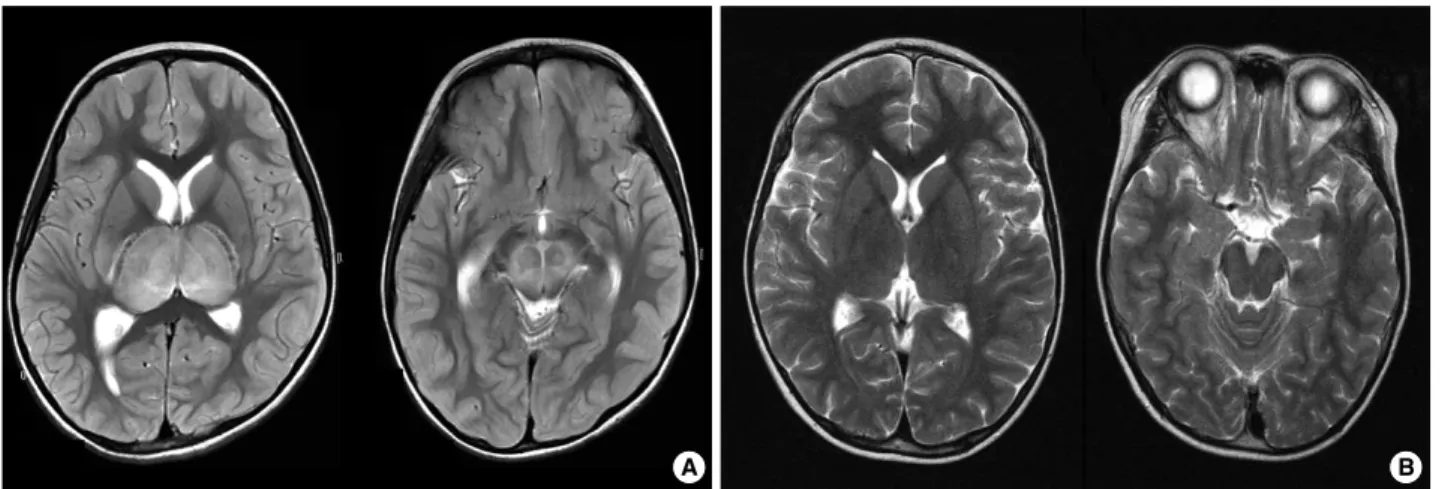
Table 4. Treatment and its outcome in the subjects with ANE Fig. 1. Radiological findings in a patient with ANE. (A) MRI shows symmetric, multifocal brain lesions involving bilateral thalami and upper brain stem tegmentum without involvement of other CNS regions. (B) Follow-up MRI shows complete resolution of previous lesions.
A B
of trasnsaminase, asymmetric thalamic involvement and no lesion in the brain stem tend to recover well (6). Considering the fact that ANE is a rare neurological condition, we expe- rienced a variety of different cases. They met the diagnostic criteria proposed by Mizuguchi et al. (2).
ANE is known to be one of the neurological complications of viral infections, such as influenza A and B, human herpes- virus 6, varicella zoster or mycoplasmal infection (7-14). In addition, it is reportedly associated with the measles virus in an animal study (15). Serological tests or polymerase chain reaction for certain viruses, and mycoplasma were unremark- able in our study. As shown in Table 2, the subjects showed various results in laboratory findings. Interestingly CSF pro- tein was within the normal range in all cases, but all had typi- cal thalamic involvement on the images. In an attempt to ex- clude other resembling encephalopathies, Herpes IgM, oligo- clonal band and myelin basic proteins in CSF were examined and shown unremarkable findings in most cases. Mutations of the mitochondrial DNA were screened to rule out mito- chondrial encephalopathies in four out of subjects, which all came back negative (16, 17). Considering the fact of their sim- ilarity to mitochondrial disorders on brain MRI, we believe that mitochondrial dysfunction is partly related in this con- dition, whether primary or not.
As is already known, ANE is a relatively uncommon form of acute encephalopathy especially in East Asia, although Euro- pean and American cases have been reported. It is thought to have some racial or geographic predilection (1, 6). In this study the HLA typing of 3 patients with ANE were evalu- ated to see genetic propensity. Genomic typing was perform- ed on their HLA DRB/HLA DQB genes and HLA-DRB1*
1401, HLA-DRB3*0202, and HLA-DQB1*0502 were found to be significant as compared with known data of HLA alleles in Korean (Table 3) (18). This is the first study using genom- ic typing of HLA DRB/HLA DQB genes that has never been done so far and the results may provide the immunogenetic background of ANE. However, further studies are needed to elucidate the condition.
A postmortem examination of the central nervous system could not be conducted, but bilateral thalamic necrosis is the histological hallmark of acute necrotizing encephalopathy.
Selective vulnerability of the thalami seems to be a determin-
as shock, multiple organ failure, and disseminated intravas- cular coagulation. This indicates that macrophage activation and hypercytokinemia may be involved in the pathogenesis of ANE (22).
As illustrated in Fig. 1, almost all subjects showed radio- logically pathognomonic findings of ANE. Based on previ- ously limited experience, the MRI of brain seems to be a very important diagnostic tool in ANE. With respect to the treat- ment for ANE, anti-inflammatory treatment may be effective (22-24). As shown in Table 4, the result of this study indicates early methylprednisolone pulse therapy seems to be partly effective for the children with ANE. However, well designed, comparative, multi-center studies are still required to evalu- ate the effectiveness of the regimen.
Regarding the outcome in patients, younger than 2 yr of age, those with high serum aminotransferase level, high pro- tein levels of cerebrospinal fluid, and those with brain stem lesions are thought to be poor prognostic factors (3-7, 10). In other reports, reversible or asymmetric brain involvement, focal neurologic signs showed a relatively fair prognosis (4, 8, 25). In this study, we experienced 6 patients with differ- ent manifestation of ANE. They ended up having relatively fair to good results except for two bad outcomes (patient 5, 6). As mentioned earlier, we believe that high dose steroid is fairly effective and can be a prognostic determinant when used in earlier stages of treatment. Findings on follow-up MRI seem to allow us to predict the outcome as shown in Table 4.
In addition, it is confirmed that normal serum aminotrans- ferase level, normal protein level of cerebrospinal fluid, and few neurological signs may be good prognostic factors for ANE.
In conclusion, ANE is an uncommon neurological compli- cation of acute infection in children and is to be further elu- cidated in many aspects. However, early detection and appro- priate treatment may lead to better neurological outcomes.
REFERENCES
1. Mizuguchi M. Acute necrotizing encephalopathy in childhood: a novel form of acute encephalopathy prevalent in Japan and Taiwan. Brain Dev 1997; 19: 81-92.
2. Mizuguchi M, Abe J, Mikkaichi K, Noma S, Yoshida K, Yamanaka T, Kamoshita S. Acute necrotising encephalopathy of childhood: a new syndrome presenting with multifocal, symmetric brain lesions.
J Neurol Neurosurg Psychiatry 1995; 58: 555-61.
3. San Millan B, Teijeira S, Penin C, Garcia JL, Navarro C. Acute necro- tizing encephalopathy of childhood: report of a spanish case. Pedi- atr Neurol 2007; 37: 438-41.
4. Yoshikawa H, Watanabe T, Abe T, Oda Y. Clinical diversity in acute necrotizing encephalopathy. J Child Neurol 1999; 14: 249-55.
5. Yagishita A, Nakano I, Ushioda T, Otsuki N, Hasegawa A. Acute encephalopathy with bilateral thalamotegmental involvement in in- fants and children: imaging and pathology findings. Am J Neurora- diol 1995; 16: 439-47.
6. Mastroyianni S, Gionnis D, Voudris K, Skardoutsou A, Mizuguchi M. Acute necrotizing encephalopathy of childhood in non-Asian patients: report of three cases and literature review. J Child Neurol 2006; 21: 872-9.
7. Olgar S, Ertugrul T, Nisli K, Aydin K, Caliskan M. Influenza A asso- ciated acute necrotizing encephalopathy. Neuropediatrics 2006; 37:
166-8.
8. Okumura A, Kidokoro H, Mizuguchi M, Kurahashi H, Hirabayashi Y, Morishima T, Watanabe K. The mildest form of acute necrotizing encephalopathy associated with influenza A. Neuropediatrics 2006;
37: 261-3.
9. Grose C. The puzzling picture of acute necrotizing encephalopathy after influenza A and B virus infection in young children. Pediatr Infect Dis J 2004; 23: 253-4.
10. Ohasaka M, Houkin K, Takigami M, Koyanagi I. Acute necrotizing encephalopathy associated with human herpesvirus-6 infection. Pedi- atr Neurol 2006; 34: 160-3.
11. Skelton BW, Hollingshead MC, Sledd AT, Phillips CD, Castillo M.
Acute necrotizing encephalopathy of childhood: typical findings in an atypical disease. Pediatr Radiol 2008; 38: 810-3.
12. Tran TD, Kubota M, Takeshita K, Yanagisawa M, Sakakihara Y.
Varicella-associated acute necrotizing encephalopathy with a good prognosis. Brain Dev 2001; 23: 54-7.
13. Kirton A, Busche K, Ross C, Wirrell E. Acute necrotizing encepha- lopathy in Caucasian children: two cases and review of the litera- ture. J Child Neurol 2005; 20: 527-32.
14. Ashtekar CS, Jaspan T, Thomas D, Weston V, Gayatri NA, White- house WP. Acute bilateral thalamic necrosis in a child with Myco- plasma pneumonia. Dev Med Child Neurol 2003; 45: 634-7.
15. Libert UG, Schneider-Schaulies S, Baczko K, ter Meulen V. Anti- body-induced restriction of viral gene expression in measles ence- phalitis in rat. J Virol 1990; 64: 706-13.
16. Chou HF, Liang WC, Zhang Q, Goto Y, Jong YJ. Clinical and genet- ic features in a MELAS child with a 3271T>C mutation. Pediatr Neu- rol 2008; 38: 143-6.
17. Qi Y, Zhang Y, Wang Z, Yang Y, Yuan Y, Niu S, Pei P, Wang S, Ma Y, Bu D, Zou L, Fang F, Xiao J, Sun F, Zhang Y, Wu Y, Wang S, Xiong H, Wu X. Screening of common mitochondrial mutations in Chinese patients with mitochondrial encephalomyopathies. Mito- chondrion 2007; 7: 147-50.
18. Oh HH, Kwon SH, Kim CW, Choe BH, Ko CW, Jung HD, Suh JS, Lee JH. Moelcular anaysis of HLA Class II-associated susceptibility to neuroinflammatory disease in Korean children. J Korean Med Sci 2004; 19: 426-30.
19. Kim JH, Kim IO, Lim MK, Park MS, Choi CG, Kim HW, Kim JE, Choi SJ, Koh YH, Yang DM, Choo SW, Chung MJ, Yoon HK, Goo HW, Lee M. Acute necrotizing encephalopathy in Korean infants and children: imaging findings and diverse clinical outcome. Kore- an J Radiol 2004; 5: 171-7.
20. Goo HW, Choi CG, Yoon CH, Ko TS. Acute necrotizing encepha- lopathy: diffusion MR imaging and localized proton MR spectro- scopic findings in two infants. Korean J Radiol 2003; 4: 61-5.
21. Ichiyama T, Endo S, Kaneko M, Isumi H, Matsubara T, Furukawa S.
Serum cytokine concentration of influenza-associated acute necrotiz- ing encephalopathy. Pediatr Int 2003; 45: 734-6.
22. Okumura A, Mizuguchi M, Kidokoro H, Tanaka M, Abe S, Hosoya M, Aiba H, Maegaki Y, Yamamoto H, Tanabe T, Noda E, Imataka G, Kurahashi H. Outcome of acute necrotizing encephalopathy in relation to treatment with corticosteroids and gammaglobulin. Brain Dev 2009; 31: 221-7.
23. Akiyoshi K, Hamada Y, Yamada H, Kojo M, Izumi T. Acute necro- tizing encephalopathy associated with hemophagocytic syndrome.
Pediatr Neurol 2006; 34: 315-8.
24. Manara R, Franzoi M, Cogo P, Battistella PA. Acute necrotizing en- cephalopathy: combined therapy and favorable outcome in a new case. Child Nerv Syst 2006; 22: 1231-6.
25. Wong AM, Simon EM, Zimmerman RA, Wang HS, Toh CH, Ng SH. Acute necrotizing encephalopathy of childhood: correlation of MR findings and clinical outcome. AJNR Am J Neuroradiol 2006;
27: 1919-23.