Received: May 7, 2020 Revised: July 20, 2020 Accepted: August 4, 2020 Trauma and InJury
Correspondence to Yang Bin Jeon, M.D., Ph.D.
Department of Traumatology, Gachon University College of Medicine, 38-13 Dokjeom-ro 3beon-gil, Namdong-gu, Incheon 21565, Korea
Tel: +82-32-460-3010 Fax: +82-32-460-2372 E-mail: [email protected]
Traumatic Tricuspid regurgitation as a Cause of failure to Wean from
mechanical Ventilation
Yang Bin Jeon, M.D., Ph.D.
1, Chul Hyun Park, M.D., Ph.D.
2, Dae Sung Ma, M.D.
31
Department of Traumatology, Gachon University College of Medicine, Incheon, Korea
2
Department of Thoracic and Cardiovascular Surgery, Gachon University College of Medicine, Incheon, Korea
3
Department of Thoracic and Cardiovascular Surgery, Dankook University Hospital, Cheonan, Korea
A 55-year-old man underwent emergent sternotomy due to cardiac tamponade occur- ring just after an accidental fall from a 10-m height. Tricuspid valve regurgitation was found on echocardiography while he was on mechanical ventilation after the operation.
The patient was weaned successfully from mechanical ventilation after tricuspid valve repair under cardiopulmonary bypass. Traumatic tricuspid valve regurgitation is a rare blunt chest injury and its symptoms occur late. Tricuspid regurgitation should be con- sidered as a reason for failure to wean from mechanical ventilation after blunt cardiac trauma.
Keywords: Tricuspid valve regurgitation; Trauma; Cardiac tamponade
INTRODUCTION
Cardiac valve injury by trauma occurs in 9% of patients with blunt cardiac trauma ac-
cording to Parmley and colleagues, and aortic valve injury is the most common type of
these injuries [1]. Injury of the tricuspid valve structures after blunt chest trauma has
rarely been reported [1-4]. Traumatic tricuspid valve regurgitation (TTR) is usually
well tolerated [5]; however, tricuspid valve replacement is the conventional treatment
in cases with delayed presentation [6]. Particularly in patients with multiple trauma,
other associated injuries may obscure cardiac symptoms and signs, thereby causing
delays in the diagnosis and treatment of cardiac valve injury. We experienced a note-
worthy case of traumatic TR in the early phase of multiple trauma.
Traumatic Tricuspid regurgitation as a Cause of failure to Wean from
mechanical Ventilation
Yang Bin Jeon, M.D., Ph.D.
1, Chul Hyun Park, M.D., Ph.D.
2, Dae Sung Ma, M.D.
31
Department of Traumatology, Gachon University College of Medicine, Incheon, Korea
2
Department of Thoracic and Cardiovascular Surgery, Gachon University College of Medicine, Incheon, Korea
3
Department of Thoracic and Cardiovascular Surgery, Dankook University Hospital, Cheonan, Korea
A 55-year-old man underwent emergent sternotomy due to cardiac tamponade occur- ring just after an accidental fall from a 10-m height. Tricuspid valve regurgitation was found on echocardiography while he was on mechanical ventilation after the operation.
The patient was weaned successfully from mechanical ventilation after tricuspid valve repair under cardiopulmonary bypass. Traumatic tricuspid valve regurgitation is a rare blunt chest injury and its symptoms occur late. Tricuspid regurgitation should be con- sidered as a reason for failure to wean from mechanical ventilation after blunt cardiac trauma.
Keywords: Tricuspid valve regurgitation; Trauma; Cardiac tamponade
CASE REPORT
A 55-year-old man was transported by helicopter with mental changes that developed after an accidental fall from a 10-m height. Soon after arrival, his systolic blood pressure was 62 mmHg, his pulse rate was 130 beats per minute, his respiratory rate was 28 breaths per minute, his body temperature was 35.5°C, and his neck vein was not distended. Initial arterial blood gas analysis showed the following findings: pH, 7.29; PCO
2, 46 mmHg; PO
2, 114 mmHg; base excess, -4.5; lactate, 5.5 mmol/L; and he- moglobin, 12.6 g/dL. We started fluid resuscitation with 2,500 mL of normal saline, two pints of packed red blood
cells, and 0.5 mg/kg/min of norepinephrine. We checked whole-body computed tomography (CT) 2 hours after the start of resuscitation because of uncertain Focused As- sessment of Sonography for Trauma (FAST) results and normal chest X-ray findings (Fig. 1A). CT showed a small to moderate amount of pericardial effusion (Fig. 2), which we thought indicated cardiac tamponade. As soon as the diagnosis was made, we went to the operating theater and opened the chest by a median sternotomy incision. When we opened the pericardial sac, we found a small amount of blood. The right atrium (RA) was bruised and there was a 1-cm laceration between the RA and the superior vena cava. The right pleura had a pinpoint-size opening.
A B C
Fig. 1. Chest X ray (CXR) shows a change of cardiac silhouette according to time sequence. (A) is the CXR at the time of arrival at the trauma bay.
(B) shows cardiomegaly and lung filtration after 5 days of emergency operation. (C) is the CXR on the first day after correction of tricuspid regurgitation.
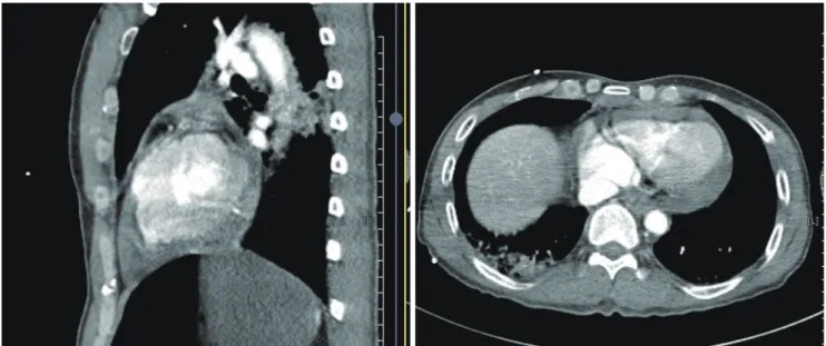
Fig. 2. Chest computed tomography shows pericardial effusion (hemopericardium) at trauma bay.
We closed the laceration with a figure-of-8 suture, extend- ed the right pleural opening, and inserted a chest tube.
After sternotomy, the patient was sedated and placed on mechanical ventilation because of hypo-oxygenation and difficulty in weaning from the ventilator. On the 5th post- operative day, the patient showed marked cardiomegaly (Fig. 1B) and we conducted cardiac echocardiography (Fig.
3). The echocardiogram showed severe TR with anterior leaflet flail movement, enlargement of the RA and the right ventricle, and pulmonary hypertension with a mean pulmonary artery pressure of 43 mmHg. We planned open heart surgery for severe TR. The second operation was performed under cardiopulmonary bypass. The heart was arrested with cardioplegic solution and the RA was explored. The anterior papillary muscle (APM) and con- necting chordae were cut from the ventricular wall and a remnant patent foramen ovale (PFO) was found (Fig. 4).
The rupture was repaired by reimplantation of APM to the ventricular septum near the tear site with two pledget- ed 5-0 Prolene sutures. Additionally, the annulus was re- duced by a modified DeVega annuloplasty, and the PFO was closed. The patient was extubated on the first postop- erative day (Fig. 1C). He underwent four operations for his combined injuries and was discharged uneventfully.
DISCUSSION
Isolated TTR has been sparsely reported and there have been no large-scale studies yet. However, in recent years, early evaluations of cardiac injury have become more
common in blunt chest trauma patients, as reported in several case reports, case series, and a literature review [1-6]. Some of these injuries are diagnosed months or years after the initial injury when a new murmur is heard [1,4-6], while others are diagnosed acutely because of he- modynamic disability [2,3].
The purported mechanism of injury is compression of the heart during late diastole or isovolemic systole [7]. At this time, the cardiac chambers are full and the valves are
Fig. 3. Echocardiography shows tricuspid valve regurgitation on 5th days after first operation. Color Doppler image shows tricuspid valve regurgitation
and the arrow is the prolapsed anterior leaflet of tricuspid valve.
Fig. 4. Right atrium is opened during cardioplegic arrest of heart. Arrow