467
DOI: 10.4046/trd.2009.67.5.467
ISSN: 1738-3536(Print)/2005-6184(Online) Tuberc Respir Dis 2009;67:467-470
CopyrightⒸ2009. The Korean Academy of Tuberculosis and Respiratory Diseases. All rights reserved.
결핵성 공동으로 오인된 외상 후 발생한 가성 폐낭종 1예
가톨릭대학교 의과대학
1내과학교실,
2영상의학교실
이현정1, 강지영1, 임선미1, 지은혜1, 김지현1, 김세원1, 이상학1, 문화식1, 이배영2
A Case of Post-Traumatic Pulmonary Pseudocyst Mimicking Pulmonary Cavitary Tuberculosis
Hyun Jeong Lee, M.D.1, Ji Young Kang, M.D.1, Sun Mie Yim, M.D.1, Eun Hye Ji, M.D.1, Ji Hyun Kim, M.D.1, Sei Won Kim, M.D.1, Sang Haak Lee, M.D.1, Hwa Sik Moon, M.D.1, Bae Young Lee, M.D.2
Departments of 1Internal Medicine, 2Radiology, The Catholic University of Korea, College of Medicine, Seoul, Korea
A traumatic pulmonary pseudocyst is a rare complication of blunt thoracic trauma. The clinical symptoms and signs are similar to other respiratory diseases, such as pulmonary tuberculosis. Therefore, a trauma history with the resulting radiologic and clinical findings is important for making a diagnosis. A 26-year-old male was admitted to our hospital due to cough for 3 days. The chest x-ray revealed diffuse infiltrations and a cavitary lesion at the left lung. His left chest had hit a tree as a result of motorcycle accident one day before admission. Initially, it was assumed that his symptoms and chest X-ray might be due to a tuberculosis infection. However, bronchoscopy revealed old blood clots at both lungs, particularly in the left lower lobe bronchus. A transbronchial lung biopsy showed alveolar hemorrhage. A traumatic pulmonary pseudocyst was diagnosed from his trauma history and these findings. Computed tomography of the chest performed 4 months later showed regression of the cavitary lesion.
Key Words: Lung Injury; Cysts; Wounds and Injuries; Cavity
Address for correspondence: Sang Haak Lee, M.D.
Department of Internal Medicine, The Catholic University of Korea, College of Medicine, 620-56, Jeonnong 2-dong, Dongdaemun-gu, Seoul 130-709, Korea
Phone: 82-2-958-2445, Fax: 82-2-968-7250 E-mail: [email protected]
Received: Sep. 16, 2009 Accepted: Sep. 29, 2009
서 론
외상으로 인한 폐 실질의 손상은 폐좌상, 폐열상, 폐혈 종, 가성 폐낭종 등이 있다. 이 중에서 외상 후 발생하는 가성 폐낭종(post-traumatic pulmonary pseudocyst)은 대 부분 비관통성 흉부 둔상이 원인이 되며 폐손상과 관련된 질환 중에서 3% 미만을 차지 하는 비교적 드문 질환이다1. 주요 증상으로는 혈담, 흉통, 기침 등으로 비특이적이며 손상된 폐에 고여 있던 혈전이 흡수 되면서 발열과 백혈구 증가 현상이 동반될 수 있다2. 이러한 환자는 병력에서 수 상력과 증상 발생의 선후 관계가 명확하여 대부분의 경우
외과 계열로 입원하게 되고 단순 흉부 촬영과 흉부 전산화 단층촬영으로 진단하게 된다. 감별이 필요한 질환은 폐암, 기관지 낭종, 결핵, 진균종, 폐농양 등이 있다.
저자들은 3일간의 기침을 주소로 내원한 25세 남자에 서 흉부 방사선상 공동 및 폐침윤 소견을 보여 폐결핵을 의심하였으나 최종적으로 외상 후 발생한 가성 폐낭종으 로 진단한 증례를 경험하였기에 문헌 고찰과 함께 보고 하는 바이다.
증 례
환 자: 남자, 25세
주 소: 내원 3일 전 발생한 기침
현병력: 환자는 내원 3일 전부터 오한을 동반한 기침 증상이 있었으며, 내원 전일 오토바이 운전 중 좌측 가슴 을 가로수에 부딪히는 사고가 발생하여 타 병원에서 촬영 한 단순 흉부 방사선에서 폐침윤 소견 보여 본원으로 전원
Image of the Month
HJ Lee et al: Post-traumatic pulmonary pseudocyst
468
Figure 1. Initial posteroanterior chest X-ray shows ill-de- fined ground glass opacity and cavitary change in left lung field.
Figure 2. Computed tomography image with a lung win- dow setting shows clustered small and large cavitary le- sions with wall thickening in left lower lobe, predominantly superior segment.
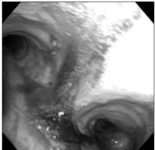
Figure 3. Flexible bronchoscopy shows old blood clot from trachea to both main bronchus, predominantly in left lower lobe bronchus.
되었다.
과거력: 특이 사항 없었다.
가족력: 특이 사항 없었다.
사회력: 비흡연자이다.
신체검사 소견: 환자는 급성 병색 소견을 보이고 있었 으며 혈압은 120/60 mmHg, 맥박 75회/분, 호흡수 20회/
분, 체온은 36.8oC이었다. 환자의 피부 및 두부 이학적 검 사에서 이상이 없었고, 경부 림프절은 촉지되지 않았다.
좌측 폐야에서 거친 호흡음이 청진되었으며, 좌측 흉곽의 왼쪽 세 번째 및 네 번째 갈비뼈의 빗장 중간선에 경미한 압통이 있었으나 육안적 이상 소견은 없었다. 심음은 정
상이었고 복부의 압통 및 반동 압통은 없었고, 장음은 정 상이었다.
검사실 소견: 말초혈액검사에서 백혈구 10,500/mm3 (호중구 78.2%, 림프구 12.7%, 호산구 0.2%), 혈색소 14.7 g/dL, 혈소판은 244,000/mm3이었고 혈청 생화학검사에 서 공복혈당 104 mg/dL, 혈중요소질소 18.5 mg/dL, 크레 아티닌 0.9 mg/dL였다. 아스파르테이트아미노전이효소 184 IU/L, 알라닌아미노전이효소 66 IU/L, 젖산탈수소효 소 938 IU/L (정상치 218∼472), 크레아틴키나아제 5,627 IU/L (정상치 55∼215)로 상승하였고 총 빌리루빈 1.8 mg/dL, 총 단백 7.4 g/dL, 알부민 4.7 g/dL, 칼슘 8.9 mg/
dL, 인 3.8 mg/dL, 적혈구 침강속도는 2 mm/h (정상치 2∼10), high sensitivity C-reactive protein 4.5 mg/dL (정 상치 <0.3)이었다. 대기 중 동맥혈 가스검사는 pH 7.41, PaCO2 38.3 mmHg, PaO2 92.2 mmHg, HCO3−
23.8 mmol/L, 산소포화도는 98.8%이었다. 심전도에서 규칙적 인 동성맥 소견 보였다.
방사선학적 소견: 단순 흉부사진에서 좌측 폐야 상부에 경계가 불분명한 젖빛 유리 음영과 공동이 관찰되었다 (Figure 1). 흉부 전산화 단층촬영에서 좌측 하엽에 무리 를 이룬 크고 작은 공동이 관찰되었고 공동벽이 두꺼워진 소견이었다. 경계가 불분명한 젖빛 유리 음영이 좌측 상 엽과 하엽에 있었고 소량의 흉수가 있었다(Figure 2).
경 과: 가래로 항산성 도말, 그람염색, 세균배양 시행
Tuberculosis and Respiratory Diseases Vol. 67. No. 5, Nov. 2009
469
Figure 4. Microscopic finding shows intra-alveolar hemor-rhage, deposit of fibrinoid fluid and focal infiltration of neu- trophils (H&E stain, ×100).
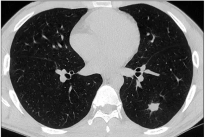
Figure 5. Follow up chest computed tomography per- formed 4 months later shows much regression of the pre- viously noted cavitary and infiltrative lesion.
하였으며 확인된 균주는 없었다. 내원 4병일에 결핵성 폐 병변 의심하에 기관지 내시경을 시행하였다. 육안적으로 기관에서 양측 주기관지까지 오래된 혈괴가 있었으나 급 성 출혈 소견은 없었고, 특히 좌측 하엽 기관지에 다량의 혈괴가 관찰되었다(Figure 3). 경기관지 폐생검, 기관지 세척 및 솔검사를 시행하였는데 결핵 및 세균, 곰팡이 등 의 도말 및 배양검사는 모두 음성이었고, 경기관지 폐생검 결과 폐포 내에 출혈 및 섬유소성 액체가 관찰되었다 (Figure 4). 환자는 외상 후에 발생한 가성 폐낭종으로 진 단되어, 보존 치료 후 증상 및 방사선 소견 호전되어 12병 일째 퇴원하였다. 수상 4개월 뒤 추적 관찰한 흉부 전산화 단층촬영에서 병변은 현저히 감소되었다(Figure 5).
고 찰
외상성 가성 폐낭종은 대부분의 경우 흉부 둔상이 원인 이 되며 수상 당일에 흉부 영상 촬영으로 진단되는 경우는 환자의 50% 정도밖에 되지 않는다1. 왜냐하면 폐실질 손 상이 가성 폐낭종으로 발전하는데 10일까지도 소요될 수 있기 때문이다. 국외 증례 보고에 따르면 낭종의 크기는 외상의 정도와 관련되어 4 cm 이상 병변일 경우는 다발 손상이 원인이 되며 기계 호흡이 필요한 정도의 중증도 환자에서 주로 관찰되었다2. 호발 연령은 30세 미만으로, 이는 외상 발생 빈도가 고령군에 비해 많고 폐실질이 상대 적으로 약해서 열상에 취약하며 흉곽의 탄력성이 높아 외 상이 폐로 쉽게 전달되기 때문으로 생각되고 있다2-5. 외상 으로 인하여 가성 폐낭종이 형성되는 과정은 세 가지 정도
로 생각할 수 있는데, 첫째로는 압축력(compressive force) 과 감압력(decompressive force) 사이의 전단 응력(shear stress)으로 인하여 폐실질이 손상되고 이로 인하여 공동 을 형성되는 경우, 둘째로는 후두가 막힌 상태에서 외상으 로 인하여 기관지 내압이 상승함으로써 소기관지가 파열 되는 경우, 마지막으로 손상된 기관지가 막힘으로써 폐포 가 파열하는 경우 등이다5,6. 가성 폐낭종은 진성 폐낭종과 는 달리 병변에 상피 세포나 기관지 벽의 구성 요소를 포 함하지 않으며 또한 소아에서 주로 보여지는 기류(pneu- matocele)처럼 감염의 합병증으로 발생하지 않는다7. 조 직학적 소견은 포식 세포가 혈철소를 포함하고 주변 조직 은 섬유화가 동반된 특징을 보인다8. 가성 폐낭종의 벽은 폐엽 사이의 결합조직에서 유래하였을 것으로 생각된다8. 방사선과적으로 유사한 형태를 보일 수 있는 질환으로는 결핵성 공동, 폐농양, 국소성 농흉, 폐격리증, 악성 종양 등이 있다. 반면, 외상성 가성 폐낭종의 경우에는 수상의 병력이 있으면서 기침, 객혈 등의 호흡기 증상과 함께 비 교적 짧은 시간 내에 병변의 크기나 형태가 변한다는 특징 이 있다.
치료로는 감염의 증거가 있을 때, 출혈이 동반되었을 때, 크기가 점차적으로 증가하여 산소 교환의 장애를 유발 할 때, 폐낭종이 파열되면서 기흉이 동반되었을 때 항생제 의 투약이나 추가 시술이 필요하다9. 예방적 항생제의 사 용은 추천되지 않으며, 감염이 동반된 가성 폐낭종은 크기 가 2 cm 이상이거나 항생제 투약 72시간 이내 반응이 없 을 경우 배액 치료하여야 한다. 또한 크기가 6 cm 이상이 거나 보존적 치료에 반응이 없을 경우에는 외과적 절제가 필요하다3. 그러나 합병증이 동반되지 않은 가성 폐낭종
HJ Lee et al: Post-traumatic pulmonary pseudocyst
470
은 시간 경과에 따라 자연 관해 되기 때문에 배액이나 수 술과 같은 불필요한 치료를 하지 않기 위해서는 낭종성 병변에 대한 정확한 감별이 무엇보다 중요하다. 본 환자 에서는 감염이 동반되거나 합병증이 발생하지 않아 보전 적 치료만으로 치유된 경우였다. 가성 낭종은 상피세포가 없기 때문에 흡수가 느려 영상학적으로 완전 관해 되는 데까지는 2∼3개월이 걸리는 것으로 알려져 있다1. 본 환자는 수상과 증상 발생의 선후 관계가 불명확하였 고 공동을 동반하는 폐침윤 소견을 보이는 흔한 질환인 결핵 및 기타 감염성 폐렴의 가능성을 우선적으로 고려하 여 기관지 내시경 검사를 시행하여 조직 검사 결과 외상성 가성 폐낭종으로 최종 진단한 경우이다. 가성 폐낭종은 외상 후 발생하는 드문 합병증으로 대부분 외과 영역에서 치료하게 되며 정확한 진단만 된다면 보존적 치료만으로 완치가 가능하다. 본 환자의 경우처럼 외관상 수상의 정 도가 심하지 않은 경우에도 폐에 공동성 병변이 발생할 수 있으므로, 수상의 병력이 있다면 영상학적으로 공동성 병변에 대한 감별 진단과 더불어 자세한 병력 청취가 무엇 보다 중요할 것으로 생각된다.
참 고 문 헌
1. Kato R, Horinouchi H, Maenaka Y. Traumatic pulmo-
nary pseudocyst. Report of twelve cases. J Thorac Car- diovasc Surg 1989;97:309-12.
2. Ganske JG, Dennis DL, Vanderveer JB Jr. Traumatic lung cyst: case report and literature review. J Trauma 1981;21:493-6.
3. Melloni G, Cremona G, Ciriaco P, Pansera M, Carretta A, Negri G, et al. Diagnosis and treatment of traumatic pulmonary pseudocysts. J Trauma 2003;54:737-43.
4. Stulz P, Schmitt HE, Hasse J, Grädel E. Traumatic pul- monary pseudocysts and paramediastinal air cyst: two rare complications of blunt chest trauma. J Trauma 1984;24:850-3.
5. Fagan CJ. Traumatic lung cyst. Am J Roentgenol Radi- um Ther Nucl Med 1966;97:186-94.
6. Fagan CJ, Swischuk LE. Traumatic lung and paramedi- astinal pneumatoceles. Radiology 1976;120:11-8.
7. Athanassiadi K, Gerazounis M, Kalantzi N, Kazakidis P, Fakou A, Kourousis D. Primary traumatic pulmonary pseudocysts: a rare entity. Eur J Cardiothorac Surg 2003;23:43-5.
8. Sorsdahl OA, Powell JW. Cavitary pulmonary lesions following nonpenetrating chest trauma in children. Am J Roentgenol Radium Ther Nucl Med 1965;95:118-24.
9. Lee SY, Lee SJ, Park HJ, Lee CS, Lee KR, Oh MH. A case of bilateral traumatic pulmonary pseudocyst. J Korean Soc Traumatol 2004;17:238-42.