Introduction
Tricuspid regurgitation (TR) can be caused by pathologic involvement of the tricuspid valve but more often occurs in the structurally normal tricuspid leaflet and chordae which is called functional TR.1)2) Functional TR can occur in several car- diovascular diseases such as left-sided heart disease, myocardial and pulmonary diseases.3) This functional TR was often ig- nored in the past because it was regarded to be simply related to other pathologic problems such as left sided valvular disease and it can improve after correction of underlying problem. How-
ORIGINAL ARTICLE J Cardiovasc Ultrasound 2015;23(3):136-142
ever, multiple recent studies have shown that TR may progess inspite of appropriate repair of the left side valve and that func- tional TR may be an important factor of adverse clinical out- come. Prior studies have shown that annular dilatation and tethering of the tricuspid valve contribute to the development of functional TR.3-6) Atrial fibrillation (AF) is suggested as one of the risk factors for development of TR.7-9) AF is often associ- ated with left and right atrial enlargement, thus causing annu- lar dilatation.4)7)10) However, not all patients with AF and dilat- ed right atrium (RA) develop significant TR. In this study, we
• Received: June 10, 2015 • Revised: August 17, 2015 • Accepted: August 17, 2015
• Address for Correspondence: Sung-Hee Shin, Division of Cardiology, Department of Internal Medicine, Inha University Hospital, 27 Inhang-ro, Jung-gu, Incheon 22332, Korea Tel: +82-32-890-2440, Fax: +82-32-890-2447, E-mail: [email protected]
• This is an Open Access article distributed under the terms of the Creative Commons Attribution Non-Commercial License (http://creativecommons.org/licenses/by-nc/3.0) which permits unrestricted non-commercial use, distribution, and reproduction in any medium, provided the original work is properly cited.
Clinical and Echocardiographic Factors Affecting Tricuspid Regurgitation
Severity in the Patients with Lone Atrial Fibrillation
Jae-Hyung Park, MD, Sung-Hee Shin, MD, PhD, Man-Jong Lee, MD, Myung-Dong Lee, MD, Hyun-Ik Shim, MD, Jaewoong Yoon, MD, Sehwan Oh, MD, Dae-Hyeok Kim, MD, PhD,
Sang-Don Park, MD, Sung-Woo Kwon, MD, Seong-Ill Woo, MD, PhD, Keum-Soo Park, MD, PhD, and Jun Kwan, MD, PhD
Division of Cardiology, Department of Internal Medicine, Inha University Hospital, Incheon, Korea
Background: Atrial fibrillation (AF) can be a risk factor for development of significant tricuspid regurgitation (TR). We in- vestigated which clinical and echocardiographic parameters were related to severity of functional TR in patients with lone AF.
Methods: A total of 89 patients with lone AF were enrolled (75 ± 11 years; 48% male): 13 patients with severe TR, 36 patients with moderate TR, and 40 consecutive patients with less than mild TR. Clinical parameters and echocardiographic measure- ments including right ventricular (RV) remodeling and function were evaluated.
Results: Patients with more severe TR were older and had more frequently persistent AF (each p < 0.001). TR severity was re- lated to right atrial area and tricuspid annular systolic diameter (all p < 0.001). The patients with moderate or severe TR had larger left atrial (LA) volume and increased systolic pulmonary artery pressure (SPAP) than the patients with mild TR (p = 0.04 for LA volume; p < 0.001 for SPAP). RV remodeling represented by enlarged RV area and increased tenting height was more prominent in severe TR than mild or moderate TR (all p < 0.001). Multivariate analysis showed type of AF, LA volume, tricus- pid annular diameter and tenting height remained as a significant determinants of severe TR. In addition, tenting height was inde- pendently associated with the presence of severe TR (p = 0.04).
Conclusion: In patients with lone AF, TR was related to type of AF, LA volume, tricuspid annular diameter and RV remodel- ing. Especially, tricuspid valvular tethering seemed to be independently associated with development of severe functional TR.
KEY WORDS: Tricuspid regurgitation · Atrial fibrillation · Tenting height.
investigated which clinical or echocardiographic parameters were related to severity of functional TR in patients with lone AF. We hypothesized that multifactorial factors such as the geo- metric change of right ventricle (RV) in addition to tricuspid annular dilatation by RA enlargement would play a role in de- termining TR severity in the patients with lone AF.
Methods
Study population
A total 89 patients with lone AF were enrolled in this study. By reviewing echocardiographic reports of the patients with lone AF between Jan 2009 and Dec 2014, 36 patients with moder- ate and 13 patients with severe TR were identified. Then con- trol group was selected from our echocardiographic database during same period and consisted of 40 consecutive patients with lone AF but no more than mild TR. For patients who had multiple echocardiograms, we used the first echocardiogram showing significant TR. The patients with pacemaker, left ven- tricular (LV) ejection fraction < 50%, rheumatic valvular dis- ease, significant left sided valvular disease, prior valvular surgery, intrinsic tricuspid valvular disease or congenital heart disease were excluded.
Echocardiography
Standard echocardiographic parameters including M-mode, two-dimensional (2D) and Doppler measurements were ana- lyzed in accordance with the American Society of Echocar- diography guidelines.11)12) Right atrial area (RAA) was mea- sured by tracing RA endocardium from the lateral aspect of the tricuspid annulus to the septal aspect at the apical 4-cham- ber view. Right ventricular end-diastolic area (RVEDA) and end-systolic area (RVESA) were obtained from a RV-focused apical 4-chamber view. RV fractional area change (RVFAC) was calculated by (RVEDA - RVESA) / RVEDA × 100.11) RV systolic area was divided by RV long-axis dimension (from the tip of the RV apex to the midpoint of tricuspid annular plane) to derive the RV spherical index.13) Tricuspid annular diameter was measured in the apical 4-chamber view as the distance be- tween the insertion of the septal leaflet and the insertion of the anterior leaflet, and tenting height was also measured as the perpendicular distance between the tip of leaflet coaptation and tricuspid annulus plane at the time of maximal systolic closure.8) Systolic pulmonary artery pressure (SPAP) was estimated from the peak systolic TR velocity using Bernoulli equation, and RA pressure was derived based on the inferior vena cava diameter and its respiratory change. Tricuspid annular plane systolic ex- cursion was acquired by placing M-mode cursor through the tricuspid annulus and measuring the distance of its systolic excursion in the longitudinal plane. Systolic excursion velocity of RV free wall was obtained with a tissue Doppler imaging at the apical 4-chamber view.12) Severity of TR was graded based on the color-flow Doppler using the ratio of TR jet area to the
RAA: mild if the ratio < 20%, moderate if 20% to 40% and se- vere if ≥ 40%.14) Vena contract width more than 7 mm was also defined as having severe TR. All echocardiographic measure- ments were averaged over 5 cardiac cycles.
Data analysis
Data were expressed as mean ± standard deviation for contin- uous variables and counts and percentages for categorical vari- ables. We divided the patients into 3 groups according to TR severity and applied one-way analysis of variance with the Scheffe post hoc test for continuous variables or Kruskal-Wal- lis analysis for categorical variables. The relationship between tenting height and other echocardiographic parameters was examined with Pearson’s correlation analysis. We assessed uni- variate regression analysis with vena contract width and other parameters. In addition, multivariate stepwise linear regres- sion analysis was performed to assess independent determi- nants among parameters with the variables which had a p value
< 0.1 from the univariate regression analysis. Vena contract width was used as a dependent variable in this multivariate linear regression analysis. To avoid collinearity, we constructed 3 different multivariate models using the variables with a vari- ance inflation factor less than 5. Logistic regression was also ap- plied to assess independent parameters in determining severe TR. All statistical analyses were performed using SPSS Statis- tics Version 19.0 (IBM Corp., Armonk, NY, USA) and STATA 14.0 (STATA Corp., College Station, TX, USA). p value of <
0.05 was considered statistically significant.
Results
Clinical characteristics
A total of 89 patients with lone AF were enrolled (age 75 ± 11 years; 48% male): 13 patients with severe TR, 36 patients with moderate TR, and 40 patients with no more than mild
Table 1. Baseline characteristics of overall patients Overall (n = 89)
Age, yr 075.2 ± 11.2
Male, n (%) 43 (48.3)
Diabetes, n (%) 19 (21.4)
Hypertension, n (%) 61 (68.5)
Type of AF, n (%)
Paroxysmal 15 (16.9)
Persistent 74 (83.2)
Duration of AF, months 059.7 ± 90.4
LVEF (%) 060.5 ± 4.3
SPAP (mm Hg) 046.0 ± 13.5
LAV (mL) 102.0 ± 40.0
RAA (cm2) 026.6 ± 9.6
AF: atrial fibrillation, LAV: left atrial volume, LVEF: left ventricular ejection fraction, RAA: right atrial area, SPAP: systolic pulmonary artery pressure
TR. Baseline characteristics of the study population are sum- marized in Table 1. When the patients were divided into three groups according to the severity of TR, they were similar in gender and underlying diseases such as diabetes and hyperten- sion. However, the patients who have more severe TR were old- er (p < 0.001). All patients with moderate or severe TR had per- sistent AF, whereas 38% of patients with no more than mild TR had paroxysmal AF (Table 2). Duration of AF was related to the severity of TR (p = 0.008).
Echocardiographic parameters according to TR severity
There was no significant difference between the groups re- garding LV size, LV mass, and LV systolic function (Table 3).
Left atrial (LA) volume was larger in the patients with moder- ate or severe TR as compared to the patients with mild TR (p = 0.04). In addition, RA area as well as tricuspid annular diame-
ter were larger in the patients with more severe TR than in the patients with milder TR (p < 0.001). RV systolic and diastolic area, RV spherical index and tenting height were larger in the patients with severe TR than with mild or moderate TR (p <
0.001), whereas they were comparable between mild and mod- erate group. SPAP was higher in the patients with moderate or severe group than with mild group (p < 0.001). RV function, reflected by several parameters such as RVFAC, systolic excur- sion velocity, and tricuspid annular plane systolic excursion, was similar between the groups.
Relationship between tenting height and right heart remodeling
Tenting height was associated with RA size, RV size and tri- cuspid annular diameter (r = 0.60, p < 0.0001 for RAA; r = 0.70, p < 0.0001 for RVEDA; r = 0.67, p < 0.0001 for RVESA;
r = 0.60, p < 0.0001 for tricuspid annular diameter). In addition,
Table 2. Clinical characteristics in patients with AF according to TR severity
Mild (n = 40) Moderate (n = 36) Severe (n = 13) p value
Age, yr 70.1 ± 12.2 78.6 ± 7.900 080.5 ± 10.2 < 0.001
Male, n (%) 24 (60) 12 (33) 07 (54) 0.060
Diabetes, n (%) 10 (25) 05 (14) 04 (31) 0.340
Hypertension, n (%) 25 (63) 27 (75) 09 (69) 0.510
Type of AF, n (%) < 0.001
Paroxysmal 15 (38) 00 (0) 00 (0)0
Persistent 25 (63) 36 (100) 13 (100)
Duration of AF, months 45.9 ± 71.8 60.2 ± 112.6 101.2 ± 61.0 0.008
AF: atrial fibrillation, TR: tricuspid regurgitation
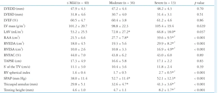
Table 3. Echocardiographic parameters in patients with AF according to TR severity
≤ Mild (n = 40) Moderate (n = 36) Severe (n = 13) p value
LVEDD (mm) 047.9 ± 4.3 47.2 ± 4.4 048.2 ± 4.3 0.700
LVESD (mm) 031.8 ± 4.6 30.7 ± 4.0 031.4 ± 3.1 0.510
LVEF (%) 060.5 ± 4.7 60.4 ± 3.8 061.2 ± 4.6 0.860
LV mass (g/m2) 101.2 ± 20.7 98.8 ± 22.3 105.4 ± 19.4 0.639
LAV (mL/m2) 053.2 ± 25.5 72.8 ± 27.2* 066.8 ± 18.0* 0.037
RAA (cm2) 021.5 ± 6.6 27.7 ± 7.8* 039.6 ± 9.5*,† < 0.001
RVEDA (cm2) 018.0 ± 4.5 19.3 ± 5.6 029.9 ± 8.2*,† < 0.001
RVESA (cm2) 010.0 ± 2.6 10.8 ± 3.3 016.9 ± 4.9*,† < 0.001
RVFAC (%) 044.0 ± 7.0 43.8 ± 6.2 043.0 ± 6.0 0.890
TAPSE (cm) 017.3 ± 4.9 16.6 ± 5.8 017.1 ± 2.2 0.830
S’ of the TV (cm/s) 011.1 ± 3.0 10.4 ± 3.6 011.8 ± 2.4 0.390
RV spherical index 001.6 ± 0.4 01.7 ± 0.5 002.7 ± 0.5*,† < 0.001
SPAP (mm Hg) 038.0 ± 11.4 52.7 ± 11.4* 052.1 ± 12.3* < 0.001
Tricuspid annulus (mm) 029.8 ± 5.1 32.6 ± 4.7* 041.3 ± 3.6*,† < 0.001
Tenting height (mm) 004.6 ± 1.0 04.7 ± 1.1 008.2 ± 1.7*,† < 0.001
*Indicates p ≤ 0.05 as compared to group with less than mild TR, †Indicates p ≤ 0.05 as compared to group with moderate TR. AF: atrial fibrillation, LAV: left atrial volume, LVEDD: left ventricular end-diastolic dimension, LVEF: left ventricular ejection fraction, LVESD: left ventricular end-systolic dimension, RAA:
right atrial area, RVEDA: right ventricular end-diastolic area, RVESA: right ventricular end-systolic area, RVFAC: right ventricular fractional area change, S’:
systolic myocardial velocity, SPAP: systolic pulmonary artery pressure, TAPSE: tricuspid annular plane systolic excursion, TR: tricuspid regurgitation, TV: tri- cuspid valve
the correlation between the tenting height and RV spherical in- dex was significant (r = 0.67, p < 0.0001) (Fig. 1).
Determinants of severe TR
Univariate analysis showed TR severity was related to age, type of AF, LA volume, RA area, RV area, RV spherical index,
Fig. 1. Relationship between tenting height and RAA (A), RVESA (B), TV annulus (C), and RV spherical index (D). RAA: right atrial area, RV: right ventricle, RVESA: right ventricular end-systolic area, TV: tricuspid valve.
4.0
3.5
3.0
2.5
2.0 1.5
1.0
0 2 4 6 8 10 12 r = 0.67 p < 0.0001
Tenting height (mm)
RV spherical index
D
50
45
40 35 30
25
20
0 2 4 6 8 10 12 r = 0.60 p < 0.0001
Tenting height (mm)
TV annulus (mm)
C
30
25
20
15
10
5
0 2 4 6 8 10 12 r = 0.67 p < 0.0001
Tenting height (mm) RVESA (cm2 )
B
Tenting height (mm)
A
60
50
40
30
20
10
0 2 4 6 8 10 12 r = 0.60 p < 0.0001 RAA (cm2)
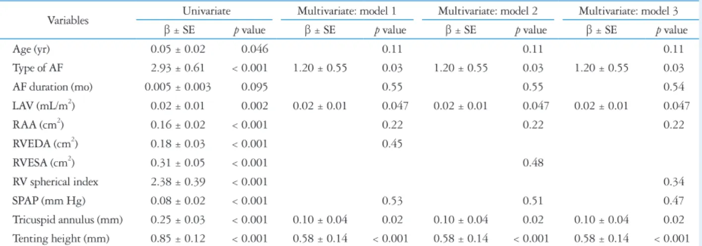
Table 4. Univariate and multivariate regression analyses of factors associated with tricuspid regurgitation severity
Variables Univariate Multivariate: model 1 Multivariate: model 2 Multivariate: model 3
β ± SE p value β ± SE p value β ± SE p value β ± SE p value
Age (yr) 00.05 ± 0.02 0.046 0.110 0.110 0.110
Type of AF 02.93 ± 0.61 < 0.001 1.20 ± 0.55 0.030 1.20 ± 0.55 0.030 1.20 ± 0.55 0.030
AF duration (mo) 0.005 ± 0.003 0.095 0.550 0.550 0.540
LAV (mL/m2) 00.02 ± 0.01 0.002 0.02 ± 0.01 0.047 0.02 ± 0.01 0.047 0.02 ± 0.01 0.047
RAA (cm2) 00.16 ± 0.02 < 0.001 0.220 0.220 0.220
RVEDA (cm2) 00.18 ± 0.03 < 0.001 0.450
RVESA (cm2) 00.31 ± 0.05 < 0.001 0.480
RV spherical index 02.38 ± 0.39 < 0.001 0.340
SPAP (mm Hg) 00.08 ± 0.02 < 0.001 0.530 0.510 0.470
Tricuspid annulus (mm) 00.25 ± 0.03 < 0.001 0.10 ± 0.04 0.020 0.10 ± 0.04 0.020 0.10 ± 0.04 0.020 Tenting height (mm) 00.85 ± 0.12 < 0.001 0.58 ± 0.14 < 0.001 0.58 ± 0.14 < 0.001 0.58 ± 0.14 < 0.001 AF: atrial fibrillation, LAV: left atrial volume, RAA: right atrial area, RV: right ventricle, RVEDA: right ventricular end-diastolic area, RVESA: right ventricular end-systolic area, SPAP: systolic pulmonary artery pressure
SPAP, tricuspid annulus and tenting height (Table 4, Fig. 2).
Multivariate regression demonstrated type of AF, LA volume, tricuspid annular diameter and tenting height were indepen- dently associated with TR severity (Table 4). By multivariate logistic analysis with tenting height, tricuspid annular diameter and LA volume, tenting height was independently associated with the presence of severe TR (p = 0.04).
Discussion
TR is a common echocardiographic finding. Although func- tional TR has somewhat been ignored in the past, it has attract- ed attention following clinical observations that functional TR itself can be an important predictor of clinical outcomes.2)15) Functional TR is known to be related to tricuspid annular dil- atation, which can be caused by RV remodeling in various con- ditions.3-5) Also, tethering of the tricuspid leaflets is often ob- served. While AF is known to contribute to the development of significant TR, the mechanism of TR development in AF is not clear. Most previous studies about functional TR included the patients with a variety of diseases and enrolled the patients with
AF as only a part of study population.3)4)16)17) In our study, we as- sessed which clinical and echocardiographic parameters would contribute to severe TR in AF, and type of AF, LA volume, tri- cuspid annular diameter and tenting height were independent- ly related to more severe TR in lone AF. The patients with more severe TR had larger RA and tricuspid annular size, greater RV size, and more globular RV than milder TR group. Tenting height was strongly related to RA and RV enlargement as well as RV spherical deformation.
AF can cause significant LA and RA enlargement, thus po- tentially leading to dilatation of mitral and tricuspid annuli lo- cated at the inferior edge of the atrium.10) But annular dilatation and valvular regurgitation are reported to be greater in the tri- cuspid valve than in the mitral valve because the fibrous skele- ton is less developed in the tricuspid valve.7) Previous data have reported aging and right heart enlargement as important patho- physiologic mechanism for developing severe TR in the patents with AF.8)9) In our study, the patients with moderate or severe TR had larger annular dilatation with more RA enlargement than the patients with no more than mild TR while the patients
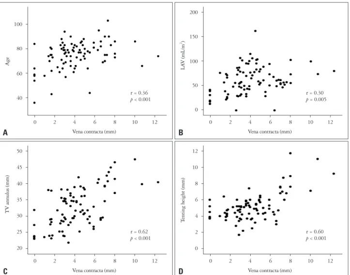
Fig. 2. Relationship between vena contracta and age (A), LAV (B), TV annulus (C), and tenting height (D). LAV: left atrial volume, TV: tricuspid valve.
12
10
8
6
4
2
0
0 2 4 6 8 10 12 r = 0.60 p < 0.001
Vena contracta (mm)
Tenting height (mm)
D
50
45
40
35
30
25
20
0 2 4 6 8 10 12 r = 0.62 p < 0.001
Vena contracta (mm)
TV annulus (mm)
C
200
150
100
50
0
0 2 4 6 8 10 12 r = 0.30 p = 0.005
Vena contracta (mm) LAV (mL/m2 )
B
100
80
60
40
0 2 4 6 8 10 12 r = 0.36 p < 0.001
Vena contracta (mm)
Age
A
with severe TR showed significantly greater RV size with glob- ular shape and more tethering of the tricuspid valve, reflected by increased tenting height. Isolated tricuspid annular dilata- tion relating to RA enlargement might not be enough to cause severe functional TR in the patients with lone AF. Further tri- cuspid annular enlargement with RV dilatation and remodel- ing might be required to develop severe TR, leading to an al- tered ventricular force balance to close the leaflets and incomplete closure of the tricuspid leaflets. However, we could not eluci- date causal relationship in our cross-sectional study design and RV enlargement can be caused by significant TR. Significant TR can dilate right heart chamber more and dilated right heart would deteriorate TR. So, TR can beget TR.
Prior studies revealed that the annular plane became larger, more planar, and circular in patients with functional TR as op- posed to saddle shape in control subjects.18) Spinner et al.19) showed that RV dilatation can result in displacement of all of the papillary muscles using three-dimensional echocardiogra- phy. They demonstrated that apical and lateral displacement of all papillary muscles was related to TR severity, with the ex- ception of lateral displacement of anterior papillary muscle, along with leaflet tethering. Displacement of the papillary muscles and annular dilatation would make the papillary mus- cle to move away from the annulus, thus leading to leaflet teth- ering. Our study showed that leaflet tethering can contribute to the presence of severe TR in AF. This finding is compatible with other previous studies, which revealed tricuspid valve teth- ering as an important geometric cause of functional TR.5)20-22) Relative positioning of the papillary muscles may also be a sig- nificant contributor to the tethering of the leaflets.
Study limitation
Several limitations of our analysis should be noted. First, our study could not elucidate the causal relationship because of cross- sectional design as pointed out previously. Second, we graded TR severity based on the ratio of TR jet area to the RAA and vena contracta width. While TR jet area and vena contracta are commonly used in assessing TR grade and most TR jet was a central jet in our population, more quantitative assessment of TR such as proximal isovelocity surface area can be recom- mended.1)23)24) Also, we used 2D echocardiography in measur- ing several parameters including tricuspid annular diameter.
Although 2D echocardiography is widely used in clinical prac- tice and also for research currently especially for the patients with AF, 2D images suffer from inherent limitations such as incom- plete comprehension of the three-dimensional tricuspid annu- lus. Three-dimensional echocardiography with one beat acqui- sition might provide more accurate measurements with more reproducible landmarks, but it is necessary to take account of its relative lower temporal and spatial resolution compared to 2D echocardiography. Finally, the study population with se- vere TR was relatively small, thus decreasing statistical power.
Conclusions
Tenting height, together with RA and RV enlargement as well as RV spherical deformation, tricuspid annular diameter and LA volume were independently associated with TR sever- ity in the patients with persistent lone AF. While the causal relationship is not elucidative in this study, tricuspid valvular tethering may predominantly contribute to development of severe TR in this population. Further large, prospective cohort studies with more quantitative methods will be required to elu- cidate the precise mechanisms and a determinant for developing severe TR in the patients with lone AF.
• Acknowledgements
This work was supported by Inha University Hospital and Inha University Research Grants (INHA-40899).
References
1. Badano LP, Muraru D, Enriquez-Sarano M. Assessment of functional tricuspid regurgitation. Eur Heart J 2013;34:1875-85.
2. Nath J, Foster E, Heidenreich PA. Impact of tricuspid regurgitation on long-term survival. J Am Coll Cardiol 2004;43:405-9.
3. Topilsky Y, Khanna A, Le Tourneau T, Park S, Michelena H, Suri R, Mahoney DW, Enriquez-Sarano M. Clinical context and mechanism of functional tricuspid regurgitation in patients with and without pulmonary hypertension. Circ Cardiovasc Imaging 2012;5:314-23.
4. Mutlak D, Lessick J, Reisner SA, Aronson D, Dabbah S, Agmon Y.
Echocardiography-based spectrum of severe tricuspid regurgitation: the fre- quency of apparently idiopathic tricuspid regurgitation. J Am Soc Echocar- diogr 2007;20:405-8.
5. Sagie A, Schwammenthal E, Padial LR, Vazquez de Prada JA, Wey- man AE, Levine RA. Determinants of functional tricuspid regurgitation in incomplete tricuspid valve closure: Doppler color flow study of 109 patients.
J Am Coll Cardiol 1994;24:446-53.
6. Kim HK, Lee SP, Kim YJ, Sohn DW. Tricuspid regurgitation: clinical importance and its optimal surgical timing. J Cardiovasc Ultrasound 2013;21:1-9.
7. Zhou X, Otsuji Y, Yoshifuku S, Yuasa T, Zhang H, Takasaki K, Mat- sukida K, Kisanuki A, Minagoe S, Tei C. Impact of atrial fibrillation on tricuspid and mitral annular dilatation and valvular regurgitation. Circ J 2002;66:913-6.
8. Najib MQ, Vinales KL, Vittala SS, Challa S, Lee HR, Chaliki HP.
Predictors for the development of severe tricuspid regurgitation with anatomi- cally normal valve in patients with atrial fibrillation. Echocardiography 2012;29:140-6.
9. Yamasaki N, Kondo F, Kubo T, Okawa M, Matsumura Y, Kitaoka H, Yabe T, Furuno T, Doi Y. Severe tricuspid regurgitation in the aged:
atrial remodeling associated with long-standing atrial fibrillation. J Cardiol 2006;48:315-23.
10. Sanfilippo AJ, Abascal VM, Sheehan M, Oertel LB, Harrigan P, Hughes RA, Weyman AE. Atrial enlargement as a consequence of atrial fibrillation. A prospective echocardiographic study. Circulation 1990;82:
792-7.
11. Lang RM, Badano LP, Mor-Avi V, Afilalo J, Armstrong A, Ernande L, Flachskampf FA, Foster E, Goldstein SA, Kuznetsova T, Lancellotti P, Muraru D, Picard MH, Rietzschel ER, Rudski L, Spencer KT, Tsang W, Voigt JU. Recommendations for cardiac chamber quantification by echocardiography in adults: an update from the American Society of Echo- cardiography and the European Association of Cardiovascular Imaging. J Am Soc Echocardiogr 2015;28:1-39.e14.
12. Rudski LG, Lai WW, Afilalo J, Hua L, Handschumacher MD, Chan-
drasekaran K, Solomon SD, Louie EK, Schiller NB. Guidelines for the echocardiographic assessment of the right heart in adults: a report from the American Society of Echocardiography endorsed by the European Association of Echocardiography, a registered branch of the European Society of Cardiol- ogy, and the Canadian Society of Echocardiography. J Am Soc Echocardiogr 2010;23:685-713; quiz 786-8.
13. Fukuda S, Gillinov AM, Song JM, Daimon M, Kongsaerepong V, Thomas JD, Shiota T. Echocardiographic insights into atrial and ventric- ular mechanisms of functional tricuspid regurgitation. Am Heart J 2006;
152:1208-14.
14. Singh JP, Evans JC, Levy D, Larson MG, Freed LA, Fuller DL, Lehm- an B, Benjamin EJ. Prevalence and clinical determinants of mitral, tricus- pid, and aortic regurgitation (the Framingham Heart Study). Am J Cardiol 1999;83:897-902.
15. Topilsky Y, Nkomo VT, Vatury O, Michelena HI, Letourneau T, Suri RM, Pislaru S, Park S, Mahoney DW, Biner S, Enriquez-Sarano M.
Clinical outcome of isolated tricuspid regurgitation. JACC Cardiovasc Imag- ing 2014;7:1185-94.
16. Kim HK, Kim YJ, Park JS, Kim KH, Kim KB, Ahn H, Sohn DW, Oh BH, Park YB, Choi YS. Determinants of the severity of functional tricuspid regurgitation. Am J Cardiol 2006;98:236-42.
17. Seo HS, Ha JW, Moon JY, Choi EY, Rim SJ, Jang Y, Chung N, Shim WH, Cho SY, Kim SS. Right ventricular remodeling and dysfunction with subsequent annular dilatation and tethering as a mechanism of isolated tri- cuspid regurgitation. Circ J 2008;72:1645-9.
18. Ton-Nu TT, Levine RA, Handschumacher MD, Dorer DJ, Yosefy C, Fan D, Hua L, Jiang L, Hung J. Geometric determinants of functional tri- cuspid regurgitation: insights from 3-dimensional echocardiography. Circu-
lation 2006;114:143-9.
19. Spinner EM, Lerakis S, Higginson J, Pernetz M, Howell S, Veledar E, Yoganathan AP. Correlates of tricuspid regurgitation as determined by 3D echocardiography: pulmonary arterial pressure, ventricle geometry, annu- lar dilatation, and papillary muscle displacement. Circ Cardiovasc Imaging 2012;5:43-50.
20. Hung J. The pathogenesis of functional tricuspid regurgitation. Semin Thorac Cardiovasc Surg 2010;22:76-8.
21. Fukuda S, Song JM, Gillinov AM, McCarthy PM, Daimon M, Kongsaerepong V, Thomas JD, Shiota T. Tricuspid valve tethering pre- dicts residual tricuspid regurgitation after tricuspid annuloplasty. Circula- tion 2005;111:975-9.
22. Mikami T, Kudo T, Sakurai N, Sakamoto S, Tanabe Y, Yasuda H.
Mechanisms for development of functional tricuspid regurgitation determined by pulsed Doppler and two-dimensional echocardiography. Am J Cardiol 1984;53:160-3.
23. Zoghbi WA, Enriquez-Sarano M, Foster E, Grayburn PA, Kraft CD, Levine RA, Nihoyannopoulos P, Otto CM, Quinones MA, Rakows- ki H, Stewart WJ, Waggoner A, Weissman NJ; American Society of Echocardiography. Recommendations for evaluation of the severity of native valvular regurgitation with two-dimensional and Doppler echocardiography.
J Am Soc Echocardiogr 2003;16:777-802.
24. Lancellotti P, Moura L, Pierard LA, Agricola E, Popescu BA, Tri- bouilloy C, Hagendorff A, Monin JL, Badano L, Zamorano JL; Eu- ropean Association of Echocardiography. European Association of Echo- cardiography recommendations for the assessment of valvular regurgitation.
Part 2: mitral and tricuspid regurgitation (native valve disease). Eur J Echo- cardiogr 2010;11:307-32.