• 검색 결과가 없습니다.

Post-traumatic tricuspid regurgitation with anterior papillary muscle rupture, corrected by papillary muscle reimplantation

N/A
N/A
Protected

Academic year: 2021

Share "Post-traumatic tricuspid regurgitation with anterior papillary muscle rupture, corrected by papillary muscle reimplantation"

Copied!
4
0
0

로드 중.... (전체 텍스트 보기)

전체 글

(1)

159

Case report

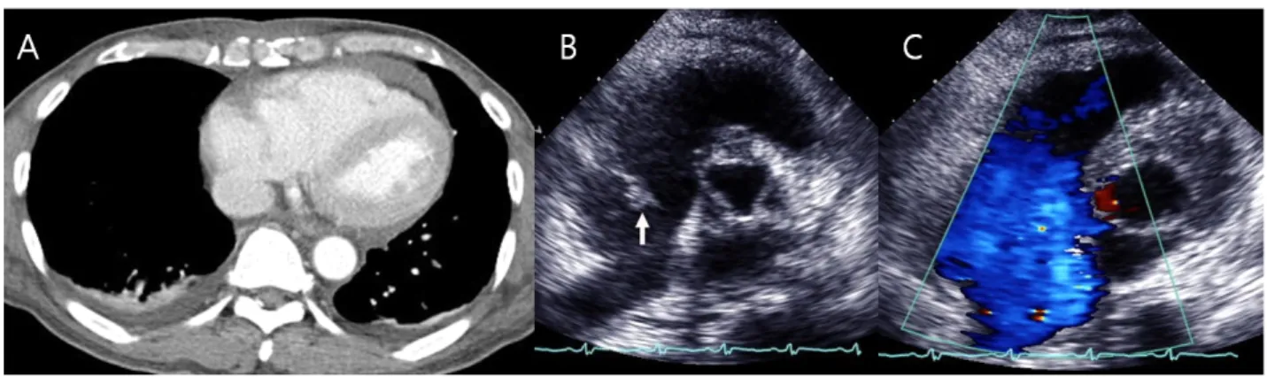
A 60-year-old male was transferred to our emergency department for evaluation and treatment for mild dyspnea and pericardial effusion detected by chest computerized tomography (CT) scan at 2 days after an anterior chest wall and left hand blunt trauma, which occurred when the patient was unwinding a bulky steel chain during sailing. Before transfer, the patient experienced an episode of hypotension and tachycardia

during orthopedic surgery to repair multiple left upper phalanges fractures at a local medical center. At admission, he complained of mild dyspnea, determined to be New York Heart Association (NYHA) functional class I, without neck vein distention or edema of the face and periphery. The patient did, however, present with multiple shallow abrasions, approximately 10 x 10 cm in size on the anterior chest wall and no chest pain or tenderness. Vital signs were recorded: blood pressure 100/60 mmHg and pulse rate 110 beats/min.

Tongue dehydration was detected. An

Kosin Medical Journal 2015;30:159-162.

http://dx.doi.org/10.7180/kmj.2015.30.2.159 KMJ

Case Report

Post-traumatic tricuspid regurgitation with anterior papillary muscle rupture, corrected by papillary muscle reimplantation

Hae Young Lee, Sung Ho Cho, Jong In Kim

Department of Thoracic & Cardiovase Surgery, College of Medicine, Kosin University, Busan, Korea

유두근 재이식수술로 교정한 외상후 삼첨판막부전증

이해영, 조성호, 김종인

고신대학교 의과대학 흉부외과학교실

A 60-year-old male patient with blunt chest trauma was transferred to our facility because of unstable vital signs and pericardial effusion. These conditions occurred after orthopedic surgery to repair multiple left finger fractures at a local medical center. Trans-thoracic echocardiography showed severe tricuspid regurgitation and he underwent papillary muscle reimplantation and tricuspid annuloplasty open heart surgery for post-traumatic tricuspid regurgitation with anterior papillary muscle rupture. We report early surgical traumatic valve disease correction without complications.

Key Words: 1. Heart valves, papillary muscles, 2. Trauma, blunt 3. Tricuspid valve insufficiency

4. Tricuspid valve, repair

Corresponding Author: Hae Young Lee, Department of Thoracic & Cardiovase Surgery, College of Medicine, Kosin University, 262, Gamcheon-ro, Seo-gu, Busan 49267, Korea

TEL: +82-51-990-6466 FAX: +82-51-990-3066 E-mail: [email protected]

Received:

Revised:

Accepted:

Apr. 17, 2014

Jan. 2, 2015

Feb. 24, 2015

(2)

Kosin Medical Journal 2015;30:159-162.

160

electrocardiogram showed only a right bundle branch block. A chest CT scan showed a small, newly developed pleural effusion, but there was no change in the pericardial effusion interval at the anterior space of the right ventricle (RV) (Fig. 1A). Transthoracic echocardiography (TTE) showed severe tricuspid regurgitation (TR) with flail anterior leaflet due to ruptured anterior tricuspid valve (TV) papillary muscle (PM) (Figs. 1B, C). The RV end-diastolic dimension was measured at 35.6mm, which was within the normal limit, and the annular size was 40mm in the apical four chamber view. The vital signs were restored immediately after appropriate fluid replacement therapy, although

there was an elevated GOT/GPT of 1437/1663 IU/L due to chronic diffuse alcoholic hepatocellular disease.

Additional hepatic dysfunction was caused by temporary hypovolemic shock during the previous surgery, in which the patient’s values declined to 58/109 IU/L, which delayed the open heart surgery on the 12

th

post-traumatic day. Initial pericardiotomy during open heart surgery showed bloody pericardial effusion and the preoperative diagnosis of severe TR due to anterior PM rupture was confirmed (Fig. 2A). The PM rupture was induced by trauma and annular dilatation; gross and saline inspections through right atriotomy were performed during a routine cardiopulmonary bypass.

Fig. 1. Chest computed tomography scan showed small amounts of pericardial effusion anterior to the right ventricle and right pleural effusion(A). Echocardiography showed that the tricuspid valve anterior leaflet had flailed papillary muscle(arrow)(B). Doppler echocardiography showed severe regurgitant flow at the right atrioventricular level during systole(C).

Fig. 2. Intraoperative gross findings: Ruptured papillary muscle (PM) of the tricuspid valve anterior leaflet(arrow)(A),

right ventricle ruptured PM stump(arrow)(B), and PM reimplantation and tricuspid annuloplasty with a 33mm

Duranring(C).

(3)

Post-traumatic tricuspid regurgitation with anterior papillary muscle rupture, corrected by papillary muscle reimplantation

161 We reimplanted the anterior PM fibrous cuff after

trimming and excision of the necrotic tissue at the healthy RV myocardial wall near the remnant stump(Fig. 2B) using a trans fixed mattress of 4-0 polypropylene sutures with a pledget. Surgery was completed with tricuspid annuloplasty (TAP) using a 33-mm Duran ring (Fig. 2C). Intraoperative trans-esophageal echocardiography (TEE) showed tricuspid leaflet coaptation, however, the TTE also showed mild regurgitation (Fig. 3) at the same level as the 7

th

postoperative day. The patient was discharged at the 10

th

postoperative day without any events.

Discussion

Post-traumatic cardiac valve injury is rare but, the TV is injured most frequently because it is anatomically located beneath the sternum. However, unlike the other valve positions, traumatic TR tends to be omitted and the diagnosis delayed because of other emergent

surgeries caused by multiple concurrent traumas or because of a relatively long asymptomatic period.

1

The literature suggests that early diagnosis followed by early surgical treatment is important because the ruptured chordae shrink over time; in addition, the PM, valve deformity and RV structure dysfunction inhibit valve repair. A valve repair is superior to valve replacement because it is associated with better long-term outcomes and is not associated with artificial valve-related complications.

2

We performed an early operation on the 12

th

post-traumatic day. Valvular heart disease due to peri-operative hypotension was confirmed by TTE;

heart murmurs combined with dyspnea and hemopericardium were confirmed with a chest CT at a local medical center.

Pathologically, tricuspid insufficiency can include chordae rupture (55.4%), rupture of the anterior PM (27.0%), leaflet tear (14.8%), an entire valve rupture or concomitant annular dilatation. In addition, only chordae shortening related to valve atrophy and annular dilatation without structural valve defects has been reported (5%).

3

Wall et al. suggested that PMrupturescouldbedetectedearlierthanchordaeshorteni ng.

4

The valve rupture mechanism was supported by increased intracardiac pressure at the time of contusion during late diastolic phase, isometric systolic phase, or due to delayed contused PM disruption.

5

Echocardiography must be performed as a routine evaluation after blunt chest trauma to evaluate cardiovascular lesions and should be considered as a single important modality that corresponds with the operative view.

6

Surgical treatment must be decided on an individual basis by evaluating the pathology and Fig. 3. Echocardiography showed well coapted tricuspid

valve leaflets(arrow) at the 7

th

postoperative day.

(4)

Kosin Medical Journal 2015;30:159-162.

162

anatomy of the injured valve and the regurgitation mechanism.

7

In many cases, concomitant annular dilatation aggravates severe regurgitation by RV dilatation.

Therefore annuloplasty has been accepted as an essential procedure to prevent dilatation induced by post-operative repair failure and to correct distorted and dilated annulus.

3

(In cases of leaflet lesion, direct repair is recommended, and chordal lesion is primarily treated with replacement because of the high rate of repair failure. However, other methods such as chordal implant, double orifice and quadrangular resection have been reported in recent studies. Chordal rupture has been associated with a better prognosis than ruptured PM repair that includes either a partial head portion rupture or an entire root rupture.) In cases of PM lesion, a PM implantation is the best surgical option when the ruptured portion can be clearly identified without RV dilatation. In addition, tethering and coaptation failure of the reimplanted PM can be prevented with minimal PM displacement.

8

Most reports have suggested that repair is associated with better outcomes than replacement, although these reports did not address the valvular injury severity or diagnostic interval. Nevertheless, early correction up to 3 months from onset was associated with preventing progressive annular enlargement.

3

We were able to diagnose traumatic TR before chordal rupture because of the acute manifestations due to the ruptured anterior PM. Accurate reimplantation was feasible because we identified a portion of healthy wall around a detached PM stump and the annulus was dilated, up to 40mm, without RV dilatation. The reimplantation was followed by TAP using a Duran

ring to minimize possible annular dilatation induced by failed reimplantation. In conclusion, we report a case of early surgical correction of a post-traumatic valvular disease with an acute status diagnosis.

References

1. Gayet C, Pierre B, Delahaye JP, Champsaur G, Fouet XA, Rueff P. Traumatic tricuspid insufficiency: An underdiagnosed Disease. Chest 1987;92:429-32.

2. WG Ma, GH Luo, HS Sun, JP Xu, SS Hu, XD Zhu.

Surgical treatment of traumatic tricuspid insufficiency:

experience in 13 cases. Ann Thorac Surg 2010;90:1934-8.

3. Maisano F, Lorusso R, Sandrelli L, Torracca L, Coletti G, Canna G, et al. Valve repair for traumatic tricuspid regurgitation. Eur J Cardiothorac Surg 1996;10:867-73.

4. Matthew J. Wall, Jr, Ernesto R. Soltero. Trauma to cardiac valves. Current opinion in cardiology 2002;17:188-92.

5. Naja I, Pomar JL, Barriuso C, Mestrea C, Mulet J. Traumatic tricuspid regurgitation. J Cardiovasc Surg (Torino) 1992;33:256-7.

6. Shapiro MJ, Yanofsky SD, Trapp J, Durham RM, Labovitz A, Sear JE, et al. Cardiovascular evaluation in blunt thoracic trauma using transesophageal echocardiography.

1991;31:835-40.

7. Bang JH, Woo JS, Choi PJ, Park KJ, Jeong SS, Yi JH.

Repair of ruptured papillary muscle of the tricuspid valve as a cause of tricuspid insufficiency following blunt chest trauma. Korean J Thorac Cardiovasc Surg 2010;43:413-6.

8. De Bonis M, Lapenna E, La Canna G, Grimaldi A, Maisano

F, Torracca L, et al. A novel technique for correction

of severe tricuspid valve regurgitation due to complex

lesions. Eur J Cardiothorac Surg 2004;25:760–5.

수치

Fig. 1. Chest computed tomography scan showed small amounts of pericardial effusion anterior to the right ventricle  and  right pleural  effusion(A)

참조

관련 문서