© 2012 The Korean Academy of Medical Sciences.
This is an Open Access article distributed under the terms of the Creative Commons Attribution Non-Commercial License (http://creativecommons.org/licenses/by-nc/3.0) which permits unrestricted non-commercial use, distribution, and reproduction in any medium, provided the original work is properly cited.
pISSN 1011-8934 eISSN 1598-6357
Late Simultaneous Presentation of Left Ventricular
Pseudoaneurysm and Tricuspid Regurgitation after Blunt Chest Trauma
A 32-yr-old man developed progressive exertional dyspnea 4 yr after blunt chest trauma due to an automobile accident. Two-dimensional echocardiography and computed- tomographic coronary angiography demonstrated a large pseudoaneurysm of the left ventricle and severe tricuspid regurgitation. The patient underwent successful surgical exclusion of the pseudoaneurysm by endoaneurysmal patch closure and repair of the tricuspid valve regurgitation. To the best of our knowledge, this is the first case of these 2 different pathologies presenting late simultaneously after blunt chest trauma and successful surgical repairs in the published literature.
Key Words: Wounds and Injuries; Tricuspid Valve Insufficiency; Aneurysm; False Ho-Ki Min1, Do Kyun Kang1,
Hee Jae Jun1, Youn-Ho Hwang1, Sang-Hoon Seol2, Kyubok Jin2, Jong Woon Song3, and Cheol Kyu Oh4 Departments of 1Thoracic and Cardiovascular Surgery, 2Internal Medicine, 3Radiology, and
4Urology, Haeundae Paik Hospital, Inje University College of Medicine, Busan, Korea
Received: 12 October 2011 Accepted: 20 January 2012 Address for Correspondence:
Ho-Ki Min, MD
Department of Thoracic and Cardiovascular Surgery, Haeundae Paik Hospital, Inje University College of Medicine, 875 Haeundae-ro, Haeundae-gu, Busan 612-030, Korea Tel: +82.51-797-3135, Fax: +82.51-797-3135 E-mail: [email protected]
http://dx.doi.org/10.3346/jkms.2012.27.4.443 • J Korean Med Sci 2012; 27: 443-445
CASE REPORT
Cardiovascular Disorders
INTRODUCTION
Blunt chest trauma may result in various cardiac defects includ- ing tricuspid valve injury, mitral valve avulsion, ventricular sep- tal rupture, pseudoaneurysm, and true aneurysm (1). We re- port a case of a left ventricular pseudoaneurysm (LVP) and se- vere tricuspid regurgitation (TR) presenting coincidentally 4 yr after a blunt chest injury. Herein we describe successful surgi- cal repair of the tricuspid valve and exclusion of the pseudoan- eurysm by endoaneurysmal patch closure between the aneu- rysm and the left ventricular cavity.
CASE DESCRIPTION
A 32-yr-old man visited our out-patient-department with par- oxysmal coughing and dyspnea on exertion in July 2011. He had suffered from multiple traumas due to an automobile acci- dent 4 yr earlier in a foreign country. At that time, he underwent hepatic resection for liver injury and thoracostomy for hemo- thorax. Blood pressure was 120/80 mmHg, heart rate was 90 beats per min, and other vital signs were normal. On ausculta- tion, systolic murmurs were noted on the right parasternal bor- der, which increased by inspiration. Chest roentgenogram
showed an enlarged heart and a circular mass-like lesion over- lapped with the cardiac silhouette. Two-dimensional echocar- diography revealed a large pseudoaneurysm arising from the inferobasal surface of the left ventricle with a neck measuring 18 mm and the sac 87 mm at its widest diameter, and severe tri- cuspid valve regurgitation due to the prolapsed anterior leaflet.
The right ventricle was dilated with a normal systolic function and the left ventricular function was preserved (Fig. 1).
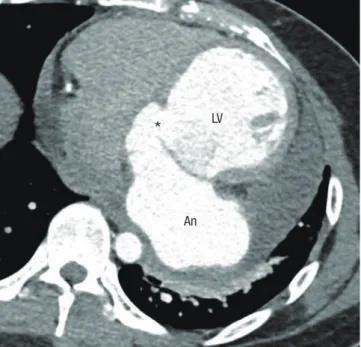
Transesophageal echocardiography confirmed these impres- sions. The coronary CT angiography showed normal coronary arteries and the presence of a large left ventricular pseudoan- eurysm (Fig. 2).
The operation was performed using cardiopulmonary bypass and cardioplegic arrest. The pericardium was free of adhesions.
The aneurysm was located close to the medial side of the left circumflex artery and near to the cephalic side of the coronary sinus. The aneurysm was entered and organized thrombi were removed. Additionally, we approached to the left atrium using transseptal approach and identified the opening of the aneu- rysm and the relation between the aneurysmal neck and sub- valvular apparatus via the mitral valve. The neck of the aneu- rysm was identified on each side of the aneurysm, both exter- nally and internally (Fig. 3). Then, a patch of Dacron was su-
Min H-K, et al. • Traumatic LV Pseudoaneurysm and TR
444 http://jkms.org http://dx.doi.org/10.3346/jkms.2012.27.4.443
tured at the base of the pseudoaneurysm by multiple pledget- buttressed 4-0 Prolene (Ethicon, Somerville, NJ, USA) and rein- forced by a single running suture of 4-0 Prolene, to close the communication between the left ventricle and the pseudoan- eurysm. The external wall of the pseudoaneurysm was resected and closed with a double row of 4-0 Prolene by placing two wide strips of Teflon felt on each side. After a saline test, the prolapsed anterior leaflet of the tricuspid valve was confirmed near to the annulus due to the rupture of chordae and the annulus was di- lated. The anterior leaflet was fused to the septal leaflet adjacent to the annulus using a simple 5-0 Prolene suture and was stabi- lized with an Edwards MC3 annuloplasty ring (Edwards Life- Sciences, Irvine, CA, USA). The patient was easily weaned off bypass, and transesophageal echocardiography showed no re- sidual communication, the achievement of a complete repair of the aneurysmal cavity, and a successful repair of the valve without detectable regurgitation.
The postoperative course was uneventful, and the patient was discharged to home on the seventh postoperative day.
Transthoracic echocardiography confirmed that the left ventri- cle had a normal size, normal shape, good systolic function,
Fig. 2. Enhanced chest CT scan with axial image shows the pseudoaneurysm (An) of the left ventricle (LV) at the level of the mitral valve, which is communicating via the small opening (*).
*
LVAn
Fig. 1. Transthoracic echocardiographic images from the apical four-chamber view. (A) The pseudoaneurysm (An) is clearly demonstrated, (B) with aberrant flow through the basal left ventricular (LV) communication on color flow mapping, and (C) the modified four-chamber view with color flow mapping reveals severe regurgitation of the tricuspid valve with a prolapsed anterior leaflet.
LV
An
V V V
10 10 -60 10
60
20 20 20
C B
A
Fig. 3. These photographs show an intra-operative view (arrows) (A) and an opened neck (*) of the pseudoaneurysm (B).
A B
*
Min H-K, et al. • Traumatic LV Pseudoaneurysm and TR
http://jkms.org 445
http://dx.doi.org/10.3346/jkms.2012.27.4.443
trivial TR, and there was no evidence of pseudoaneurysm re- currence before discharge. Clinical follow-up 4 month later showed a healthy young man with improved exercise capacity.
DISCUSSION
Blunt chest trauma may result in various cardiac defects includ- ing tricuspid valve injury, mitral valve avulsion, ventricular sep- tal rupture, pseudoaneurysm, and true aneurysm (1).
Traumatic LVP is an uncommon complication of chest trau- ma. The possible mechanisms of pseudoaneurysm formation after chest injury include a contusion of the myocardial wall, a coronary artery lesion leading to ischemic necrosis, and an in- tramyocardial dissecting hematoma (2-4).
The clinical course of an untreated LVP is relatively poor (5).
They have a high tendency for rupture and sudden death, un- like true ventricular aneurysms, due to their comparatively thin- ner wall containing the hematoma (6). A “steal” of blood into the aneurysm during systole reduces cardiac output and causes dyspnea and lethargy, which our patient suffered.
Regarding the surgical technique, surgical repair of post-trau- matic LVP presents several options (6). With large defects near the base of the heart, a patch closure at the neck of the aneu- rysm may be preferable to avoid excessive traction on the myo- cardium or distortion of the coronary artery and coronary si- nus. A patch closure is probably more often recommended to preserve the correct shape and function of the left ventricle. For a small defect with the absence of myocardial disease, direct primary suture repair may be an option. In our case, we used a patch closure with excision and oversewing the sac because the defect size was as large as 20 mm and the lesion was adjacent to the left circumflex artery and the coronary sinus.
Post-traumatic TR is a rare occurrence, but due to its anterior location, it remains the most frequently reported valve injury following blunt chest trauma (7, 8). Major mechanisms for trau- matic tricuspid valve injury include rupture of the papillary muscles, chordal disruption, free rupture of the leaflets or com- plete destruction of the valve (7, 8). The surgical strategy should be adapted individually to the patient’s specific pathology. Be- sides the general consensus to repair the valve whenever feasi- ble, various approaches and techniques for effective surgical
treatment have been described. In terms of repair, tricuspid an- nuloplasty is mandatory because the annulus is dilated in most cases. In our case, we used a fusion of the antero-septal com- missure followed by the implantation of an annuloplasty ring to reduce and stabilize the annular size.
In this report, the LVP combined with severe TR with the prolapsed anterior leaflet was noted coincidentally 4 yr after blunt chest trauma. To the best of our knowledge, this is the first case of these 2 different pathologies presenting late simultane- ously after blunt chest trauma and successful surgical repairs in the published literature.
In conclusion, we report an unusual case of a post-traumatic LVP combined with TR presenting late simultaneously. Clini- cians must be aware of this rare, but important pathology, and that it may present late after blunt chest trauma. Repair is tar- geted safely and effectively with an appropriate technique ac- cording to specific pathologies.
REFERENCES
1. Currie A, Venugopal P, Hayes N, Qureshi S, Austin C. Delayed presenta- tion of a post-traumatic left ventricular pseudoaneurysm in a child. Ann Thorac Surg 2010; 89: 1633-5.
2. Grieco JG, Montoya A, Sullivan HJ, Bakhos M, Foy BK, Blakeman B, Pi- farré R. Ventricular aneurysm due to blunt chest injury. Ann Thorac Surg 1989; 47: 322-9.
3. Pliam MB, Sternlieb JJ. Intramyocardial dissecting hematoma: an un- usual form of subacute cardiac rupture. J Card Surg 1993; 8: 628-37.
4. Maselli D, Micalizzi E, Pizio R, Audo A, De Gasperis C. Posttraumatic left ventricular pseudoaneurysm due to intramyocardial dissecting he- matoma. Ann Thorac Surg 1997; 64: 830-1.
5. Frances C, Romero A, Grady D. Left ventricular pseudoaneurysm. J Am Coll Cardiol 1998; 32: 557-61.
6. Stewart S, Huddle R, Stuard I, Schreiner BF, DeWeese JA. False aneurysm and pseudo-false aneurysm of the left ventricle: etiology, pathology, diag- nosis, and operative management. Ann Thorac Surg 1981; 31: 259-65.
7. Emmert MY, Pretre R, Suendermann S, Weber B, Bettex DA, Hoerstrup SP, Falk V. Severe traumatic tricuspid insufficiency detected 10 years after blunt chest trauma. Clin Res Cardiol 2011; 100: 177-9.
8. Son KH, Son HS, Chung JH, Chung WJ, Sun K, Lee SH. Repair of post- traumatic tricuspid regurgitation using artificial chordae and an annu- loplasty ring. Korean J Thorac Cardiovasc Surg 2008; 41: 489-91.