• 검색 결과가 없습니다.

Survivin, Possible Marker and Prognostic Factor in Oral Squamous Cell Carcinomas

N/A
N/A
Protected

Academic year: 2021

Share "Survivin, Possible Marker and Prognostic Factor in Oral Squamous Cell Carcinomas"

Copied!
12
0
0

로드 중.... (전체 텍스트 보기)

전체 글

(1)

INTRODUCTION

Oral squamous cell carcinoma (OSCC) is the most com- mon malignant head and neck tumor and is responsible for more than 90% of head and neck cancers; a class of cancers that afflict estimated 500,000 patients annually

worldwide

1)

. The social and medical impacts of these lesions are greater than those of other more common tumors because of their site of involvement. Despite recent advances in radiation therapy and improvements in surgical techniques, OSCC has a relatively unfavor- able prognosis with 5-year survival rate of only 35%-50%

2)

. Although improved imaging systems and intensive research efforts have enhanced our basic mechanistic understanding of OSCC, five-year survival statistics have not changed in the past twenty years

2,3)

. Although this lack of progress can be explained by several obser- vations, including failure to respond to multiple thera-

* Corresponding author Hoon Myoung

Department of OMFS, College of Dentistry, Seoul National Univ.

28-2, Yun-Gun dong, Chong-No gu, Seoul, Korea Tel: +82-2-2072-3059 Fax: +82-2-766-4948 E-mail: myoungh@snu.ac.kr

Survivin, Possible Marker and Prognostic Factor in Oral Squamous Cell Carcinomas

Young-Youn Kim

1

, Myung-Jin Kim

1

, Keum-Kang Choi

1

, Seong-Doo Hong

2

, Hoon Myoung

1

Department of Oral and Maxillofacial Surgery and

1

Department of Oral Oncology Dental Research Institute, College of Dentistry, Seoul National University, Korea

Survivin is a member of the inhibitors of apoptosis (IAP) family that have been known to inhibit activated caspases in apoptosis. In contrast to most IAP family members, survivin mRNA is expressed during fetal development, is not found in normal adult tissues and is overexpressed again in the cancer. Though survivin expression has been documented in most human cancers, little is known about its expression in OSCC and its potential value as a predictor of cancer survival.

The purpose of this study was to investigate survivin expression in OSCC and to evaluate its value as a prognostic marker. We evalu- ated survivin expressions in cancer lines and OSCC samples and investigated the relationships between survivin expressions and clini- co-pathological parameters including stage, differentiation, proliferation, lymph node metastasis, blood vessel density, and gelatinolytic activity.

With immunohistochemistry, we analyzed survivin expression in 38 OSCCs. Patients’clinico-pathological parameters and their sur- vival rate were analyzed to reveal their correlations with Survivin expressions. We cultured oral cancer cell lines and evaluated the cor- relation between gelatinolytic activities and survivin expressions of them.

Survivin protein was observed both in nuclei and cytoplasm of tumor specimens while little or not observed in normal gingival mucosal tissues. Additionally, survivin expressions were correlated with lymph node metastasis, tumor proliferation and survival rate.

Survivin expression was observed in 100% of 38 samples of OSCC and its expression levels are statistically associated with the prolif- erative activity of the tumors, lymph node metastasis and the survival of the patients. Based on these results, survivin is commonly expressed in OSCC and may thus provide valuable prognostic information related with lymph node metastasis, proliferation and sur- vival rate as well as a potential therapeutic target in OSCC.

Key words

Oral squamous cell carcinoma, Survivin, IAP, Apoptosis, Prognostic marker Abstract

※This work was supported by the Korea Research Foundation, Grant KRF-2005-E00251.

(2)

pies and the proximity of tumors to vital and complex structures that limits the availability of surgical ablation, the primary reason for the low OSCC survival rate is the lack of appropriate tumor markers to assess risk and prognosis.

Apoptosis is a process by which senescent or damaged cells are eliminated. It is a multi-step cascade regulated by proteins that promote or inhibit cell death. Apoptosis is thought to be an important mechanism by which chemotherapy and radiation therapy remove cancer cells.

Aberrant inhibition of apoptosis interferes with normal cell regulation and promotes tumor development

4)

.

Survivin regulates cell division and inhibits apoptosis

5)

. It is a member of the inhibitor of apoptosis (IAP) family, which has been shown to directly 6-8 and indirectly

4,9)

inhibit activated caspases. In contrast to most IAP family members, survivin mRNA is expressed diffusely during fetal development but not generally found in normal adult tissues. In the majority of cancers studied to date, survivin expression is associated with poor prognosis.

Survivin is overexpressed in most human cancers whose occurrences include bladder

10)

, blood

11)

, colon

12)

liver

13)

, brain

14)

, lung

15)

, pancreas

16)

, prostate

17)

, and kidney

18)

. And in some tumors, survivin appears to have a primarily nuclear localization

13,19)

. However, there is a paucity of reports dealing with survivin as a survival predictor and clinico-pathological investigations on the role of survivin for OSCC have been limited. In this study, we investigat- ed the value of surviving as a marker aggressiveness of OCCC and prognostic significance of survivin in a con- secutive, retrospective series of 38 primary oral squa- mous cell carcinomas.

The purpose of this study is to investigate survivin expression in OSCC and to assess its value as a prognos- tic marker.

We analyzed survivin expressions in OSCC cell lines and tumor samples and investigated the relationship between survivin expressions and several clinico-patho- logical parameters including stage, proliferation, lymph node metastasis, blood vessel density, and gelatinolytic activity.

MATERIALS AND METHODS 1. Patients and Data Collection

Forty-two patients were gathered and 4 cases out of them were excluded because chemotherapy or radiother-

apy was used alone as a primary treatment modality in three cases and other simultaneous primary tumors were present in one case. Thirty-eight patients with OSCC from January 1996 to December 2001 were selected.

Clinical information including cancer stage was collected from the chart records and cancer staging was based on the American Joint Committee for Cancer (AJCC) cTNM staging system, 5th edition. The patients’survivals were evaluated on June 2004.

2. Histo-pathological Data (Differentiation and Lymph Node Metastasis) and proliferation

Tumor differentiation and pathologically diagnosed lymph node metastasis were reviewed from the patho- logic reports. Tumor differentiations were classified as well differentiation, moderate differentiation and poor differentiation.

Immunohistochemistry and proliferation index using PCNA were as follows. Tissues were fixed in 10%

buffered formalin and processed with standard paraffin wax embedding procedures. Sections of four-?m in size were taken and went through alcohol and immunostain with streptavidin biotin technique. In brief, deparaffiniz- tion and rehydration were performed in xylene and graded alcohol and then phosphate buffered saline.

Slides were immersed in 0.3-3% H

2

O

2

for 10 min at room temperature to quench endogenous peroxidase, applied with one or two drops of blocking agent and allowed to remain in place for 10-20 minutes. Then specimens were covered with one or two drops of diluted primary anti- body and incubated at room temperature for 12-24 hours in a closed incubation chamber. After incubation, the excess antiserum was removed from the slide with gentle stream of cold PBS from a wash bottle. DAKO monoclon- al mouse anti-proliferating cell nuclear antigen, clone PC10 was used for PCNA. Color reaction was developed using 3-30-diaminobenzidine (DAB) tetrachloride with 0.02% hydrogen peroxide in Tris buffer (pH 7.4). Sections were counterstained with Harris hematoxylin, dehydrat- ed in alcohol, cleared in xylene and mounted. Two observers independently evaluated immunostaining results without knowledge of patient clinical data.

Assessment of staining reaction was carried out by counting 10

3

cancer cells in each sample (magnification

×400) and by calculating the labeled cell percentage of

them. Samples with their proliferation indexes were cate-

gorized into high proliferating group and low proliferat-

(3)

ing group arbitrarily determined by 50% and 25% of cut- off point

20

.

3. Immunohistochemistry for micro vessel density and survivin

Four-μm serial sections were taken from formalin- fixed, paraffin-embedded tissue blocks and placed on tri- ethoxysilane-coated glass slides. Immunohistochemistry was performed using the streptavidin-biotin-peroxidase method. To prevent tissue peroxidase activity, tissue sec- tions were treated with 3% H

2

O

2

for 30 min. To assess vessel density, we used the JC70 monoclonal antibody (DAKO, Copenhagen, Denmark) recognizing CD31 (platelet/endothelial cell adhesion molecule; PECAM-1) and the Ab469 anti-survivin polyclonal antibody (DAKO, Copenhagen, Denmark). The JC70 (1:20) and Ab469 (1:400) were applied at room temperature for 30 min followed by conjugation with the streptavidin- biotin-peroxidase complex. The sections were stained with 3,3’-diaminobenzidine hydrochloride (DAB) and counterstained with Mayer’s hematoxylin.

Two oral pathologists reviewed each stained section.

Blood vessel density was assessed with the method described by Bosari et al.

21

. Specimens where endothelial cells were immunostained with an anti-CD31 monoclon- al Ab scanned and three areas of the highest vasculariza- tion were chosen at low power (×40 and 100). The num- ber of micro vessels of each area was counted at high microscopic field (×200). The average value of the three fields was used to evaluate the significance of micro ves- sel density. Micro vessels adjacent to necrotic areas were excluded from the appraisal. T-test was done to evaluate the relation between survivin and micro vessel density.

To evaluate survivin immunoreactivity, we used the method previously described by Kennedy et al.

22

. Survivin immunoreactivity was evaluated semiquantita- tively according to the percentage of cells demonstrating distinct nuclear and/or diffuse cytoplasmic immunohis- tochemical reactivity. Five areas were chosen in each slide, nuclear and cytoplasmic tumor cell immunoreac- tivities were not separately assessed at ×40 magnifica- tion. Arbitrary scores were assigned as follows grade 0:

<5%; grade 1: 5-20%; grade 2: 21-50%; grade 3: 51-75%;

grade 4: >76%. A cutoff value of >20% was established as positive result and tumors with score of 0 or 1 were con- sidered as negative. As a positive control, MCF-7 (a breast carcinoma cell line) was used because of strongest

immunoreactivity to survivin of all the cell lines. As a negative control, duplicate sections stained without exposure to primary antibody was used.

4. Cell Culture

HSC-3 (a highly invasive OSCC cell line), KB (a well- known OSCC cell line), A253 (originating from a salivary gland carcinoma cell line), HT1080 (osteosarcoma cell line) and two kinds of breast carcinoma cell lines (MCF7 and MDA-MB-231) were used in this study. As a nega- tive control, primary cultured normal oral gingival fibroblasts were used. The cells were obtained from ATCC (USA) and JCRB (Osaka, Japan) and cultured at 37

℃ in a humidified atmosphere of 5% CO

2

. Cells were maintained with Dulbecco’s Modified Eagle’s Medium (DMEM: Gibco, NY, USA) supplemented with 10% fetal bovine serum (Gibco, NY, USA), 100 U/ml penicillin, and 100μg/ml streptomycin (Gibco, NY, USA). For sub- culture and re-suspension, the cells were detached from the flask after being treated with 0.1% trypsin-EDTA, and then diluted with media to a final concentration of 5

×10

7

cells/ml.

5. Gelatin Zymography

Gelatin zymography was done to evaluate the activity of MMP-2 by which invasiveness of tumors could be analyzed indirectly.

Gelatin was added to 10% polyacrylamide separating gel to final concentration of 0.1 mg/ml. 10 ml of the con- ditioned medium was acquired and filtered with a 0.22- μm filter. Twelve μl of the conditioned medium from each cell was mixed with sample buffer (10% SDS, 50%

glycerol, 25 mM Tris-HCl, pH 6.8, and 0.1% bromophe- nol blue) and loaded onto the gel. Following elec- trophoresis, the gels were washed twice in 2.5% Triton X- 100 for 30 minutes at room temperature to remove the SDS. The gels were then incubated at 37℃ overnight in substrate buffer containing 50 mM Tris-HCl and 10 mM CaCl

2

at pH 8.0. The gels were stained with 0.15%

Coomassie blue R250 in 50% methanol, 10% glacial acetic

acid for 20 minutes at room temperature and destained

in the same solution without stain. HT1080 conditioned

medium was used as a positive control because it

showed strongest gelatinolytic activity of all the cell

lines, and fresh serum free medium was used as a nega-

tive control. The gelatinolytic activities of the latent (72

(4)

kDa) and activated (62 kDa) forms of MMP-2 were ana- lyzed as previously described

23-24)

. The activities of the latent MMP-2 present in 10 μl (0.5 μg total protein) of the primary cultured normal oral mucosal fibroblasts condi- tioned media were defined as 10 arbitrary units of MMP- 2. The activities of the latent and activated forms and the total enzymatic activity were measured by two-dimen- sional densitometric analysis using the BioRad scanning densitometer and Adobe software.

6. Western blotting

The samples were lysed in RIPA buffer containing pro- tease and phosphatase inhibitors. Equal amounts of pro- tein extracts were electrophoresed on 10.18% SDS poly- acrylamide gels and electrophoretically transferred to nitrocellulose membranes, which were then incubated in 5% BSA for 3 hours followed by overnight incubation with antibodies against survivin. The membranes were then washed twice with Tris-buffered saline (TBS) con- taining Tween-20 and then with TBS alone for 10 min- utes each time. Anti-rabbit and anti-mouse alkaline phosphatase-conjugated antibodies were then added to the blot and incubated for 2 hours. After washing the membrane, color was developed using the 5-bromo-4- chloro-3-indolyl phosphate/nitroblue tetrazolium (BCIP/NBT) substrate from Promega (Madison, WI, USA). The bands were analyzed and quantitated using a BioRad scanning densitometer. The HT1080 cell line was used as a positive control. The normal gingival fibroblast was used as a negative control. Protein expression was expressed as relative units (RU). One hundred relative units is the ratio of the density of a positive control to that of a negative control.

7. Statistical Analysis

Statistical analysis was performed using the SPSS sys- tem (release 10.0 software). The correlation test and Chi- square test were used to investigate the relationship between survivin expression and other clinico-pathologi- cal parameters. Correlation was also estimated by Spearman’s rank correlation coefficient (rho) in the case of in vitro studies. T-test was done to evaluate the rela- tion between survivin expression and blood vessel densi- ty. Survivin expression results were also analyzed rela- tive to patient outcome using the survival test. Time was calculated from the day of surgery to the study endpoint

(June, 2004). Survival curves were estimated by the Kaplan-Meier test and log rank test. Nominal or numeric variable influence on survival was assessed by Univariate analysis. P value <0.05 was considered statis- tically significant.

RESULTS

1. Survivin expression and its relationship with Gelatinolytic activity in tumor cell lines (Fig. 1-a,b)

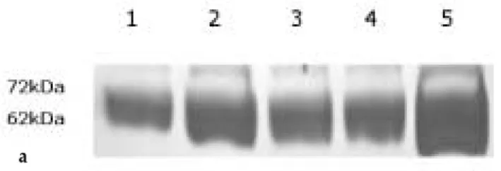
The latent and activated forms of MMP-2 were observed at positions of 72kDa and 62 kDa respectively.

As shown in Fig. 1, the gelatinolytic activities in the can- cer cell lines were higher than in normal mucosal gingi- val fibroblasts, the latter producing only a vague band on the zymogram gel plate. Significant differences were also observed in the gelatinolytic activities among the different tumor cell lines. The total gelatinolytic activity of HT1080 was 155.6±9.9U, the highest among the group. Those of HSC-3 and MCF-7 were 103.1±4.9U and 99.7±7.0U, respectively, and belonged to a homoge- neous subset. The next homogeneous subset of cancer cell lines included KB, MDA-MB-231, and A253. These lines showed the lowest gelatinolytic activity (84.9±

3.3U, 72.9±10.0U, 65.4±5.5U, respectively). By western blot, survivin expressions were detected in all tumor cell lines and normal gingival fibroblast cells. Although, like its gelatinolytic activity, HT1080 survivin expression was higher than any other cell lines; the differences of sur- vivin expressions did not vary much among other cell lines. In statistical analysis using correlation testing with Spearman’s rho (0.6), there was no significant correla- tion between gelatinolytic activity and survivin expres- sion (p=0.208). Survivin expressions were variable in repeated experiments, and these phenomena were reflected in the expression data and by the high standard deviation (SD) (Fig. 1-c).

2. Clinicopathological profile of patient and immuno- histochemical localization of survivin in oral squamous cell carcinomas

Patients’ages ranged from 40 to 75 with a median val-

ue of 58.5 years. Twenty-five patients (65.8%) were male

and fourteen (34.2%) female. Histopathological examina-

tion of lymph nodes demonstrated metastasis in 21 of 38

patients who had undergone curative surgical neck dis-

(5)

section (Table 1). In this study, most cases were well dif- ferentiated. There were 30 (78.9%) cases of well differen- tiation, 5 (13.2%) of moderate differentiation, and 3 (7.9%) of poor differentiation. The moderately and poor- ly differentiated groups were therefore combined into a group for statistical analyses. Cancer stage based on TNM was evaluated and the numbers of stage I, stage II, stage III, and stage IV cases were 6, 5, 10, and 17, respec- tively. There were no cases showing distant metastasis.

Proliferating indexes were obtained and cutoff points were determined at 50% and 25% stained percentage and at each cutoff point were determined two groups: a high- ly proliferating group and a lowly proliferating group.

At 50% cutoff point, 21 cases belonged to the lowly proliferating group and 17 cases to the highly proliferat- ing group. At 25% cutoff point, 10 cases belonged to the lowly proliferating group and 28 cases to the highly pro-

liferating group. In the 38 OSCC biopsy specimens, immunohistochemical analysis revealed that survivin was present in the nuclei and cytoplasms of tumor cells.

Survivin stained had a granular appearance in the cyto- plasm of cancer cells (Fig. 2). Eight tumors (21.0%) did not express survivin above the cutoff value of 20% and 30 tumors (79%) showed survivin expression above the cutoff value. The distribution of survivin in tumors was as follows: 0% grade 0, 21.0 % grade 1; 39.5% grade 2;

7.9% grade 3; and 31.6% grade 4.

3. Relationship between expression of survivin and histological lymph node metastasis (Table 2)

Twenty cases of 21 OSCCs (95.2%) with lymph node metastasis had high levels of survivin expression. On the other hand, only 10 cases of 17 OSCCs (58.8%) without Fig. 1-a. Gelatinolytic activity

lane 1: positive control (HT1080), lane 2: MCF7, lane 3: HSC3, lane 4:KB, lane 5: negative control (normal oral mucosal fibrob- last)

Fig. 1-b. Western blotting

Western blotting for survivin in cultured cell lines. Two parallel gel images at different experimental timing were shown. As shown, there were many differences in survivin expression at each experiment. In all cases, lysates were normalized for total protein content before SDS-PAGE/immunoblot assay. Single bands of various intensities around 16 kDa indicated the expression of sur- vivin at various levels.

lane 1: HT1080, lane 2: MCF7, lane 3: HSC3, lane 4:KB, lane 5: nor- mal oral mucosal fibroblast, lane 6: A253 lane, 7: MDA-MB-231

Fig. 1-c. The relative levels of expression of survivin and gelatinolytic activity in each cell line.

No correlation was shown between survivin and gelatinolytic activity.

(Spearman’s rho=0.6, P>0.05).

a

c

b

Gelatinolytic activity

Survivin expression

(6)

lymph node metastasis had high levels of Survivin expression. In contrast, only 1 case of 21 OSCCs (4.8%) with lymph node metastasis had low levels of Survivin expression whereas 7 cases of 17 OSCCs (41.2%) without

lymph node metastasis had low levels of survivin.

Statistical analysis using the Chi-square test revealed a significant correlation between survivin expression and lymph node metastasis (P = 0.006).

Table 1. Clinicopathological profiles and immunohistochemical expression of survivin in 38 OSCCs case age sex proliferation stage differentiation lymph node Micro vessel survivin im

25% 50% metastasis density munoreactivity

1 40 M H H 4 W + 72 2

2 41 M H L 3 W + 65 2

3 41 F H L 4 W + 47 4

4 41 M H H 4 W + 26 4

5 43 F L L 2 W - 23 1

6 46 F H H 3 W - 27 1

7 47 F H L 4 P + 41 4

8 48 F H H 1 W - 52 2

9 49 M H H 3 W + 64 3

10 49 f H H 4 W + 71 4

11 50 M L L 3 W + 68 2

12 51 F L L 4 W - 62 1

13 52 F H H 3 W + 59 2

14 53 M L L 2 M - 50 4

15 53 M H H 1 W - 82 4

16 55 F H H 4 M - 26 1

17 55 M H L 3 W + 68 4

18 58 M H L 1 W - 128 2

19 58 M H H 2 W - 33 2

20 59 M L L 4 W + 51 1

21 59 M H L 3 W + 12 2

22 60 M H H 1 W - 27 2

23 60 M H L 4 P + 23 3

24 62 F L L 4 W + 68 4

25 62 M H H 4 W + 68 4

26 64 M L L 4 W - 45 1

27 65 M L L 4 W - 25 1

28 65 M H L 3 W + 36 4

29 66 M H L 1 W - 50 2

30 66 F H H 3 M + 29 2

31 66 F H L 3 W + 28 4

32 67 F H H 2 W - 26 2

33 67 M H H 4 P + 50 4

34 68 M H H 2 M - 40 2

35 68 M L L 4 W + 69 2

36 71 M H L 1 W - 68 2

37 72 F H H 4 M + 68 3

38 75 M L L 4 W - 68 1

W: well differentiated, M: moderately differentiated, P: poorly differentiated

H: highly proliferating or high micro vessel density, L: lowly proliferating or low micro vessel density

(7)

Table 4. Correlation between survivin expression and cancer stage

Immunoreactivity Number of Cases P value

of survivin Stage 1 Stage 2 Stage 3 Stage 4 (χ

2

)

High 6 (100) 4 (80.0) 10 (100) 11 (64.7)

(grade 2 or 3 or 4)

P=0.091>0.05

Low 0 (0) 1 (20.0) 0 (0) 6 (35.3)

(grade 0 or 1)

total 6 (100) 5 (100) 10 (100) 17 (100)

Table 2. Correlation between survivin expression and lymph node metastasis

Immunoreactivity Number of Cases

P value

of survivin Without lymph node With lymph node

2

) involvement (%) involvement (%)

High

(grade 2 or 3 or 4) 10 (58.8) 20 (95.2)

P=0.006<0.05 Low

(grade 0 or 1) 7 (41.2) 1 (4.8)

Total 17 (100) 21 (100)

Table 3. Correlation between survivin expression and histological differentiation

Immunoreactivity Number of Cases P value

of survivin Well (%) Moderate or Poor (%) (χ

2

)

High

(grade 2 or 3 or 4) 23 (76.7) 7 (87.5)

P=0.504>0.05 Low

(grade 0 or 1) 7 (23.3) 1 (12.5)

Total 30 (100) 8 (100)

a. Infiltrating tumor cells showing distinct nuclear pos- itivity with polyclonal antibody to survivin Original magnification ×100.

b. Representative immunohistochemical data showing nuclear staining of survivin in OSCC. Note both nuclear and cytoplasmic staining. Original magnification ×200.

Fig. 2. Immunohistochemical expression of survivin.

a b

(8)

4. Relationship between expression of survivin and histological differentiation, microvessel density and cancer stage (Table 3, 4, Fig. 3)

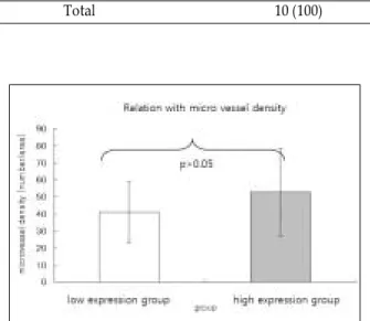
As shown in Tables 3, 4 and Fig. 3, we found no statisti- cally significant results between the expressions of sur- vivin and histological differentiation, micro vessel densi- ty, or cancer stage (Table 3, 4. P = 0.504, and 0.091, respectively, Fig. 3. P=0.6).

5. Relationship between expression of survivin and proliferation of OSCCs (Table 5)

Using the 50% cutoff point for the PCNA staining index, there was no significant correlation between sur- vivin expressions and proliferation of OSCCs (P = 0.241).

However, survivin expression was significantly related with the subgroup of proliferation at 25% cutoff point.

Statistical analysis using the Chi-square test revealed a significant correlation between survivin expression and the proliferation (P = 0.000<0.05). 26 cases of 28 OSCCs

(92.5%) with high proliferating index had high levels of survivin. In contrast, only 4 cases of 10 OSCCs (40.0%) with low proliferating index had high levels of survivin.

Table 5. Correlation between survivin expression and proliferation

Immunoreactivity Number of Cases P value

of survivin Low (under 25%) (%) High (over 25%) (%) (χ

2

)

High

(grade 2 or 3 or 4) 4 (40.0) 26 (92.9)

P=0.000<0.05 Low

(grade 0 or 1) 6 (60.0) 2 (7.1)

Total 10 (100) 28(100)

Fig. 3. Relationship between survivin expression and blood vessel density.

No relationship between survivin expression and blood vessel density (P>0.05)

Fig. 5. Effect of survivin expression on overall survival in OSCCs.

Kaplan-Meier overall survival curves for OSCC pa- tients stratified according to survivin levels (dotted line: low survivin expression group, solid line: high survivin expression group)

Fig. 4. Kaplan-Meier estimation of overall survival.

As marked in the graph, 3-year and 5-year survivals are

estimated to 68.7% and 21.5% respectively.

(9)

6. Effect of survivin expression on patient survival (Fig. 4, 5)

Period of survival varied from 39 to 76 months with its median of 47.06 months. The overall 3-year survival rate was 68.0% and 5-year survival rate, 21.5% (Fig. 4).

Univariate analysis demonstrated that survivin overex- pression had a significant negative effect on survival rate. The Kaplan-Meier survival curve showed signifi- cant separation (Fig. 5). Log rank tests showed signifi- cant survival difference between the high survivin expression group and the low survivin expression group (P = 0.0441).

DISCUSSION

Many clinical risk factors of OSCC have been suggest- ed as useful prognostic factors. Unfortunately, the prog- nostic value of these risk factors has been called into question by several clinicians and researchers.

Identification of better diagnostic factors is necessary to assist the clinicians in making more accurate lesional staging and to aid in predicting prognosis. Improved prognostication would be clinically valuable, particularly where initial therapy might be tailored to tumor aggres- siveness because advanced surgical reconstruction could be applied in particularly difficult cases.

The inhibitor of apoptosis family (IAPs) of proteins may have a greater potential to inhibit apoptosis than any other family of apoptotic inhibitors, including the bcl-2 family

6)

. Humans possess eight IAP family mem- bers: NAIP, cIAP1, cIAP2, XIAP, Ts-XIAP, ML-IAP, Apollon, and survivin

4)

.

Survivin was recently identified as a tumor marker that regulates cell division and inhibits apoptosis

24)

. Additionally, survivin has been shown to directly and indirectly inhibit activated caspases

6,9)

.

Among the IAP family members, survivin has several peculiar characteristics. First, the structure of survivin is unique in that it contains a single baculovirus IAP repeat and no zinc-binding domain (RING finger). Second, sur- vivin is expressed selectively in common human cancers.

Additionally, while fetal tissues express survivin, normal adult tissues do not express survivin except in thymus and placenta

25)

. Thus, it has been postulated that survivin is one of the most tumor-specific gene products. In fact, several studies have demonstrated that survivin is over- expressed in many types of human cancer

13)

. Also, previ-

ous articles even reported that survivin expression was a molecular prognostic marker predicting survival

10-12,22)

and lymph node metastasis

26)

. But there were reports with other opinion that survivin expression cannot be considered a prognostic indicator of disease outcome

28)

.

A lack of consensus exists regarding the importance of the intracellular location of survivin expression in com- mon epithelial tumors. Previous studies analyzing sur- vivin expression by immunohistochemistry have shown a predominantly cytoplasmic distribution

25,29)

. However, nuclear accumulation of survivin has been recently described in other human tumors

19,30)

. The reason and meaning for these differences in cell distribution between tumors are not clear. It has been proposed that the subcellular distribution of survivin is regulated by active import into the nucleus and by CRM1-mediated export to the cytoplasm, suggesting that survivin may be considered a nuclear shuttling protein. Therefore, the almost exclusively cytoplasmic localization in a high number of tumors may be the result of a higher rate of nuclear export

31)

. Most of the previous studies analyzing the prognostic significance of survivin expression were performed without accounting for the subcellular loca- tion of the protein. In the present study, meaningful dif- ferences of nuclear and cytoplasmic stainging could not be found in most specimens.

The first aim of our study was to investigate the expres- sion of survivin protein in OSCC by immunohistochem- istry and to investigate its association with the histologi- cal parameters and proliferative activity of the tumors as well as with the survival rates of patients. Survivin pro- tein was detected with a variable number of positive cells ranging from 5% to 95%, and survivin was detected in both nuclear and cytoplasmic fraction of tumor cells.

In five normal gingival mucosal tissues, survivin expres- sions were sparse or not observed. These results suggest that survivin is commonly expressed in OSCC and that immunohistochemically verified expression of survivin could be an important cancer marker for OSCC.

Survivin was originally reported to be present during

fetal life but undetectable in differentiated adult tissues

5)

.

Subsequent studies have shown that survivin is

expressed in the normal endometrium, a highly prolifer-

ating tissue

32)

. These results revealed the close correlation

with proliferation and differentiation. Expression of the

gene occurs in the G2/M phase in a strict cell cycle-

dependent manner

33)

, thus potentially explaining prefer-

ential expressions of survivin in poorly differentiated

(10)

and metastatic OSCC cases which likely to exhibit high proliferative potential. It has recently been suggested that the protective anti-apoptotic effect of survivin in proliferating malignant cells might be a mechanism to stabilize tumor cells with chromosomal abnormalities, favoring the survival of these cells and the progression of the tumors

34)

. In the present study, survivin was mostly expressed in the proliferating basal cell layer of tumor mass. In the normal squamous cell epithelium of the gin- gival mucosa survivin was also mainly localized in the basal layer. The restriction of survivin expression to the basal layer of the normal squamous cell epithelium sug- gests that survivin may be related to cell proliferation also in OSCC. Statistical analysis revealed that survivin expression significantly correlated with the proliferating index. Survivin has been implicated in a dual role in con- nection of suppression of apoptosis to regulation of chro- mosomal segregation and cell division

35)

. Targeting experiments using antisense RNAs against survivin or dominant-negative mutants resulted in spontaneous apoptosis, increased caspase activity, and inhibition of cell proliferation

36)

. A role for survivin in blocking apop- tosis has also been demonstrated in vivo

37)

. However, the mechanisms of antiapoptotic effect by survivin are not completely clear.

By contrast, we observed no correlation between sur- vivin expression and differentiation in the present study.

Survivin is the only member of the IAP family for which a function in the nucleus has been identified 38.

Therefore, it might be assumed that dedifferentiation of normal epithelium as carcinogenesis and tumor progres- sion cause re-expression of survivin

39)

. Unfortunately, in the case of OSCC, correlation between these events can- not be confirmed because of the naturally heterogeneous biological characteristics of OSCC and because of the dif- ficulty of determining differentiation states.

Invasion into adjacent structure via basement mem- brane destruction and subsequent lymph node metasta- sis contributes to poor OSCC prognosis

40)

. The relation- ship between survivin expression and metastasis may also arise from an essential function of survivin: when survivin inhibits apoptosis, the proportion of cancerous cells in a tissue increases with continued growth.

Previous studies have indicated that when abnormal cells that would otherwise be removed by apoptosis con- tinue to grow, their potential for invasion and metastasis increases

41,42)

. The present study revealed a significant correlation between lymph node metastasis, a condition

related to the invasion ability of OSCC, and survivin expression. Therefore, it seems likely that increased lymph node metastasis in OSCC with high survivin lev- els might stem from increase in invasiveness.

Based on the results of the proliferation and lymph node metastasis, we postulated that clinical cancer stages based on TNM staging system might be closely related with survivin expression because TNM system reflects tumor growth through proliferation (T-score) and lymph node metastasis (N-score). The advanced stage of disease in OSCC may reflect the rapid growth and aggressive- ness of the tumor itself rather than a late diagnosis of a slowly growing tumor. However, there was no signifi- cant correlation between stage and survivin expression.

This suggests that clinical TNM staging system be severely limited because it might only crudely reflect the severity or the clinical aspects of OSCC. For example, tumor size (T-stage) and lymph node metastasis (N- stage) are changed with time, although survivin expres- sion could not be thought determined much by time fac- tor. Another example is small sized tumor with bony erosion. It should be categorized into stage 4 in cTNM system regardless of its size.

To identify the primary protein responsible for tumor invasiveness, we used an in vitro model involving cell lines and zymography. The target was MMP-2; an enzyme that is important for extracellular matrix turnover and that has recently been implicated in inva- sion

26)

. The assumption was that the upregulated inva- siveness of OSCC cells by MMP-2 might be closely linked to survivin expression.

The molecular interaction between survivin and MMPs is still unclear. However, recent studies have demon- strated that highly metastatic cancers exhibit a higher resistance to apoptotic cell death compared with low metastatic forms

43,44)

, and that survivin expression is closely associated with the invasive phenotype of esophageal

45)

and ovarian

46)

cancers. Yoshida et al.

46reported that the expression of survivin enhanced

invasive activity of ovarian cancer cells by up-regulation

of MMP-2. Interestingly, there was a no close correlation

between survivin and MMP-2 activity in the tumor cell

lines examined. These results suggest that overexpres-

sion of both survivin and MMP-2 in the tumor cell lines

could not be directly correlated, and invasive OSCC cells

might escape apoptosis not by expressing a higher level

of survivin. Additional studies are needed to clarify the

molecular events that co-regulate the expression of sur-

(11)

vivin and the MMP genes in OSCC.

In the present study, we investigated the relation between micro vessel density and survivin expression.

Angiogenesis is essential for cancerous tissues to receive adequate nutrition necessary for their continuous growth. Tumors exhibiting high numbers of micro ves- sels have been shown to possess high metastatic poten-

tial

47,48)

. In one study, VEGF stimulation caused increased

expression of survivin

49)

. Conversely, survivin may pre- vent new blood vessels formed by VEGF from disap- pearing by apoptosis. Thus, it might be assumed that VEGF and survivin appear to function cooperatively to increase and maintain newly formed blood vessels. In the present study, however, no significant relation was found between survivin expression and vessel density.

Although no significant relation was detected in this study, a larger-scale study will be needed to prove that newly formed blood vessels are apparently retained, pro- viding greater blood flow to the cancerous tissue and promoting metastasis in patients with high survivin expression.

It has been reported that survivin expression might provide a strong advantage factor for tumor progression by affording protection from broad apoptosis-inducing stimuli and by maintaining proper mitotic progression of the proliferating and invasive population

26)

. However, the prognostic value of survivin expression in OSCC remains a controversial issue. Our study suggests that survivin levels could have prognostic value for the pro- gression of OSCC. OSCC patients with high survivin expression had a shorter overall survival. In this regard, it is of note that survivin overexpression in other human cancers has been associated with unfavorable prognostic features, poor response to therapy, high relapse rate, and shortened survival

12,15,35)

. Furthermore, survivin has been considered to be an independent prognostic factor in some cancers

11)

.

Although present results support survivin expression in tumor as a powerful prognostic factor for the progres- sion of OSCC and patient survival, the present study has some limitations. First, because this study included only 38 patients, larger prospective studies will be required to confirm the role of survivin expression in OSCCs.

Second, studies of additional molecular markers, includ- ing bcl-2 proteins and other IAP family members, will assist in a better understanding of OSCC. Future studies should also investigate tumor response to treatments such as radiotherapy and chemotherapy. Conclusively,

the results from this study indicate that survivin is com- monly expressed in OSCC and that its expression levels are strongly associated with tumor proliferation, lymph node metastasis, and patient survival rate. These results suggest a potential role in cell cycle regulation and tumor progression. Survivin may provide prognostic information and have great potential as a therapeutic tar- get in OSCC.

REFERENCES

1. Pisani P, Parkin DM, Bray F, Ferlay J: Estimates of the worldwide mortality from 25 cancers in 1990. Int J Cancer 1999;83:18-29.

2. Schliephake H: Prognostic relevance of molecular markers of oral cancer - a review. Int J Oral Maxillofac Surg 2003;32:233-45.

3. Friedlander PL: The use of genetic markers in the clinical care of patients with head and neck cancer. Arch Otolaryngol Head Neck Surg 2003;129:363-6.

4. Reed JC: The survivin saga goes in vivo. J Clin Invest 2001;108:965-9.

5. Ambrosini G, Adida C, Sirugo G, Altieri DC: Induction of apoptosis and inhibition of cell proliferation by survivin gene targeting. J Biol Chem 1998; 273: 461-6.

6. LaCasse E, Baird S, Korneluk RG, MacKenzie AE: The in- hibitor of apoptosis (IAPs) and their emerging role in can- cer. Oncogene 1998;17:3247-59.

7. Deveraux QK, Reed JC: IAP family proteins - suppressors of apoptosis. Genes Dev 1999;13:239-52.

8. Shin S, Sung BJ, Cho YS, Kim HJ, Ha NC, Hwang JI, Chung CW, Jung YK, Oh BH: An anti-apoptotic protein human survivin is a direct inhibitor of caspase-3 and ?7.

Biochemistry 2001; 40:1117-23.

9. O’Connor DS, Grossman D, Plescia J, Li F, Zhang H, Villa A, Tognin S, Marchisio PC, Altieri DC: Regulation of apop- tosis at cell division by p34cdc2 phosphorylation of surviv- ing. Proc Natl Acad Sci USA, 2000;97:13103-7.

10. Swana HS, Grossman D, Anthony JN, Weiss RM, Altieri DC: Tumor content of the antiapoptosis molecule survivin and recurrence of bladder cancer. N Engl J Med 1999;341:452-3.

11. Adida C, Haioun C, Gaulard P, Lepage E, Morel P, Briere J, Dombret H, Reyes F, Diebold J, Gisselbrecht C, Salles G, Altieri DC, Molina TJ: Prognostic significance of survivin expression in diffuse large B-cell lymphomas. Blood 2000;96:1921-5.

12. Kawasaki H, Altieri DC, Lu CD, Toyoda M, Tenjo T, Tanigawa N: Inhibition of apoptosis by survivin predicts shorter survival rates in colorectal cancer. Cancer Res 1998;58:5071-4.

13. Ito T, Shiraki K, Sugimoto K, Yamanaka T, Fujikawa K, Ito M: Survivin promotes cell proliferation in human hepato- cellular carcinoma. Hepatology 2000;31:1080-5.

14. Nakagawara A: Molecular basis of spontaneous regression of neuroblastoma: role of neurotrophic signals and genetic abnormalities. Hum Cell 1998;11:115-24.

15. Monzo M, Rosell R, Felip E, Astudillo J, Sanchez JJ, Maestre J, Martin C, Font A, Barnadas A, Abad A: A novel anti- apoptosis gene: reexpression of survivin messenger RNA as a prognosis marker in nonsmall-cell lung cancers. J Clin Oncol 1999;17:2100-4.

16. Satoh K, Kaneko K, Hirota M, Masamune A, Satoh A,

Shimosegawa T: Expression of survivin is correlated with

(12)

cancer cell apoptosis and is involved in the development of human pancreatic duct cell tumors, Cancer 2001;92:271-8.

17. Xing N, Qian J, Bostwick D, Bergstralh E, Young CY:

Neuroendocrine cells in human prostate over-express the anti-apoptosis protein surviving. Prostate 2001;48:7-15.

18. Takamizawa S, Scott D, Wen J, Grundy P, Bishop W, Kimura K, Sandler A: The survivin : fas ratio in pediatric renal tumors J Pediatr Surg 2001;36:37-42.

19. Okada E, Murai Y, Matsui K, Isizawa S, Cheng C, Masuda M, Tacano Y: Survivin expression in tumor cell nuclei is predictive of a favorable prognosis in gastric cancer pa- tients. Cancer Lett 2001;163:109-16.

20. Noguchi N, Kawashiri K, Tanaka A, Kato K, Nakaya H:

Effects of fibroblast growth inhibitor on proliferation and metastasis of oral squamous cell carcinoma. Oral Oncol 2003;39:240-7.

21. Bosari S: Microvessel quantitation and prognosis in inva- sive breast carcinoma. Hum Pathol 1992;23:755-61.

22. Kennedy SM, O’Driscoll L, Purcell R, Fitz-Simons N, McDermott EW, Hill AD: Prognostic importance of sur- vivin in breast cancer. Br J Cancer 2003;88:1077-83.

23. Hong SD, Hong SP, Lee JI, Lim CY: Expression of matrix metalloproteinase-2 and -9 in oral squamous cell carcino- mas with regard to the metastatic potential. Oral Oncol 2000;36:207-13.

24. Myoung H, Hong SP, Lee JI, Lim CY: Anti-cancer effect of genistein in oral squamous cell carcinoma with respect to angiogenesis and in vitro invasion. Cancer Sci 2003;94:215- 20.

25. Ambrosini G, Adida C, Altieri DC: A novel anti-apoptosis gene, survivin, expressed in cancer and lymphoma. Nat Med 1997;3:917-21.

26. Muzio LL, Staibano S, Pannone G, Mignogna MD, Mariggio A, Salvatore G, Chieffi P, Tramontano D, Rosa GD, Altieri DC: Expression of the Apoptosis Inhibitor sur- vivin in Aggressive Squamous Cell Carcinoma.

Experimental and Molecular Pathology 2001;70:249-54.

27. Miyachi K, Sasaki K, Onodera S, Taguchi T, Nagamachi M, Kaneko H, Sunagawa M: Correlation between survivin mRNA expression and lymph node metastasis in gastric cancer. Gastric Cancer 2003;6:217-24.

28. O’Driscoll L, Linehan R, Kennedy M, Cronin D, Purcell R, Glynn S: Lack of prognostic significance of survivin, sur- vivin-deltaEx3, survivin-2B, galectin-3, bag-1, bax-alpha and MRP-1 mRNAs in breast cancer. Cancer Lett 2003;201:225-36.

29. Adida C, Recher C, Raffoux E, Daniel MT, Taksin AL, Rousselot P, Sigaux F, Degos L, Altieri DC, Dombret H:

Expression and prognostic significance of survivin in de novo acute myeloid leukaemia. Br J Haematol 2000;111:196-203.

30. Frost M, Jarboe EA, Orlicky D, Gianani R, Thompson LC, Enomoto T, Shroyer TS: Immunohistochemical localization of survivin in benign cervical mucosa, cervical dysplasia, and invasive squamous cell carcinoma. Am J Clin Pathol 2002;117:738-44.

31. Rodriguez JA, Span SW, Ferreira CG, Kruyt FA, Giaccone G: CRM1-mediated nuclear export determines the cyto- plasmic localization of the antiapoptotic protein surviving.

Exp Cell Res 2002; 275:44-53.

32. Konno R, Yamakawa H, Utsunomiya H, Ito K, Sato S, Yajima A: Expression of survivin and Bcl-2 in the normal human endometrium. Mol Hum Report 2000;6:529-34.

33. Li F, Ambrosini G, Chu EY, Plescia J, Tognin S, Marchisio PC, Altieri DC: Anti-apoptosis gene, survivin, and progno-

sis, Control of apoptosis and mitotic spindle checkpoint by surviving. Nature 1998;396:580-4.

34. Uren AG, Wong L, Pakusch M, Fowler KJ, Burrows FJ, Vaux DL, Choo KH: Survivin and the inner centromere protein INCENP show similar cell-cycle localization and gene knockout phenotype. Curr Biol 2000;10:1319-28.

35. Altieri DC: The molecular basis and potential role of sur- vivin in cancer diagnosis and therapy. Trends Mol Med 2001;7:542-7.

36. Hedley DW, Friedlander ML, Taylor IW, Rugg CA, Musgrove EA: Method for analysis of cellular DNA content of paraffin-embedded pathological material using flowcy- tometry. J Histochem Cytochem 1983;31:1333-5.

37. Grossman D, Kim PJ, Schechner JS, Altieri DC: Inhibition of melanoma tumor growth in vivo by survivin targeting.

Proc Natl Acad Sci USA 2001;98:635-40.

38. Suzuki A, Hayashida M, Ito T, Kawano H, Nakano T, Miura M, Akahane K, Shiraki K: Survivin initiates cell cycle entry by the competitive interaction with Cdk4/

p16(INK4a) and Cdk2/cyclin E complex activation.

Oncogene 2000;19:3225-34.

39. Adida C, Crotty PL, McGrath J, Berrebi D, Diebold D, Altieri DC: Developmentally regulated expression of the novel cancer anti-apoptosis gene survivin in human and mouse differentiation. Am J Pathol 1998;152:43-9.

40. Liotta LA, Rao CN, Barsky SH: Tumor invasion and the ex- tracellular matrix. Lab Invest 1983;49:636-49.

41. Wyllie AH, Bellamy COC, Bubb AR, Clarke AR, Corbet S, Curtis L: Apoptosis and carcinogenesis. Br J Cancer 1999;80:34-7.

42. Sierra A, Castellsague X, Escobedo A, Lloveras B, Ramnrez MG, Moreno A: Bcl-2 with loss of apoptosis allows accu- mulation of genetic alterations: a pathway to metastatic progression in human breast cancer. Int J Cancer 2000;89:142-7.

43. Glinsky GV, Glinsky VV: Apoptosis and metastasis: a supe- rior resistance of metastatic cancer cells to programmed cell death. Cancer Lett 1996;101:43-51.

44. Glinsky GV, Glinsky VV, Ivanova AB, Hueser CJ:

Apoptosis and metastasis: increased apoptosis resistance of metastatic cancer cells is associated with the profound defi- ciency of apoptosis execution mechanisms. Cancer Lett 1997;115:185-93.

45. Kato J, Kuwabara Y, Mitani M, Shinoda N, Sato A, Toyama T, Mitsui A, Nishiwaki T, Moriyama S, Kudo J, Fujii Y:

Expression of survivin in esophageal cancer: correlation with the prognosis and response to chemotherapy. Int J Cancer 2001;95:92-5.

46. Yoshida H, Ishiko O, Sumi T, Matsumoto Y, Ogita S:

Survivin, bcl-2 and matrix metalloproteinase-2 enhance progression of clear cell- and serous-type ovarian carcino- mas. Int J Oncol 2001;19:537-42.

47. Xiangming C, Hokita S, Natsugoe S, Tanabe G, Baba M, Takao S: Angiogenesis as an unfavorable factor related to lymph node metastasis in early gastric cancer. Ann Surg Oncol 1998;5:585-9.

48. Maeda K, Chung YS, Ogawa Y, Takatsuka S, Kang SM, Ogawa M: Prognostic value of vascular endothelial growth factor expression in gastric carcinoma. Cancer 1996; 77:858- 63.

49. Tran J, Rak J, Sheehan C, Saibil SD, LaCasse E, Korneluk

RG: Marked induction of the IAP family antiapoptotic pro-

teins survivin and XIAP by VEGF in vascular endothelial

cells. Biochem Biophys Res Commun 1999;264:781-8.

수치

Fig. 1-c. The relative levels of expression of survivin and gelatinolytic activity in each cell line
Table 1. Clinicopathological profiles and immunohistochemical expression of survivin in 38 OSCCs  case age sex proliferation stage differentiation lymph node  Micro vessel  survivin im
Table 3. Correlation between survivin expression and histological differentiation
Fig. 5. Effect of survivin expression on overall survival in OSCCs.

참조

관련 문서

Examination of such materials led to the conclusion that Pak Chi-wŏn originally intended to have the five texts including “ Yangmaesihwa” in Yŏrha ilgi

The proposal of the cell theory as the birth of contemporary cell biology Microscopic studies of plant tissues by Schleiden and of animal tissues by Microscopic studies of

The index is calculated with the latest 5-year auction data of 400 selected Classic, Modern, and Contemporary Chinese painting artists from major auction houses..

In the present paper, L-shaped curves are general- ized and it is shown that every knot in the minor subfamilies, called sporadic examples of Berge’s lens space surgery, is

Concentration-Dependent inhibition of cell viability by adenosine in human oral fibroblast and FaDu human head and neck squamous cell carcinoma · · ·

The most important factor to level-differentiated instruction was to give attraction to student and let them have motivation themselves, and after finishing

Taylor Series

Objective: The purpose of this study was to analyze recent trend in incidence of basal cell carcinoma and squamous cell carcinoma in patients from the Gwangju City