• 검색 결과가 없습니다.

A Case of Radiation Bronchitis Induced Massive Hemoptysis after High-Dose-Rate Endobronchial Brachytherapy

N/A
N/A
Protected

Academic year: 2022

Share "A Case of Radiation Bronchitis Induced Massive Hemoptysis after High-Dose-Rate Endobronchial Brachytherapy"

Copied!
6
0
0

로드 중.... (전체 텍스트 보기)

전체 글

(1)

Tuberc Respir Dis 2012;73:325-330

CopyrightⒸ2012. The Korean Academy of Tuberculosis and Respiratory Diseases. All rights reserved.

A Case of Radiation Bronchitis Induced Massive Hemoptysis after High-Dose-Rate Endobronchial Brachytherapy

Seok Jeong Lee, M.D.1, Jong-Young Lee, M.D.2, Soon Hee Jung, M.D.3, Shun Nyung Lee, M.D.1, Ji-Ho Lee, M.D.1, Chong Whan Kim, M.D.1, Saehyun Jung, M.D.1, Ye-Ryung Jung, M.D.1, Won-Yeon Lee, M.D.1 Departments of 1Internal Medicine, 2Radiation Oncology, and 3Pathology, Yonsei University Wonju College of Medicine, Wonju, Korea

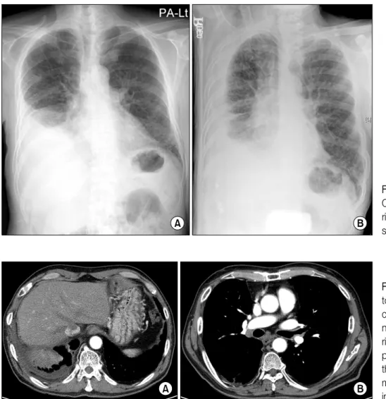
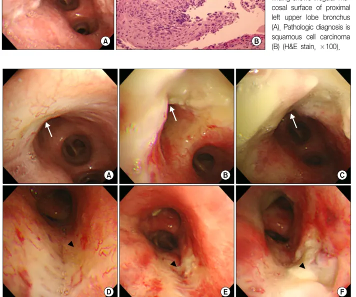
High-dose-rate endobronchial brachytherapy (HDREB) have been used as the treatment of early endobronchial cancer, as well as for palliation of advanced cancer. However, fatal hemoptysis can occur after HDREB at the rate of 7∼32%. We report a case of massive hemoptysis due to radiation bronchitis developed after HDREB. A 67-year-old man was treated with HDREB for early endobronchial cancer on the left upper lobe bronchus. He complained of persistent cough from 4 weeks after completion of HDREB. Radiation bronchitis was observed on the bronchoscopy at 34 weeks, and it was progressed from mucosal swelling and exudate formation to necrosis and ulceration without local relapse. In addition, he died of massive hemoptysis after 15 months. The patient had no sign or radiologic evidences to predict the hemoptysis. This case implies that HDREB directly contributes to an occurrence of a fatal hemoptysis, and follow-up bronchoscopy is important to predict a progression of radiation bronchitis and fatal hemoptysis.

Key Words: Radiation; Bronchitis; Hemoptysis; Brachytherapy

Address for correspondence: Won-Yeon Lee, M.D.

Department of Internal Medicine, Yonsei University Wonju College of Medicine, 162, Ilsan-dong, Wonju 220-701, Korea Phone: 82-33-741-0926, Fax: 82-33-741-0928

E-mail: [email protected] Received: Apr. 22, 2012 Revised: May 24, 2012 Accepted: Jul. 3, 2012

CCIt is identical to the Creative Commons Attribution Non-Commercial License (http://creativecommons.org/licenses/by-nc/3.0/).

Introduction

High-dose-rate endobronchial brachytherapy (HDREB) alone or with external beam radiotherapy is used in the treatment of endobronchial lung cancer for the main pur- pose of symptom relief

1,2

. In addition to, it can bring lo- cal treatment and life prolongation in inoperable cases of early endobronchial cancer

3-5

.

However, HDREB can cause massive hemoptysis in 7∼32% of patients, which is a serious complication of brachytherapy

1,6-8

. Most of investigators explained the pathogenesis of hemoptysis is due to cancer progression

or recurrence

2,7

. But, a recent study reported about complication of HDREB in localized and early endo- bronchial cancer and showed the result that fatal he- moptysis after HDREB was a complication of the treat- ment itself

5

.

According to several reports about outcome of HDREB in Korea, occurrence of fatal hemoptysis has been reported as about 2.5% to 16%

9,10

. However, in all of them, the purpose of HDREB was to relieve symp- tom in advanced lung cancer which was previously treated with surgery or external beam radiation therapy.

Therefore, we report a case of patient with massive hemoptysis who was diagnosed radiation bronchitis af- ter HDREB for localized endobronchial squamous cell carcinoma.

Case Report

A 67-year-old man visited for cough and sputum lasting

for 5 months. He was a current smoker with a history of

40 pack-year smoking and had a coronary arterial disease.

(2)

Figure 1. Chest X-ray.

Chest poteroanterior (A) and right decubitus view (B) show right pleural effusion.

Figure 2. Chest computed tomography scans. Chest computed tomography sca- ns show consolidation in right lower lobe and right pleural effusion (A) but, there is no evidence of tu- mor or endobronchial lesion in left bronchial tree (B).

Right pleural effusion was seen in a chest X-ray (Figure 1).

The character of pleural effusion was exudates with many lymphocytes and low level of adenosine deaminase. A consolidation was seen in computed to- mography (CT) after the effusion had been drained (Figure 2A) and no endobronchial lesion was found on left upper lobe in chest CT (Figure 2B). Bronchoscopy was performed to differentiate causes of the right lung lesion.

There was no endobronchial lesion in right bronchial tree.

Incidentally, mucosal nodularity was observed in the left upper lobe bronchus (Figure 3A) and was diagnosed with squamous cell carcinoma with biopsy (Figure 3B). A pul- monary wedge resection and pleural biopsy were per- formed to evaluate right lower lobe and pleural lesions through video assisted thoracoscopic surgery, and the results were chronic organizing pneumonia and sub-

pleural fibrosis with chronic inflammation. The lesions of right lower lobe and right pleura were remained without progression until the last follow-up radiologic examination. Lymph node involvement and distant metastasis were not observed in the staging work-up.

Despite the clinical stage was IA, the patient had moder- ate chronic obstructive pulmonary disease (post-broncho- dilator forced expiratory volume in 1 second, 1.74 L) and his endobronchial cancer involved the left main bron- chus. The patient might be intolerable for surgical treat- ment, such as pneumonectomy or sleeve lobectomy, and we decided to do curative local therapy.

HDREB was performed with 6 fractions of 5 Gy for

6 weeks. After the treatment, the patient complained of

persistent cough, but bronchoscopy and chest CT had

not shown the progression of lung cancer until 4 weeks

(3)

Figure 3. Bronchoscopic and pathologic findings at initial visit. Bronchoscopic finding shows irregular mu- cosal surface of proximal left upper lobe bronchus (A). Pathologic diagnosis is squamous cell carcinoma (B) (H&E stain, ×100).

Figure 4. Bronchoscopic findings following high-dose-rate endobronchial brachytherapy. Radiation bronchitis was de- tected in left upper lobe bronchus at 34 weeks (A, arrow) and in left mainstem bronchus at 50 weeks after last brachyther- apy (D, arrowhead). Bronchoscopic findings show mucosal edema and fibrosis. The bronchitis progressed to necrosis and ulceration at 54 weeks after brachytherapy (B, E). The necrosis and ulceration became larger and deeper despite a treatment of oral corticosteroids (C, F).

after HDREB. Further follow-up bronchoscopies were performed at 34, 50, 54, and 59 weeks after the HDREB.

Grade 1 radiation bronchitis was observed in the left upper lobe bronchus which was previous cancer lesion

at 34 weeks (Figure 4A) and was also observed in the

medial wall of the left mainstem bronchus which was

not cancer lesion but contact area of guide sheath for

radiation source at 50 weeks after HDREB (Figure 4D).

(4)

On the bronchoscopic finding at 54 weeks (12 months), mucosal necrosis of the left upper lobe bronchus was advanced (Figure 4B) and ulcer lesions began to appear in the left main bronchus (Figure 4E). The patient start- ed taking oral steroids at 55 weeks, but the area of the left upper bronchial lesions extended (Figure 4C) and left main bronchus ulcers also increased in the depth and extent at 59 weeks (14 months) (Figure 4F). Bron- chial biopsies for the bronchitis were performed four times among five follow-up bronchoscopic examina- tions, and it showed acute or chronic inflammation and no cancer cells were observed. The patient died sud- denly due to massive hemoptysis at 15 months after HDREB.

Discussion

HDREB have been undergone for the purpose of cu- rative treatment for early lung cancer as well as the re- lief of symptoms of advanced lung cancer, which is ob- taining a positive result and is becoming increasingly performed. When HDREB is applied to patients with lo- calized endobronchial cancer, it is usually performed using a protocol of 6 fractions of 5 Gy at intervals of one week. The two-year locoregional control rate is 60% to 85% and two-year overall survival rate is 47%

to 78% in localized early lung cancer

3-5

. However, this treatment approach can lead to massive hemoptysis, which leads to high mortality

1,6-8

.

Speiser and Spratling

11

categorized bronchial inflam- mation and stenosis that occurs after HDREB into four grades and defined them radiation bronchitis. According to their classification, Grade 1 consists of a slight in- flammation with swelling and thin whitish membrane.

Grade 2 consists of an increase in white fibrous mem- brane with great exudation. Grade 3 consists of a severe inflammatory response with a marked membranous exudate. The final progression, Grade 4 bronchitis is greater fibrosis with resulting circumferential stenosis.

The mean time to developing Grade I and IV lesions were 16 weeks and 55 weeks. Taulelle et al.

12

divided radiation bronchitis into 5 grades. Grade 5 consists of

bronchial wall necrosis which cause fistula formation.

They explained that late fistula formation is the ultimate phase of radiation bronchitis, while early fistulae are re- sults of massive tumor shrinkage. Interestingly there were two separated ulcerations in this case. One was at the targeted cancer lesion of left upper lobe, and the other was developed on the medial wall of left main- stem bronchus. We supposed the ulceration of left mainstem bronchus was developed by radiation effect during in and out of radiation source on the area that contacted guide sheath for radiation source. On the bronchoscopic findings of this case swelling and exu- dates as the Grade 1 to 3 of radiation bronchitis were the early changes, and then it progressed to necrosis and ulcer about one year after brachytherapy. We have assumed that fistula developed after last bronchoscopy and was followed by massive hemoptysis.

The mortality due to hemoptysis after HDREB for treatment of early endobronchial cancer is 0% to 4.4%

3-5

. Even if it is less frequent than in case of pallia- tive HDREB, predicting occurrence of hemoptysis, pre- venting and treatment of hemoptysis are crucial.

The risk factors of massive hemoptysis after HDREB

are radiation dose

2,8

, laser treatment underwent in the

same region

6,8

, repeated brachytherapy in the same re-

gion

2,8

, treatment for palliative aim

2,7

, endobronchial tu-

mor size

7

, developing radiation bronchitis

11

, local treat-

ment failure and contact of brachytherapy applicator

with the bronchial wall that close to major blood ves-

sels

6

. Hennequin et al.

7

reported a patient who died

from massive hemoptysis after HDREB for lung cancer

in right upper lobe bronchus and suggested a relation-

ship between the location of the cancer and occurrence

of massive hemoptysis. Hara et al.

6

reported that mas-

sive hemoptysis occurs frequently in patients whose

used applicator touched directly bronchial wall facing

the major blood vessels in CT. In the post-mortem ex-

amination in 2 patients, fistula formations were identi-

fied between superior vena cava and trachea in one

case and between left pulmonary artery and left main

bronchus in another case. But, in this study, all patients

with massive hemoptysis failed to control cancer by lo-

(5)

cal treatment, so it is unclear which is more involved to occurrence of massive hemoptysis either local failure or relationship with major blood vessels. In our case, no treatment but HDREB was performed for cancer treatment and there was no recurrence after macro- scopic and microscopic remission. According to the pre- vious reports, only risk factor expected in this patient was the location of the cancer lesion including some of the left upper lobe bronchus that was close to left pulmonary artery, the one of the major blood vessels.

And it suggests the possibility of fistula formation be- tween the bronchial lumen and left pulmonary artery due to radiation bronchitis.

Hemoptysis can be developed by other treatment mo- dalities for inoperable early endobronchial cancer. Inci- dences of hemoptysis are 7.8% in photodynamic ther- apy

13

, and 4% in cryotherapy

14

. But, there is no inves- tigation about direct comparison of the treatment mod- alities.

Since HDREB-treated patients experiencing massive hemoptysis cannot usually be managed by any special methods and most of them cannot survive, which is the fatal weakness of HDREB. In our case, despite the pa- tient has taken steroids, prescribed frequently in radia- tion pneumonitis, depth and width of necrosis and ul- ceration had been worse in follow-up bronchoscopy.

Matsumoto et al.

15

proposed the surgery as a way to treat ongoing bronchial necrosis. They performed omentum wrapping around left main bronchus via lapa- roscopic and open thoracotomy for a treatment of pro- gressive ulcer. And the bronchial ulcers became a little worse after the surgery, it has shown an improvement with normal mucosa. In addition, they mentioned that bronchial artery embolization for endobronchial ne- crosis and hemoptysis is not effective because they failed to treat hemoptysis occurred after HDREB and ex- perienced the patient’s death. In our case, bronchial le- sions progressed from small range of radiation bronchi- tis to deep necrosis which caused massive hemoptysis without microscopic recurrence of cancer. That suggests massive hemoptysis could be occurred after HDREB as a direct complication of the treatment regardless of local

control. The only symptom that we predict endobron- chial change was persistent cough. But there was no symptom that can predict massive hemoptysis. In addi- tion, radiologic imaging of the treated area remained unchanged. Eventually, follow-up bronchoscopy is the only diagnostic modality to predict fatal hemoptysis es- pecially in patients with upper lobe bronchus lesions which show proximity to the major blood vessels. The median time of the onset of massive hemoptysis after HDREB is approximately 3∼12 months

1,2,4,5,7,8,10

. During the periods, patients should be monitored stringently.

Once radiation bronchitis is observed, physicians should keep in mind the lesion may progress to ulceration and massive hemoptysis may occur. However, specific treat- ment for radiation bronchitis and HDREB related he- moptysis has not been established, the more inves- tigation for development of medical or surgical treatment will be needed.

References

1. Bedwinek J, Petty A, Bruton C, Sofield J, Lee L. The use of high dose rate endobronchial brachytherapy to palliate symptomatic endobronchial recurrence of pre- viously irradiated bronchogenic carcinoma. Int J Radiat Oncol Biol Phys 1992;22:23-30.

2. Ozkok S, Karakoyun-Celik O, Goksel T, Mogulkoc N, Yalman D, Gok G, et al. High dose rate endobronchial brachytherapy in the management of lung cancer: re- sponse and toxicity evaluation in 158 patients. Lung Cancer 2008;62:326-33.

3. Marsiglia H, Baldeyrou P, Lartigau E, Briot E, Haie- Meder C, Le Chevalier T, et al. High-dose-rate brachy- therapy as sole modality for early-stage endobronchial carcinoma. Int J Radiat Oncol Biol Phys 2000;47:665-72.

4. Hennequin C, Bleichner O, Trédaniel J, Quero L, Sergent G, Zalcman G, et al. Long-term results of endo- bronchial brachytherapy: a curative treatment? Int J Radiat Oncol Biol Phys 2007;67:425-30.

5. Aumont-le Guilcher M, Prevost B, Sunyach MP, Peiffert D, Maingon P, Thomas L, et al. High-dose-rate brachy- therapy for non-small-cell lung carcinoma: a retro- spective study of 226 patients. Int J Radiat Oncol Biol Phys 2011;79:1112-6.

6. Hara R, Itami J, Aruga T, Kozuka T, Nakajima K,

Yamashita H, et al. Risk factors for massive hemoptysis

(6)

after endobronchial brachytherapy in patients with tra- cheobronchial malignancies. Cancer 2001;92:2623-7.

7. Hennequin C, Tredaniel J, Chevret S, Durdux C, Dray M, Manoux D, et al. Predictive factors for late toxicity after endobronchial brachytherapy: a multivariate ana- lysis. Int J Radiat Oncol Biol Phys 1998;42:21-7.

8. Gollins SW, Ryder WD, Burt PA, Barber PV, Stout R.

Massive haemoptysis death and other morbidity asso- ciated with high dose rate intraluminal radiotherapy for carcinoma of the bronchus. Radiother Oncol 1996;39:

105-16.

9. Chung MP, Kwon OJ, Choi DR, Huh SJ, Lim DH, Kim MK, et al. Short-term results of endobronchial brachy- therapy for malignant airway obstructions. J Korean Soc Ther Radiol 1996;14:299-306.

10. Park YJ, Kim CY, Kim KT, Yang DS, Lee S. The pallia- tive effect of endobronchial brachytherapy for pre- viously irradiated patients with lung cancer. J Korean Soc Ther Radiol Oncol 2007;25:177-84.

11. Speiser BL, Spratling L. Radiation bronchitis and steno- sis secondary to high dose rate endobronchial irra- diation. Int J Radiat Oncol Biol Phys 1993;25:589-97.

12. Taulelle M, Chauvet B, Vincent P, Félix-Faure C, Buciarelli B, Garcia R, et al. High dose rate endobron- chial brachytherapy: results and complications in 189 patients. Eur Respir J 1998;11:162-8.

13. Edell ES, Cortese DA. Bronchoscopic phototherapy with hematoporphyrin derivative for treatment of lo- calized bronchogenic carcinoma: a 5-year experience.

Mayo Clin Proc 1987;62:8-14.

14. Maiwand MO, Asimakopoulos G. Cryosurgery for lung cancer: clinical results and technical aspects. Technol Cancer Res Treat 2004;3:143-50.

15. Matsumoto I, Oda M, Imagawa T, Yachi T, Fujimori H,

Watanabe G. Management of tracheobronchial ulcer-

ation induced by high-dose brachytherapy. Ann Thorac

Surg 2009;87:1301-3.

수치

Figure  1.  Chest  X-ray.
Figure  3.  Bronchoscopic  and  pathologic  findings  at initial  visit.  Bronchoscopic  finding shows irregular  mu-cosal  surface  of  proximal  left  upper  lobe  bronchus  (A)

참조

관련 문서

* Often the configuration of atoms that has the minimum free energy after mixing does not have the same crystal structure as either of the pure components. In such cases

In the analysis of the dose constraint of nuclear medical radiation workers using chi-square test statistics, the dose constraint at (the time of) the highest

Development of the Most Appropriate Algorithm for Estimating Personal Dose of Radiation Workers under High Exposure Rate and Non-homogeneous Radiation Field in Nuclear

~ In steady flow, Lagrangian path lines are the same as the Eulerian streamlines, and both are the same as the streak lines, because the streamlines are then fixed in space

verify that USF2 interacts with BRCA1 in HeLa cells, and investigate changing interaction between.. BRCA1 and USF2 after treatment of ionizing radiation (IR), we

The index is calculated with the latest 5-year auction data of 400 selected Classic, Modern, and Contemporary Chinese painting artists from major auction houses..

results of a phase II study of high-dose radiation therapy and neoadjuvant plus concomitant 5-fluorouracil with CDDP chemotherapy for patients with anal

1 John Owen, Justification by Faith Alone, in The Works of John Owen, ed. John Bolt, trans. Scott Clark, "Do This and Live: Christ's Active Obedience as the