빽…
mm
학
mL
의
@o
칙
C
냉
N
내
&
”
U「 rd이 /,J‘
-
Effects of the Chungsimyonjatang Water Extract on the
Rat Myocardial Cells in Cultures
Han 버ung- 앓m* . 깨u Oo-gon** . Lee Si-woo* . Kim Kyung-yo*
I Abstract I
淸心運子揚 前‘場減이培養 心觸細뼈에 미치는 影響
한병삼
.
류도곤**. 이시우.
김경요원광대학교 대학원 사상의학교실
.. 원광대학교 한의과대학 생리학교실
ADR 유발성 십근독성에 대한 심근세포의 손상기전을 규명하기 위해 ADR 의 독성올 Mπ정량, NR 정량, LDH 활성도 및 심박동을 측정하였다. 배양된 십근세포에서 청심연자탕 전탕액의 심근세포 보호효과는 LDH 활성도 측정과 심박동 측정을 통해 관찰할 수 있었다. 이 실험을 통해 다음과 같은 결과를 얻올 수 있었다 .
1. ADR 은 배양심근세포에서 세포의 생존능력을 떨어뜨렸고, LDH 의 활성도를 높였으며 , 심박동수를 감소 시켰다.
2. 청심연자탕 전탕액은 배양심근세포에서 때R에 의해 증가된 LDH 활성도를 유의하게 감소시켰다.
3 청심연자탕 전탕액은 배양심근세포에서 때R에 의해 감소된 심박동올 유의하게 증가시켰다 .
이상의 결과를 통해 ADR 은 신생 마우스에서 적출해낸 배양 심근세포에서 독성효과를 나타냈음올 알 수 있었으며 , 청심연자탕 전탕액은 ADR 에 의해 유발된 심근세포독성에 매우 효과적으로 방어효과를 나타냉을 알 수 있었다
key word Chungsimyonjatang, MTI assay, NR assay, LDH aCtivity assay, Heart beating rate, Adriamycin(ADR),
Myocardial cell
I. INTRODUCTION pitation, nOCturnal emission, according to modern me-
dical research
According to [Dongyi-soose-bowon] I) Chungsim-yonj-
atang(CY η is known as a remedy for interiot-overh-
eated-dis 짧e of taeumin. Also, it is a medication for he-
art related dis 얹ses such as fright, continuous violent pal-
Research for CYT were followed; Kim, the effeCt of
the CYT on the myocardial ischemia2) ’ Kim, the influen-
3)
ce of immune and hype ←allergenic reaCtion"; Hong and
others, the effeCt of anti- s【ress"; 4) Ok and others, the
. Departmenr of Orienral Medicine Graduate Sch ∞1 of Wonkwang Univmiry
.. Deparrmenr of Orienral Medicine of W 。버<wang Universiry
교신저자 한병상 주소)
경기도 부천시 오정구 고강 l 동 473-1(2 충)
수생현의원 전화)031-( 얘 3-3330 E-maiJ)hhbbss@ 매 tel ‘ net
- 효뺑상 외 3 : 찌心훌子;U!t ‘회;aol 냉톨 心없훌m에 미치는 톨-
effeCt of cerebral neuron); Park and others, effeCt on bl-
∞d pr- 야sure and local blo 여 mass of cerebrum of ra냥,
Park and others, clinical exper 띠1ent in the use of CYT7).
However, this paper emphasizes the application
berween the CYT and the heart disease symptom, which
conduCted in the artificially d입naged myo 뎌rdial cells.
An anti-cancer medication such as adrian1ycin (ADR)
interferes the growing and the separation of the cancer
cell by control of composition of DNA, RNA and
8-10)
protein"-OV'. however, these anti-cancer medicine gives
serious dan1age to the heart and the res 띠ts are suppo-
rted by myocardial cell system test. 1\012)
To examine the mechanism of myocardial damage 쟁없t
때riamycin(ADR)-induced myocar. φotoxicity, cytOtO 잉city of
ADR was observed by MTT assay, NR assay, LDH
activity and heart beating rate. 까1e cardioproteCtive
effeCt of CYT water extraet in cultured myocardial cells
are investigated in LDH activity assay and heart beating
rate.
II. Materials and Methods
1. Materials
1) Experimental Animal
ICR types of healthy rats were used.
2) Prescription of Experiment
Prescription of CYT was based on {Dongyi-soose-
bowon] The description and an10unrs were followed:
Botanical Name
&이x OphiopoJ:onis
Rhizoma Dioscoreae
Rhizoma Aeori Graminei
Raφx Sαltellariae
WeW1t{J:) 4 8 4 4 4 4 4 4 4 4 8 1.3
’3.3
Semen Zizyphi Spinosae
Semen Biotae
Ar 피 us LonJ:an R때α Asparaj!i
Semen Raphani
Radix PolYJ:alae Semen Nelumbinis Flos Chrysanthemi
Total amount
Prescription of CYT
2. Methods
1) Cell Cultivation
까1e myocardial cell of the heart tissue was treated
for 20 minutes, after it washed Ca, Mg free Hank ’s
balanced salt solution (HBSS, Gibco). Put above produCt
in the mixture of Eagle ’s minimwn essential mediwn
(MEM, Gibco), 10% fe 때 bovine serum (FBS, Gibco),
and penicillin G (25 unit/ml). Then set 96-multiwell
plate (Gibco) in Ix 10 celVwell. 까1e cells were
exchanged with new culrured fluid within three days
and compated with none adrian1ycin culrured fluid.
2) Making of Water Extract
Fiπt, prepare 197.2g of CYT and 1.8 L of third
distilled Water in round-based flask. Second, after c∞피19
down 야lOve 빠ds for 3 hours, centrifuge at 3,αm φm for 20 min 따es. 까lird, decompressional concentration with
vaccwn concentration. Fourth, lyophilization for 24 hαIrS.
Final ProduCt: powder san1ple of 45.29g
3) The Use of ADR
Prepare the 1 mg/ml, 100 g/ml, 10 밍ml and 1 g/ml
of adrian1ycin (ADR, Sigma) in the refrigerated storage
and use them whnever they need. To study the effect
of ADR in cultured myocardial cells, rinse the
myocardial cell 3 t띠1es with phosphate buffered saline
(PBS) and culture for 9 - 72 hours in the cultured solu-
tion of 1 to 100 g/ml of ADR.
4) The Use of CYT Water Extract
Set up various concentrations of CYT Water Extract.
Before exposure to cultured myocardial cells in ADR, be
treated CYT Water ExtraCt for 3 hours. After the expo-
sure to ADR solution for 36 hours and investigate the
effeCt of CYT Warer Extraet in cultured myocardial cells.
5) Cardiotoxicity and Protection Effect
(1) MTT Assay
Rinse the MTT <3-(4,5 깅띠1ethylthiazol-2-yl) -2,5- di-
phenylretrazoliwn bromide (Sigma» and cultured my-
cκardial cell of ADR or CYT Water ExtraCt with PBS 3
-
「l’
- 사상채짙의학회지 제 13 권 제 1 효때1 -
times. Pour 50 mglm! MTI in above solution and Hear
them at 37C, 5% CO. After the completion of culti-
varion, process dime 다1Yls 띠foxide(DMSO, Merk) and me-
asure fluorometer with spearo-photOmeter ar 590 nm
(2) NR Assay
Rinse 3 times my’。cardia! cells with various concen-
trations of CYT Water Extraa in PBS. After the com-
plete reaaion, pour 5 mglm! NR in previous solution
and culture for 3 hours at 37C, 5% CO. Rinse the
cultured cell with PBS, and process in I % formalin and
I % glacial acetic acid. Finally, measure and evaluate
fluorometer with microelisa rearler at 540nm.
(3) Lactate Dehydrogenase(LDH) Activity
Place the enzyme substance of kit Oapan) in 1.0 m!
tube (Palcon) and combine them with cultured solution.
Study the reaaion of the combination at 37 C. After
complete reaaion, compound them with 3.0 ml
inhibitOr solution of dilution. Measure fluorometer with
spearophotOmeter at 540nm.
(4) Heart Beating Rate (SR)
For heart beating rate compare the cultured myo-
cardi 외 cell and 36 hours of cultured ADR solution. To
analyze the effect of CYT Warer Extraa, process the
cultured myocardial cells in various concentrations of
α'T Water Extra α for 3 hours and expose them in the
ADR solution.
(5) Statistics
The statistic results are based on ANOV A and
Tukey-Krarner Multiple Comparison Test.
III. Raul 야
1. Toxic Consequence of Adriamycin
(ADR)
1) Cell Viability Analysis
(1) MTT Assay
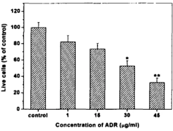
For the effea of ADR, cultured myocardial cells are
treated with various concentrarions (1 glm! to 45 glml)
of ADR for 36 hours. ADR-induced cardiotoxicity is
m얹S따ed by MTT assay in cultured myocardial cells
Cell viability 잉 decreasing, as the ADR concentrarion is
getting bigger. Es 야cially, when cultured myocardial
cells are exposed to 30 glm! (p<0.05) and 45 glm!
(p<O.OI) ADR, the decreasing rates are similar compare
to other results (Table I, Fig. I).
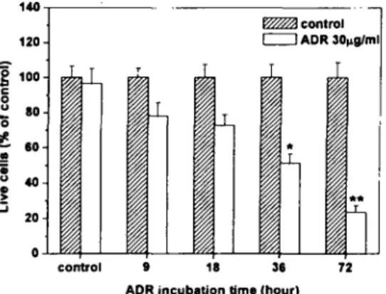
For the time-response relationship of ADR, cult 따ed
myocardi 꾀 cells a.!e treated with 30 g/m! ADR, the
value of MCV, for 9 to 72 hours. ADR-induced
cardiotoxicity is measured by MTT assay in cultured
myocardial cells. As time goes cell viability are deer-
easing; in particular, when the cultured cells are exposed
for 36 hours (p<0.05) and 72 hours (p<O.OI), the
decreasing rates are sin 피ar compare to other time
inte π따s (Table 2, Fig. 2).
Table 1. Absorbance (% of ∞ntrol! at 또KJnm Wavelength
for the Mπ assay on ADR in Cuijured 빼∞a- rdial Cells
ADR( μrI뼈) Decrease race of
ceU viab 파ry(%)
M1T absorbanc 이 59 αuo)
0 1.7 ’ :to.IS
1.44:t0.16 17.3
η 1.29 :to.09 26.3
30 0.92 :to.OS. 47.3
45 0.57:t0.04" 67.4
Culcured myocardial ceUs we πrreated with vari α15 concentrations of
ADR for 36 hours. The values are the mean:t SE for 6 experimenrs
Significant dilfeπores from the comrol are marked wirh asrerisks
.p<0.05; ..p<O.OI
120
훌 100
S 80
-。
효 80 드g
@ ‘。
흐 20
。
con ν。l 1 11 30
Concentration of ADR (
μglml ’
‘
Fig.!. D∞e검ependency of ADA.
ADR-induced cardiotoxicicy was meas 띠ed by M1T assay in cultured
myocardial αUs. Culcures were e휴:>osed to I. 15. 30 삐d 4’ μrime
ADR for 36 hours. res야aively. Other legends are the same as table
1. .p<O.05; ..p<O.OI
ADR M1T absorbance(S90run)
(¼mt) Ohr 9hr 18 hr 36 hr 12hr
0 1.47 :t 1.46 :t 1.43 :t !.36:t !.32:!:
0.19 0.16 0.18 0.17 O.IS
Decrease rate of
AD¼¿) NR absor¼¼ncc(S40run)
cell viabÕ<
0 I.S3I0.16
1.21:t0.IS 20.9
IS 1.l5:!:0.12 B
30 0.77 :to.07. G.7
4S OA3:tQÅì.. 71.9
- 효뺑심 외 3 : 째心흩子갱 몽;a 이 IS 톨 心inllll! 생에 미치는 Ii 톨 -
Table 2. Time-response Relationship of ADR by MTT
assay in Cultured My ∞ardial Cells
IA2:t 1.14:t I.여 :t 0.70:t 0.31:t
30 O.IS 0.12 0.08 O.OS. 0.02"
c‘urured myocardi 매 cells were creared
"따h 30 μmt ADR for
various cime inrervals. The values are the mean :t SE for 6
experimencs. S밍띠£icanc differences berween groups are marked wich
asterisks. ‘p<O.OS; "p<O.OI
’‘。
1N
흥 100
-
。u
o 호 .。-
g- ω
.
N
0
control 9 18 n
ADR incubation 히me (hour ’
Fig. 2. Time-dependancy of ADR in αJltured my, α;ardial
∞
Cultures were exposed co 30 J1mt ADR for 9, 18, 36 and 72
houπ, res 야ctively. Ocher legends are the same as cable 2. .<O.OS;
..p<QOl
(2) NR Assay
While the myocardial cells are culrured, rinse them
with Ca, Mg free Hank ’s Balanced Salt Solution (HBSS,
Gibco) 3 times. Cultured myocardial cells are grown in
media containing various concentrations of ADR for 36
hours. Decreasing cell viability is captured as ADR con-
centration was increasing. S야d,rulsili
ro 밍ml and 45 g/mI are showed 50.3%
(p<0.05) and 28.1 %m (p<O.Ol) of similar decrease
(Table 3, Fig. 3).
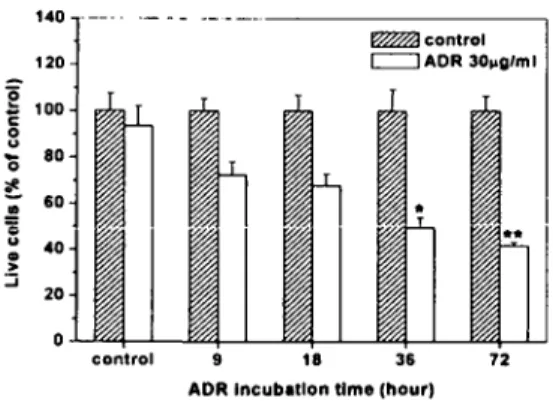
To study the time-response relationship of ADR,
rured myocardial cells are incuba 뼈 with 30 밍
ADR for 9 to 72 hours. It shows the decre 앓=
cell viability response to various 띠1e mte πaIs; parti-
arly, ADR incubion time of 36 and 72 hours dem-
onstrates the similar decreasing rates (Table 4, F핑.4).
Table 3. Absorbance (% of ∞nη01) at 540nm Wavelength
for the NR assay on ADR in My α 띠 ial Cells
Culrured myocardial cells were grown in media concainig various
concentratt 。띠 of ADR for 36 hours. The values represenc che mean
:t SE for 6 experimenrs. Significanc 버fferences from che concrol are
marked with asterisks. .p<0.05 ‘ "p<O.OI
1 기
§
’ 00
s
효
.
5 ‘0
-
그
control ’ 5
ADR concentration (
μg1 ’
‘s
Fig. 3. Dose-response relationship of ADR in cultured
myocardial cells.
Cyrocoxiciry was measured by NR 잉say. CultUres were exposed CO
αconcrol), I, IS, 30 and 45 μmt ADR for 36 hours, respeaively
Ocher legends are the same as cable 3. .p<O.OS; "p<O.OI
Table 4. Time-response Relationship of ADR by NR assay
in Culturl 때 ∞a띠ial Cells
AD μm ‘”
NR φsorbance(S40nm)
%hr 72 hr
9hr 18 hr
Ohr
1.38:t 1.34:t 1.31 :t 1.27:t 1.2s:t
0.14 0.11 0.13 0.15 0.12
1.29:t 0.97:t 0.89:t 0.63:t 0.52:t
O.IS 0.08 0.07 0.06. 0.02"
Culrured myoca 뼈외 cells weπ incubaced wich 30 μmt ADR for
various time intervals. 까 v피 ues represent che mean :tSE for 6
ape 띠nenrs. Significanc 며fferences berween groups the control are marked with asteri 화<S. .p<O.O ’ ; "p<O.OI
0
30
73-
- 사상채질의학회지 얘 13 권 째 1 호때1 -
’ 40 . --. .- - - ----
120
o:. 100
s9
'0
80
*
-;; 60
=s
a ‘0
그-
α control
c:::::J ADR 30 μg/ml
.
20 o
control 9 Ie 36
ADR Incubation time (h。‘Ir’
72
Fig. 4. Time-dependancy of ADR in cultured myocardial
cells.
Culrures were exposed to 30 μwme ADR for 9, IS, 36 and 72
hours, respecrively αII viabiliry was measured by NR assay. Other
legends are the same as table 4. *p<0.05; **p<O.OI
2. Effect of CYT W 이er Extract
1) LDH Activity
(1) ADR-induced Cardiotoxicity
To measure che LDH Aaivi η in various concen-
cration of ADR culcured mouse myocardial ceIJs are
exposed co various concencrations of ADR for 36 hou.
As a result, the concentration of ADR and LDH acci-
vicy are increased proportionalJy and indicaced che car-
diocoxicicy; specifically, when che culcured cells are
compared co none ADR-creaced cell, chey showed a
similar increasing race in 35 and 50 glml. The value of
MCV (midcycocoxicicy) presented during 35 glml ADR
process (Table 5, Fig. 5).
Table 5. Dose-response Relationship of ADR on LDH
activity in Cultured Mouse My ∞ardial Cells
ADR(JiWme) control 20 35 ,。
Amount
12.S:t 16.8:t
L2 1.5
IS.3:t 20.6:t 24.7:t
2.1 2.S* 3.6**
of
LDH Release
Cultured mouse myocardiac cells were exposed to vario 띠
concentrations of ADR for 36 hours. LDH release was measured at
wavelength of 540nm. The values are the mean :tSE for 5
experiments. Significant differences from the control are marked with
asterisks. *p<0.05; "p<O.OI
250
빼빼빼빼
--ef。“-。
*-ts-6
xa」
control 5 20 35
Concentration of ADR IμgIml ’
50
Fig. 5. Dose-res ∞nse relationship 이 ADR on LDH act-
ivity in cultured mouse my∞ardiac ∞lis. Other
legends are the 잃me as table 5. 'p<O.05, up 이 01
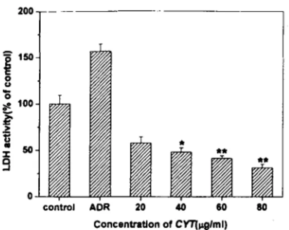
(2) 깨e Protection 이 CYT
To report che relacionship becween che ADR-induced
cardiotoxiciry and che effea of CYT Wacer Excracc
respecc to che LDH Activiry, cultured mouse myocardial
cells were preincubated wich various concentracions of
CYT water excract for 3 hou, and then exposed to 35
glml ADR for 36 hou.
As a result, the cell that only created with CYT
Water Exrracc showed nontoxic compare co che cell that
creaced in ADR. In che case of preincubaced CYT
Wacer Extraa, che concentration of ADR and LDH
Accivity races were diminishing proportionally. Scaci-
calJy, the cell that created in 40 glml (p<0.05), 60
g/ml (p<O.Ol), 80 glml (p<O.Ol) CYT Water Extract
revealed similar diminishing effea compare co che cell
chac only treaced in ADR (Table 6, Fig. 6).
Table 6. Dose-response Relationship of CYT Water Ex-
t떠이 for LDH a이vity in Cultured Mouse Myo-
ca 띠ial Cells
Amount of LDH Release
Concentration of CYT( 뼈me)
o Jiwme 20 μglm ¬ 40 Jlglme 60 Jlglme SO Jlwme
1O.6:t 1O.4:t 10.1 :t 9.6:t 9.3:t
In QS Q6 Q5 Q7
16.6:t 6.1:t 5.1:t 4.4:t 3.3:t
O.S 0.7 0.5* 0.3.. 0.4"
Culrured mouse myocardial cells were prein κubated with various
concentrations of CYT water extract for 3 hours, and then exposed
10 35 Jlglme ADR for 36 hours. LDH πlease was measured at
wavelength of 540nm. The values rep πsent the mean:t SE for ’
experiments. Significant differences from 【he ADR-treated group are
marked with asterisks. .<0.05; ..p<O.OI
ADR(jJglme)
0
”
ADR(¼wme) Beating rate Dectease of
(N·`nber/min) Bcatin rate (% of control)
0 118:t15
99 :t8 16.1
10 89:t5 24.6
30 56:t 3. 52.5
50 31 :t6.. 73.7
- 효뺑심 외 3: 찌心훌子훌 Jl!{iIOI i훌톨 心 빼IJ 에 미I 는 톨-
200- .---
기
o
con ν。I ADR 20 40 60
Concentration of CYT( μg/ml)
60
Fig. 6. Dose-response relationship of CYT water extract
for LDH activity in cunured mouse my α:ardial ∞lis.
Significant difference ftom the ADR-rreated group are marked with
asterisks. Othet legends arc the same as table 6. .<0.05; "p<O.OI
2) Heart Beating Rate
(1) ADR-induced Cardiotoxicity
To determine heart-beating rate, cultured mouse my-
ocardial cells were created with various concentrations of
ADR for 36 hours. As for the result, the beating rate
was decreasing at 1 glm! and 10 g/ml; however, they
did not show the statistical similariry. In the process of
30 g/ml and 50 glm! ADR they indicated the similar
decreasing rate of 47.5% (p<0.05) and 26.3%
(p<O.OI). The value of MCV (midcycotoxiciry) presented
during 35 g/m! ADR process (Table 7, Fig. 7).
Table 7. Dose-res ∞nse Relationship 이 ADR on SR in
Cultured Mouse Myocardiac Cells
Cui ruted mouse myocardial cells were treared with various
concentrations of ADR for 36 hours. BR was measured by count of
beating number per minute, compared with control. The values are
the mean:t SE for 5 experiments. Significant differences from the
control arc marked with asterisks. .p<0.05; ..p<O.OI
120
훌뼈
su 80'0
;!.
"::" 60
훌g 빼
겉
.JI 20
0
contr 여 1 10 30
Concentration of ADR Illg/ml)
60
Fig. 7. Dose-response relationship of ADR on beating rate
in cunured mouse my, ∞ardiac cells.
Other legends arc the same as table 7. .p<O.O ’, ..p<O.OI.
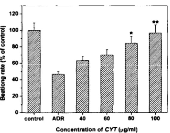
(2) The Protection of CYT
To report the relationship between heart beating rate
of ADR and the effect of CYT Water Extract respect co
the beating rate, cultured mouse myocardial cells were
preincubated with the concentrations of 40 to 100 g/ml
of CYT water extract for 3 hours, and then exposed to
30 glml ADR for 36 hours
As a result, the cell that only treated with CYT
Water Extract did not showed significant difference
compare to the cell that created in ADR In case of
preincubated CYT Water Extract, the concentration of
ADR and the beating rate are esc 외 ating proportionally
Statically, the cell that created in 60 glm! (p<0.05) and
80 glml (p<O.OI) CYT Water Extract reveals similar
escalating effect compare to the cell that only treated in
ADR (Table 8, Fig. 8).
Table 8. Dose-response Relationship of CYT Water Ex-
tract for SR in Cultured Mouse My, α::ardial Cells
ADR ωwme)
Bcatin 땅 rate (N 띠nber/min)
Concentration of CYT(Pwme)
0 μwme 40 μ밍뼈 60 JlWme 80 μwme 100 μwme
0 30
120:tll 122:t14 121:t13 123:t12 124:t1 ’
56:t4 76:t 6 84:t8 101:t1O. 116:t12"
Culrured mouse myocardial cells were prein α bated with various
concentrations of CYT water extract for 3 hours, and then exposed
to 30 μ밍me ADR for 36 hours. BR was measured by count of
bearing number per minute. The values represent the mean :tSE for
5 experimentS. Significant differences from the ADR-treated group
arc marked with asterisks. .p<0.05 ’ "p<O.OI
75
- 사상채짙의학회지 재 13 권 재 1 호 axJ\ -
120
g ω
o
control ADR ‘。 60 80 100
Concentration of CYT Il'gIml)
Fig. 8. Dose-response relationship of CYT water extract
for SR in cuured mouse myocardial ∞lis.
Significanc diffecence from che ADR-creaced group are marked wich
asrerisks. Ocher legends are che same as table 8. .p<O.05.
*‘p<O.OI
N. Further Research
According £0 [Dongyi-soose-bowon] CYT was known
as a remedy for interior-overheated-disease of tae 띠run.
However, the b∞k did not disclose the prescription or
symp£Oms. One of the recipes for the CYT is semen
nelumbinis which is not a regular formula for taumin
and uses for palpitation, fidgetiness and insomnia, invo-
luntary ejac 띠ation, leucorrhea.13) In addition, eight other
recipes of CYT such as radix ophiopogonis, rhizoma
acari graminei, radix scutellariae, semen zizyphi spinosae,
semen biotae, arillus longan and radix polygalae are
used as a treatment for various heart diseases
While many other studies are reported in heart
diseases fields, Kim declared the relationship berween
the myocardial blood and CYT.2) He stated that the
benefit of CYT as the thrombocyte growth, the amount
of fibrinogen increase, and the reduction of prothrombin
tune.
Therefore this paper considered different point of
view from other dissertations. In this paper the myo-
cardial cell was treated in the CYT Water Extraa, then
examined the protection of CYT Water Extraa against
the damaged myocardial cell.
The £oxic induced objects in the myocardial cells are
a heavy metal, ADR, dichloromethane and a che-
mical.lO, 11) ADR is the one of anthracycline and obt외nd
from the streptomyces peucetius var. caesius. It has a
same toxic influence in the myocardial cell such as
dichloromethane and ca따es the heart failure and the
18-21)
myocardial infraction.
As the know-how in the cultivation fields are
established in many areas, numerous destructive or
22.2)
nontoxic objects are discovered and explored:.'''' Also,
many studies are examined using the myocardial cell as
a model of various diseases to search for mech μusm,
10.26.27)
process, method of treatment.
Therefore this paper analyzed the CYT Water Extract
against the ADR damaged myocardial cell.
First of all, it 앉posed the cultured mouse myocardial
cell to various time and concentration of ADR. Cell
viabiliry was measured by MTf 양say and NR assay.
As a result, cell viabiliry was decreasing as the
concentration of ADR and ADR incubation times were
increasing (Table 1-4, Fig. 1-4). This result proved
that ADR induced the cardiotoxiciry that reported in
Chung and othe π.
To explore the prevention of CYT in ADR-induced
cardiotoxicity, cultured myocardial cells were pre in-
cubated with the concentrations of 0 g/ml, 20 g/ml, 40
glml, 60 gl 찌, and 80 밍찌 of CYT water extract for 3
hours, and then exposed to ADR for 36 hours. From
the result, LDH activity and heart beating rate were
determined.
LDH activity experiment indicated the proportional
relationship between the concentration of ADR and
cardiotoxicity. Among the results, ADR concentration of
35 glml and 50 g/ml increased in similar rate compare
£0 none ADR treated cell. The value of MCV presented
during 35 g/ml ADR process (Table 5, Fig. 5). For
heart beating rate when the concentration of ADR
increased, the heart beating rate deer, 얹sed. Statisti 때
similarities in ADR concentration were 30 g/ml and 50
glml, respectively. ηIe value of MCV pr 얹nted during
35 g/ml ADR process (Table 7, Fig. 7). The conclusion