Langerhans cell histiocytosis is a rare disease of unknown pathogenesis involving clonal proliferation of Langerhans cells.1The term “Langerhans cell histiocytosis” was intro- duced by the Writing Group of the Histiocyte Society,2 replacing histiocytosis X, which had been proposed in 1953 by Lichtenstein3and encompassed 3 disorders: eosi- nophilic granuloma, Hand-Schüller-Christian disease, and Letterer-Siwe disease.
Eosinophilic granuloma is the most benign and com- mon form of Langerhans cell histiocytosis, and it invol- ves localized lesions predominantly of the bones.4 It accounts for 60-70% of all cases of Langerhans cell histio- cytosis and can be seen as solitary or multifocal bone de- fects.5,6The radiographic appearance of eosinophilic gran- uloma in the jaw is quite variable and not specific7altho- ugh the lesions usually appear as radiolucent lesions with
well-defined borders.5,8The lesions might resemble peri- odontal diseases, odontogenic cysts, ameloblastoma, and malignancies.9
This case may serve to illustrate the various radiogra- phic features of eosinophilic granuloma, and the import- ance and the difficulties of an early diagnosis by plain radiographs of the jaw. The radiographic features of this case mimicked radicular cyst. However, careful interpre- tation of the radiograph revealed the non-corticated border and floating tooth appearance suggesting a more aggres- sive lesion.
This mandibular lesion underwent multifocal disse- mination, involving the femur after a one-year disease- free period. The purpose of this case report was to describe characteristic radiographic features of eosinophilic granu- loma in an adult.
Case Report
In May 2008, a 39-year-old man visited Wonkwang Dental Hospital in the city of Daejeon with pain in the
Eosinophilic granuloma in the anterior mandible mimicking radicular cyst
Byung-Do Lee, Wan Lee, Jun Lee*, Hyun-Jin Son**
Department of Oral and Maxillofacial Radiology and Wonkwang Dental Research Institute, College of Dentistry, Wonkwang University, Iksan, Korea
*Department of Oral and Maxillofacial Surgery, Wonkwang Bone Regeneration Institute, College of Dentistry, Wonkwang University, Iksan, Korea
**Department of Pathology, School of Medicine, Eulji University, Daejeon, Korea ABSTRACT
Eosinophilic granuloma is a common expression of Langerhans cell histiocytosis and corresponds with typical bone lesions. The radiographic appearance of eosinophilic granuloma in the jaw is variable and not specific. It may resemble periodontitis, radicular cyst, or malignancies. The purpose of this report is to describe the characteristic radiographic features of eosinophilic granuloma of a 39-year-old male. The lesion in the anterior mandible was first diagnosed as radicular cyst because the radiographic findings were ovoid radiolucent lesion with well-defined border. However, careful interpretation revealed a non-corticated border and floating tooth appearance that were the characteristic radiographic features for the differential diagnosis. Early clinical signs of eosinophilic granuloma can occur in the jaw and a bony destructive lesion might be mistaken for periodontitis or an odontogenic cystic lesion;
therefore, careful interpretation of radiographs should be emphasized. (Imaging Sci Dent 2013; 43: 117-22) KEY WORDS: Eosinophilic Granuloma; Histiocytosis, Langerhans-Cell; Mandible; Radicular Cyst
Received January 28, 2013; Revised February 26, 2013; Accepted March 6, 2013 Correspondence to : Prof. Byung-Do Lee
Department of Oral and Maxillofacial Radiology, College of Dentistry, Wonkwang University, #460 Iksan-daero, Iksan City, Jeonbuk 570-749, Korea
Tel) 82-63-859-2912, Fax) 82-63-857-4002, E-mail) [email protected]
Copyright ⓒ 2013 by Korean Academy of Oral and Maxillofacial Radiology
This is an Open Access article distributed under the terms of the Creative Commons Attribution Non-Commercial License (http://creativecommons.org/licenses/by-nc/3.0) which permits unrestricted non-commercial use, distribution, and reproduction in any medium, provided the original work is properly cited.
Imaging Science in Dentistry∙pISSN 2233-7822 eISSN 2233-7830
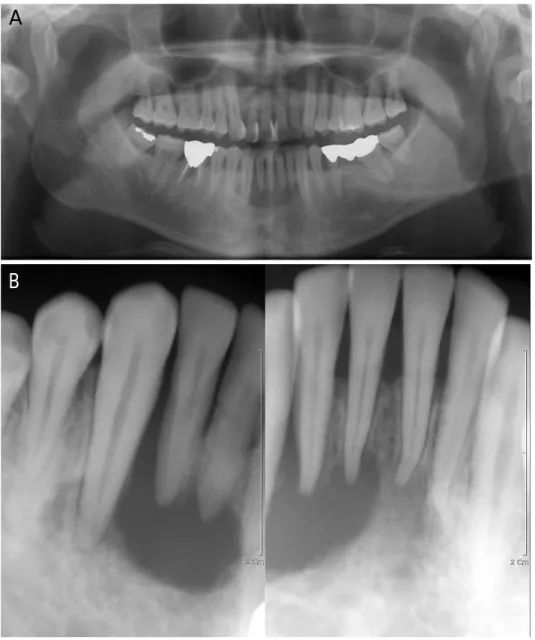
anterior region of the mandible originating several months earlier. Intraoral examination revealed a slight gingival swelling and mobility of the mandibular anterior teeth. The past history of the patient was not contributory. There was no cervical or axillary lymphadenopathy. A panora- mic radiograph showed an ovoid shaped, unilocular radio- lucent lesion with a well-defined margin in the alveolar bone from the left lower lateral incisor to the right lower canine, with involvement of the apices of the mandibular incisors (Fig. 1A). An intraoral radiograph revealed that the border of the lesion was non-corticated with beveled edges. The distal part of the right lower lateral incisor showed a typical floating tooth appearance. However, there was no tooth displacement and no root resorption (Fig.
1B). The patient underwent root canal treatments of the mandibular left central incisor and right canine for his
dental pain (Fig. 2). However, the pain did not subside, and the patient was referred to our department. We suspected an aggressive lesion rather than a cystic lesion due to the non-corticated border and floating tooth appearance. A biopsy of the mandibular anterior lesion was performed, and the patient was diagnosed with eosinophilic granu- loma.
About one month later, Cone beam Computed Tomo- graphy (CBCT), magnetic resonance (MR) imaging, and whole body bone scintigraphy with a Tc-99m MDP were performed at the dental hospital of Wonkwang University and the medical hospital of Eulgi University in the city of Daejeon. An axial CT showed destruction of the buccal and lingual cortex and a focal bony destructive lesion in the anterior mandible (Fig. 3). An MR image showed a focal high signal intense lesion in the left femur trochan-
Fig. 1.A. A panoramic radiograph shows a radiolucent lesion with a well-defined margin from the left lower lateral incisor to the right lower canine. B. A periapical radio- graph reveals a radiolucent lesion with a non-corticated border and beveled edges. The distal part of the right lower lateral incisor shows a “floating tooth” appearance.
A
B
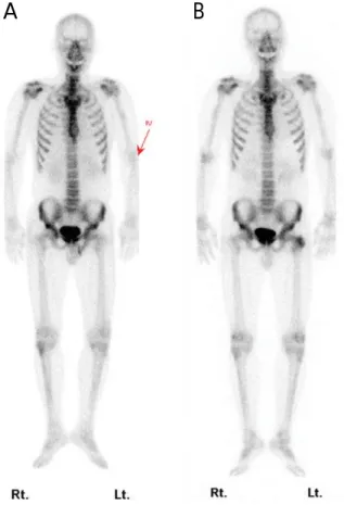
ter with adjacent bone marrow edema (Fig. 4). Bone scin- tigraphy revealed that there was no uptake of radiotracer in the trochanteric portion of the left femur on the initial study (Fig. 5A); however, a new active bone lesion show- ing increased radiotracer uptake with a central photopenic finding in the left femur was detected in a one year follow-
up study (Fig. 5B). A biopsy specimen from the left femur was also taken, and the result showed the same lesion, eosinophilic granuloma. Another examination and labora- tory tests, consisting of blood and serum biochemical studies, were within normal ranges. The treatment of the femur was carried out by surgical curettage.
Two years later, the patient revisited our dental hospital for the treatment of eosinophilic granuloma in the ante- rior mandible. A panoramic radiograph showed a slight increase in the size of the lesion in the anterior part of the mandible (Fig. 6) compared with radiographs at the initial visit. The lesion was found to extend from the left lower first premolar to the right lower canine. Alveolar bone resection of the lesion was performed for complete re- moval of the lesion, and a biopsy specimen was taken.
Fig. 2.A periapical radiograph shows root canal treatment of the mandibular left central incisor and right canine.
Fig. 3.An axial CT image shows a focal bony destructive lesion in the anterior mandible. Destruction of the buccal and lingual cortex is also observed.
Fig. 4. Fat saturated coronal T2 weighted MR image (TR/TE:
3750/103 milliseconds) shows a focal high signal lesion (arrow) in the left femur tronchanter with adjacent bone marrow edema.
Microscopically, the hematoxylin-eosin stained sample showed numerous Langerhans cells, eosinophils, and small lymphocytes. Immunohistochemically, these cells showed positivity for S-100 protein and CD1a protein (Fig. 7).
Based on the clinical, radiological, and histopathological findings, the final diagnosis was an eosinophilic granu-
loma in the mandible and femur. The patient was perio- dically recalled for a follow-up check.
Discussion
Eosinophilic granuloma, characterized by a prolifera- tion of Langerhans cells, with eosinophils, histiocytes, neutrophils, and scattered plasma cells,10 is the common form of Langerhans cell histiocytosis and is mostly seen between the first and third decades.11 It is rarely seen in adults, and affects only 1-2/million people.12
The clinical presentation of eosinophilic granuloma is different in different age groups.13Symptoms range from none to pain, swelling, loosening of teeth, and limitation of mouth opening. Adults are usually confined to a single organ and may be asymptomatic and spontaneously re- gress.13,14In the present case, the patient, a male who was 39 years old, which is an unusual age for eosinophilic gra- nuloma, presented with loosening teeth and gingival swell- ing.
Radiographically, eosinophilic granuloma appears as an osteolytic lesion with a well-demarcated border, but the borders are diffuse or poorly defined in some cases. If the lesions have invaded the alveolar crest, these cases may show the characteristic feature of a “scooped out” appear- ance.8,12,15 In many instances, the radiographic features showed selective destruction of the alveolar bone, caus- ing the tooth to have a “floating tooth” appearance.8
The radiographic features vary widely with the phase of the disease.10Poorly delimited borders at the initial stage and a more sharply delineated border in the middle phase are characteristic, whereas in the late phase a thick rind of sclerotic tissue occasionally presents. It is possible to ob- serve an osteolytic process, usually located in the medull-
Fig. 5.Bone scintigraphy. A. The initial study shows normal up- take of the radiotracer in the trochanteric portion of the left femur.
B. The one year follow-up study demonstrates an uptake lesion in the left femur.
A B
Fig. 6. Panoramic radiograph (2 years later) shows a slight increase in the lesion size in the anterior part of the mandible compared with the initial radiographs.
ary cavity, but occasionally in the cortex,16and the cortical bone may be perforated or periosteal bone formation may occur.16,17 In the present case, the mandibular lesion was observed in the medullary space, with a perforated buccal cortex but no periosteal bone formation was observed (Fig. 3).
The variable radiographic appearance makes the dif- ferential diagnosis of eosinophilic granuloma more diffi- cult. The lesion may be mistaken for an odontogenic cyst, osteomyelitis, or a malignancy.8Given its scooped out appearance, the differential diagnosis included the alter- natives of Ewing’s sarcoma, lymphoma, and metastatic disease.12In this case, a radicular cyst was suspected in the differential diagnosis because the jaw lesion showed an oval radiolucency with a well-defined border at the peri- apical region (Fig. 1). According to Hartman,18the monosto- tic form of eosinophilic granuloma is more prevalent than the polyostotic type. The bony sites most often affected by eosinophilic granuloma are the skull, femur, ribs, ver- tebrae, and mandible.8When the jaws are affected, the mandible is a more common site than the maxilla.19When multiple lesions occur, the new osseous lesions appear within 1-2 years.4 With the aid of skeletal scintigraphy, the additional lesion at the femur as well as the anterior mandible could be suspected on one year follow-up study (Fig. 5).
Routine diagnosis is usually made on the basis of histo- logical examination, and the presence of S-1007,20 and CD1a6,21by immunohistochemical examination could con- firm eosinophilic granuloma.
Treatment includes surgery, radiotherapy, chemothe- rapy, and intra-lesional injection of corticosteroids.8Sur- gery is the preferred method of treatment for a single and localized manifestation of the disease. Radiotherapy is
suggested in cases of local recurrence or if surgical treat- ment is not possible.21The patient in this case was treated with surgical resection of the mandible site and surgical curettage of the left femur.
Early clinical signs of eosinophilic granuloma can occur in the jaw and a bony destructive lesion might be mista- ken for periodontitis or a cystic lesion. The radiolucent lesion in this case was first diagnosed as a radicular cyst because of the unilocular lesion with well-defined border.
However, careful interpretation of the radiograph revealed its non-corticated border and floating tooth appearance, resulting in suspicion of a more aggressive lesion.
In conclusion, we insist on the fact that accurate radio- graphic differential diagnosis of eosinophilic granuloma is critical to both recognition and proper management of this condition.
References
1. Komp DM. Langerhans cell histiocytosis. N Engl J Med 1987;
316: 747-8.
2. Chu T, D’Angio GJ, Favara BE, Ladisch S, Nesbit M, Pritchard J. Histiocytosis syndromes in children. Lancet 1987; 2: 41-2.
3. Lichtenstein L. Histiocytosis X: integration of eosinophilic granuloma of bone, Letterer-Siwe disease, and Schüller-Chri- stian disease as related manifestations of a single nosologic entity. AMA Arch Pathol 1953; 56: 84-102.
4. Parihar A, Newaskar V. Management of polyostotic eosino- philic granuloma. Dent Res J (Isfahan) 2012; 9: 821-5.
5. Esen A, Dolanmaz D, Kalayci A, Günhan O, Avunduk MC.
Treatment of localized Langerhans’ cell histiocytosis of the mandible with intralesional steroid injection: report of a case.
Oral Surg Oral Med Oral Pathol Oral Radiol Endod 2010; 109:
e53-8.
6. Key SJ, O’Brien CJ, Silvester KC, Crean SJ. Eosinophilic granuloma: resolution of maxillofacial bony lesions following minimal intervention. Report of three cases and a review of the Fig. 7.A. Numerous Langerhans cells, recognized by their grooved, folded, or indented nuclei with fine chromatin, inconspicuous nucleoli, and thin nuclear membranes, with a few scattered eosinophils and small lymphocytes are seen (H&E stain, 400×). The tumor cells are strongly immunoreactive for S-100 protein (B, 200×) and CD1a (C, 200×) in the cytoplasm.
A B C
literature. J Craniomaxillofac Surg 2004; 32: 170-5.
7. Holzhauer AM, Abdelsayed RA, Sutley SH. Eosinophilic gra- nuloma: a case report with pathologic fracture. Oral Surg Oral Med Oral Pathol Oral Radiol Endod 1999; 87: 756-9.
8. Ardekian L, Peled M, Rosen D, Rachmiel A, Abu el-Naaj I, Laufer D. Clinical and radiographic features of eosinophilic granuloma in the jaws: review of 41 lesions treated by surgery and low-dose radiotherapy. Oral Surg Oral Med Oral Pathol Oral Radiol Endod 1999; 87: 238-42.
9. Jones LR, Toth BB, Cangir A. Treatment for solitary eosino- philic granuloma of the mandible by steroid injection: report of a case. J Oral Maxillofac Surg 1989; 47: 306-9.
10. Piattelli A, Paolantonio M. Eosinophilic granuloma of the man- dible involving the periodontal tissues. A case report. J Perio- dontol 1995; 66: 731-6.
11. Uckan S, Gurol M, Durmus E. Recurrent multifocal Langer- hans cell eosinophilic granuloma of the jaws: report of a case.
J Oral Maxillofac Surg 1996; 54: 906-9.
12. Rees J, Paterson AW. Langerhans cell histiocytosis in an adult.
Br J Oral Maxillofac Surg 2009; 47: 52-3.
13. Broadbent V, Egeler RM, Nesbit ME Jr. Langerhans cell his- tiocytosis - clinical and epidemiological aspects. Br J Cancer Suppl 1994; 23: S11-6.
14. Leavey P, Varughese M, Breatnach F, O’Meara A. Langerhans
cell histiocytosis - a 31 year review. Ir J Med Sci 1991; 160:
271-4.
15. dos Anjos Pontual ML, da Silveira MM, de Assis Silva Lima F, Filho FW. Eosinophilic granuloma in the jaws. Oral Surg Oral Med Oral Pathol Oral Radiol Endod 2007; 104: e47-51.
16. Fechner RE, Mills SE. Tumors of the bones and joints. Atlas of tumor pathology. 3rd series. Washington: Armed Forces Institute of Pathology; 1993. p. 211-5.
17. Mirra JM. Bone tumors: clinical, radiologic, and pathologic correlations. Philadelphia: Lea and Febiger; 1989. p. 1023-45.
18. Hartman KS. Histiocytosis X: a review of 114 cases with oral involvement. Oral Surg Oral Med Oral Pathol 1980; 49: 38- 54.
19. Dagenais M, Pharoah MJ, Sikorski PA. The radiographic characteristics of histiocytosis X. A study of 29 cases that in- volve the jaws. Oral Surg Oral Med Oral Pathol 1992; 74:
230-6.
20. Lam KY. Langerhans cell histiocytosis (histiocytosis X). Post- grad Med J 1997; 73: 391-4.
21. Kessler P, Wiltfang J, Schultze-Mosgau S, Neukam FW. Lan- gerhans cell granulomatosis: a case report of polyostotic mani- festation in the jaw. Int J Oral Maxillofac Surg 2001; 30: 359- 61.