Introduction
The prevalence of a forward head posture (FHP) has increased due to the increased numbers of people today who sit at a desk for extended periods (Yip et al, 2008). With a FHP, the head is in an anterior po- sition in relation to the plumb line, which is vertical
to a horizontal line through the center of gravity of the body (Griegel-Morris et al, 1992). A FHP is one of the most common types of poor head posture in patients with neck disorders (Yip et al, 2008).
Johnson (1998) suggested that a prolonged FHP might increase pressure on the posterior cranio-cer- vical structure. Subjects with a FHP show an in- Corresponding author: Sung-min Ha [email protected]
Comparison of the Thickness of the Neck Flexor Muscles of Subjects With and Without a Forward Head Posture on the Two Initial
Head Positions During Cranio-Cervical Flexion Exercise
Sung-hoon Jung1,2, BPT, PT, Oh-yun Kwon1,3,4, PhD, PT, Kyu-hwan Choi5, PhD, PT, Sung-min Ha6, PhD, PT, Su-jung Kim7, PhD, PT, In-cheol Jeon1,2, MSc, PT,
Ui-jae Hwang1,2, BPT, PT
1Kinetic Ergocise Based on Movement Analysis Laboratory
2Dept. of Physical Therapy, The Graduate School, Yonsei University
3Dept. of Physical Therapy, College of Health Science, Yonsei University
4Dept. of Ergonomic Therapy, The Graduate School of Health and Environment, Yonsei University
5Dept. of Physical Therapy, Ansan University
6Dept. of Physical Therapy, College of Health Science, Sangji University
7Dept. of Physical Therapy, College of Health Sciences, Youngsan University
Abstract
1)This study compared the effects of the initial head position (i.e., a HHP versus a relaxed head position) of subjects with and without a FHP on the thickness of the deep and superficial neck flexor muscles during CCF. The study recruited 6 subjects with a FHP and 10 subjects without a FHP. The subjects performed CCF in two different head positions: a HHP, with the head aligned so that the forehead and chin formed a horizontal line, and a relaxed head position (RHP), with the head aligned in a self-selected comfortable position. During the CCF exercise, the thickness of the longus colli (LCo) and the thickness of the sternocleidomastoid (SCM) were recorded using ultrasonography. The thickness of each muscle was measured by Image J software. The statistical analysis was performed with a two-way mixed-model analysis of variance. The thickness of the SCM differed significantly (p<.05) between the subjects with and without FHP. According to a post hoc independent t-test, the change in thickness of the SCM increased significantly during CCF in the subjects with FHP while adopting a HHP compared to that in the subjects without FHP. The change in thickness of the SCM was not significantly different between the two positions in subjects without FHP, and there was no significant change in thickness of the LCo muscle during the CCF exercise according to the initial position in both subjects with and without FHP.
The results suggest that CCF should be performed in RHP to minimize contraction of the SCM in subjects with a FHP.
Key Words: Cranio-cervical flexion; Deep neck flexor; Initial head position; Superficial neck flexor.
creased degree of upper cervical extension in com- parison to that of the lower cervical spine. Other ef- fects of a FHP are shortness of the suboccipital ex- tensors, superior obliques, inferior obliques, and rectus capitis (Page et al, 2010; Sahrmann, 2010). Ishida et al (2015) demonstrated that the muscle thickness of the longus colli (LCo) of subjects with a FHP was reduced compared to that of subjects without a FHP.
Cranio-cervical flexion (CCF) exercise aims to ac- tivate the deep cervical flexor muscles (DCFMs).
DCFMs consist of LCo, which provide support to the cervical curve, segments, and longus capitis (LC) works in synergy with the LCo in CCF (Gong et al, 2012; Jull et al, 2004b). In CCF exercise, a pressure biofeedback unit (PBU) is used to monitor cra- nio-cervical movement caused by progressive flat- tening of the cervical lordosis (Mayoux-Benhamou et al, 1997). The exercise is composed of five pro- gressive stages from a baseline pressure of 20 ㎜Hg to 22, 24, 26, 28, and 30 ㎜Hg. According to previous studies, the electromyography activity (Falla et al, 2003) and the change in the thickness (Jesus et al, 2008) of the DCFM increased when the stage of the PBU was increased during CCF. However, many studies have shown that the superficial cervical flex- or muscles (SCFMs), such as the sternocleidomastoid (SCM) and anterior scalene, of subjects with neck pain were overactivated during CCF (Falla et al, 2004; Jull et al, 2004b; Jull et al, 2008). Jull et al (2004b) suggested that the increased activity of the SCFMs during CCF could be to compensate for the poor activation of the LCo. To prevent such com- pensatory movements during CCF, the activity of the SCFMs should be monitored (Falla et al, 2004; Jull
et al, 2004b; Jull et al, 2008).
In previous studies (Jesus et al, 2008; Jull et al, 2008; Lluch et al, 2013), the head and neck were placed in a horizontal position before CCF exercise, with the subject’s forehead and chin in a horizontal position and in a midposition. However, subjects with a FHP usually have posterior neck extensor muscles tightness and lengthened DCFMs (Sahrmann, 2010).
When a subject with a FHP performs CCF exercise in a horizontal head position (HHP), the subject re- quires more torque than does a subject without a FHP. Such torque is difficult in FHP subjects be- cause of changes in the length-tension curve of the DCFMs. The change may result in greater activity of the SCFMs during CCF exercise with a FHP.
Therefore, the purpose of this study was to de- termine the effects of the initial head position on the thickness of the LCo (DCFM) and SCM (SCFM) during CCF exercise in subjects with and without a FHP. It was hypothesized that a HHP would in- crease the change in thickness of the SCM during CCF exercise in subjects with a FHP.
Methods
Subjects
Sixteen volunteers (12 males, 4 females) aged 18∼
30 years were recruited from Yonsei University stu- dents (Table 1). The inclusion criteria were young age (18∼30 years) and the presence or not of a FHP. A FHP was defined as a cranio-vertebral an- gle of 40.3°∼47.5° (Subjects with FHP). No FHP was defined as a cranio-vertebral angle of 49.2°∼
Characteristics With FHPa (n1=6) Without FHP (n2=10)
Age (year) 23.8±2.0b 22.2±1.3
Height (㎝) 177.2±4.4 170.6±8.4
Weight (㎝) 84.8±9.0 67.2±6.8
Cranio-vertebral angle (˚) 43.3±3.0 52.5±3.1
aforward head posture, bmean±standard deviation.
Table 1. General characteristics of the subjects (N=16)
55.8° (Subjects without FHP) (Cheung Lau et al, 2009). The exclusion criteria included a history of fracture injury at the cervical and shoulder region or vertebral column, scoliosis, severe thoracic kyphosis, spasmodic torticollis, rheumatic disease, tempor- omandibular joint dysfunction, neurological motion dis- orders or back pain, or loss of standing balanced. All the subjects signed written informed consent forms, and the study was approved by the Institutional Review Board of Yonsei University Wonju Campus (approval number: 1041849-201510-BM-068-01).
Instruments
2D analysis (Image J): Measurement of FHP
Image J imaging software (U.S. National Institutes of Health, Maryland, USA) was used to measure the FHP angle, which is the angle between a line drawn from the tragus of the ear to the sev- enth cervical (C7) spinous process and a horizontal line from the bottom. First, adhesive reflexive mark- ers were placed over the C7 spinous process (i.e., the midpoint of the most prominent point and tragus of the left ear). The subjects were asked to stand, face forward, and then extend and flex their neck three times until they were in a comfortable position (Yip et al, 2008).
Ultrasound
Ultrasound images of the LCo and SCM were re-
corded using the Accuvix A30 Ultrasound (Medison Co. Ltd., Seoul, Korea) system. The transducer (7.5-
㎒) was placed to the right of the neck about 5 ㎝ from thyroid cartilage and parallel to the direction of the trachea. In this position, the LCo and SCM, right carotid artery, vertebral lamina, and boundary of the SCM (benchmark) could be visualized clearly on the left side of the ultrasonography image (Jesus-Moraleida et al, 2011).
Procedure
The subjects were positioned in a supine position, with their knees bent (hips 45˚ flexion, knees 90˚
flexion), and their arms crossed on their chests.
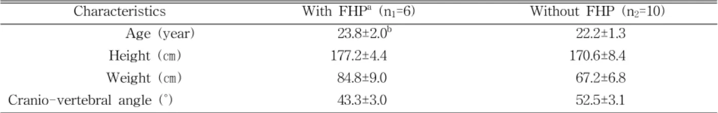
Before the CCF exercise, the subject’s head and neck were placed in the two initial head positions (Figure 1). In this study, HHP and relaxed head position (RHP) are used as initial head positions. The two terms are explained below.
-HHP: In a supine position, the subject’s forehead and chin were horizontal by anterior gliding of the head in the frontal-axis measured using a hand-made smartphone based measurement tool.
-RHP: In a supine position, the subject performed the CCF five times. After the CCF, the subject’s head was maintained in a comfortable position.
A PBU (Healience, Seoul, Korea) was placed sub- occipitally and inflated to a baseline pressure of 20
㎜Hg. The PBU is a sensitive apparatus for record- ing increases in pressure with cervical nodding. A manometer provided visible feedback on the pressure.
A B
Figure 1. Two initial positions (A: relaxed head position, B: neutral head position).
To familiarize the subjects with the CCF exercise, the subjects were shown how to perform the ex- ercise from the two initial positions. They then per- formed the exercise for 5 min. During this exercise period, the examiner verbally discouraged neck re- traction to minimize superficial muscle contraction.
As the subjects performed the CCF, the pressure was increased from a baseline pressure of 20 ㎜Hg to a maximum of 30 ㎜Hg for 10 sec, with a 2-min rest between trials. Ultrasonography images were obtained both in the resting position and when the exercises were performed at pressures up to 30 ㎜Hg (Jesus et al, 2008).
Data management
To determine the thickness of the LCo and SCM, three lines (broken lines in Figure 2) of equal length were drawn from the midline of the ultrasound image to the right side (edge) of the image. The thickness of the LCo muscle was calculated from the inferior of the carotid boundary to the superior of the echogenic vertebral lamina. The thickness of the SCM was cal- culated from the superficial to deep boundaries of the SCM. The thickness of each muscle was calculated by using the average of the three dash lines. The changes in the muscle thickness during the CCF ex- ercise were expressed as a proportion of the muscle thickness at rest (Jesus et al, 2008).
Statistical analysis
Statistical analyses were conducted using SPSS ver. 21.0 (SPSS Inc., Chicago, IL, USA). After a nor- mal distribution was examined using the Kolmogorov-Smirnov Z-test, a two-way mixed-model analysis of variance (ANOVA) was used to compare the between-group (with FHP vs without FHP) dif- ference in the muscle thickness (SCM/LCo) when the exercise was performed at the different positions (HHP/RHP). When a significant difference was found, post hoc independent t-tests were performed. An paired t-test was used to compare the between head position difference in the muscle thickness (SCM/LCo) in each group. The level of statistical significance (α) was chosen as .05.
Results
The two-way ANOVA showed that there was a significant difference (F=19.797, p<.001) between subjects with and without FHP in the change in thickness of the SCM and there was no significant difference between group in the change in thickness of the LCo (p=.814). A summary table for the two-way analysis of variance is presented in Table 2. The results of the post hoc independent t-tests demonstrated that the thickness of the SCM was
A B C
Figure 2. PBU measurements using a hand-held manometer and ultrasonography recordings (A: measurement setting, B: ultrasonography image in the resting position while adopting a HHP, C: ultrasonography image while performing CCF at a pressure of 30 ㎜Hg, PBU:
pressure biofeedback unit, SCM: sternocleidomastoid, LCo: longus colli).
significantly increased (p<.001) in the subjects with FHP in the HHP compared to that of the subjects without FHP (Table 3). There was no significant between-group difference in the change in thickness of the LCo muscle during the CCF exercise in the HHP (p=.916). A paired t-test showed that there was no significant difference except for change in thick- ness of the SCM between initial head positions in the Table 4.
Discussion
This is the first study to compare the change in thickness of the LCo and SCM during CCF exercise at two initial positions in subjects with and without FHP. The study suggests that for subjects with a FHP, CCF exercise in a RHP is more effective than CCF exercise in a HHP for minimizing changes in
the thickness of the SCM. The results of this study showed that the change in thickness of the SCM during CCF exercise in a HHP significantly in- creased in the subjects with FHP compared to that of the subject without FHP (Table 3). There are various possible explanations for this findings.
First, muscular adaptations in a FHP include shortness of the suboccipital muscles (extensors, su- perior/inferior oblique, and rectus capitis) because of the increased degree of upper cervical extension (Sahrmann, 2010). The shortness of the suboccipital extensor muscles may lead to more torque of the LCo and LC during CCF because the suboccipital extensor muscles are activated during cranio-cervical extension (Neumann, 2010). Therefore, subjects with a FHP may require more torque of the LCo and LC during CCF. Second, a FHP is a biodynamical pos- ture in which the suboccipital flexors, such as the LCo and LC, are elongated (Sahrmann, 2010). In a
Muscle Sum of squares df Mean square F p
Group SCMa .249 1 .249 19.797 <.001*
LCob .001 1 .001 .056 0.814
asternocleidomastoid, blongus colli, *statistically significant at the level of p<.012.
Table 2. Summary table for the two-way analysis of variance
Muscle Position With FHPa Without FHP T p
SCMb RHPc 1.18±.13d 1.10±.09 1.589 .134
HHPe 1.36±.10 1.08±.13 4.534 <.001
LCof RHP 1.15±.09 1.11±.13 .539 .598
HHP 1.14±.08 1.15±.19 -.108 .916
aforward head posture, bsternocleidomastoid, crelaxed head position, dmean±standard deviation, ehorizontal head position, flongus colli, *statistically significant at the level of p<.05.
Table 3. Between-group comparison of the change in the muscle thickness at the different positions
Muscle Position RHPa HHPb T p
With FHPc SCMd 1.18±.13e 1.36±.10 -5.384 <.001*
LCof 1.15±.09 1.14±.08 .150 .887
Without FHP SCM 1.10±.09 1.08±.13 .700 .502
LCo 1.11±.13 1.15±.19 -.920 .381
arelaxed head position, bhorizontal head position, cforward head posture, dsternocleidomastoid, emean±standard deviation, flongus colli, *statistically significant at the level of p<.05.
Table 4. Between-initial head position comparison of the change in the muscle thickness at the different groups
FHP, the length of the LCo and LC muscles may increase beyond physiological limits but not beyond the normal range of motion. Changes in the length-tension curve result in “stretch weakness”
(Kendall et al, 2005). When a subject with a FHP is placed in a HHP, the LCo and LC is shortened. This condition may lead to compensative movement be- cause subjects with a FHP cannot make maximal torque of the LCo in the shortened range position.
As a result, the SCM is overactivated during CCF in a HHP by subjects with a FHP. Third, in a FHP, the SCM is overactivated, and the LCo is underused (Ishida et al, 2015). Adopting a FHP for extended intervals may result in overactivation of the SCM and underactivation of the LCo during CCF. The ac- tivation of the SCM and LCo may also be altered by a prolonged FHP caused by sitting at a desk in front of a computer for long periods on a daily basis.
It is similar to the changed pattern of muscle re- cruitment observed between the DCFMs and SCFMs in young subjects with neck pain (Jull et al, 2004a;
Uthaikhup and Jull, 2009).
The results of this study suggest that CCF in a RHP is effective for preventing compensative move- ment with a FHP. Previous studies used electro- myography for monitoring the recruitment of the SCM (Kwon et al, 2011) and a force measuring apparatus for monitoring neck retraction (Falla et al, 2003).
These methods are complex and time consuming and so are difficult to apply in the clinical setting. The modified head positions (HHP and RHP) adopted in the present study may offer a convenient and effec- tive measurement method of CCF exercise for FHPs.
The present study has some limitations. First, it included only young subjects. Therefore, the results cannot be generalized to the overall population.
Second, the study included only subjects without pain. Thus, the results have limited applications to patients with neck pain. A further study is necessary to determine the effect of the initial head position on the thickness of the neck flexor muscles during CCF exercise in subjects with neck pain.
Conclusion
This is the first study to compare the change in thickness of the LCo and SCM among subjects with and without a FHP. The change in thickness of the SCM significantly increased in the subjects with FHP compared to that of the subjects without FHP during the CCF exercise with a HHP, and this change was significantly greater in the subjects with FHP. The results suggest that the initial head posi- tion is important for minimizing superficial muscle activity during CCF exercise in subjects with a FHP.
Future studies are needed to determine whether long-term CCF exercise in different positions (RHP and HHP) can minimize changes in thickness of the SCFM. Therefore, individual with FHP should select RHP during performing CCF exercise for minimizing superficial muscle activity.
References
Cheung Lau HW, Wing Chiu TT, Lam TH. Clinical measurement of craniovertebral angle by elec- tronic head posture instrument: A test of reli- ability and validity. Man Ther. 2009;14(4):
363-368.
Falla DL, Campbell CD, Fagan AE, et al.
Relationship between cranio-cervical flexion range of motion and pressure change during the cranio-cervical flexion test. Man Ther. 2003;8(2):
92-96.
Falla DL, Jull GA, Hodges PW. Patients with neck pain demonstrate reduced electromyographic ac- tivity of the deep cervical flexor muscles during performance of the craniocervical flexion test.
Spine (Phila Pa 1976). 2004;29(19):2108-2114.
Gong W, Kim C, Lee Y. Correlations between cer- vical lordosis, forward head posture, cervical ROM and the strength and endurance of the deep neck flexor muscles in college students. J Phys Ther Sci. 2012;24(3):275-277.
Griegel-Morris P, Larson K, Mueller-Klaus K, et al.
Incidence of common postural abnormalities in the cervical, shoulder, and thoracic regions and their association with pain in two age groups of healthy subjects. Phys Ther. 1992;72(6):425-431.
Ishida H, Suehiro T, Kurozumi C, et al. Correlation between neck slope angle and deep cervical flexor muscle thickness in healthy participants. J Bodyw Mov Ther. 2015 (Article in Press).
Jesus FM, Ferreira PH, Ferreira ML.
Ultrasonographic measurement of neck muscle recruitment: A preliminary investigation. J Man Manip Ther. 2008;16(2):89-92.
Jesus-Moraleida FR, Ferreira PH, Pereira LS, et al.
Ultrasonographic analysis of the neck flexor muscles in patients with chronic neck pain and changes after cervical spine mobilization. J Manipulative Physiol Ther. 2011;34(8):514-524.
http://dx.doi.org/10.1016/j.jmpt.2011.08.006
Johnson GM. The correlation between surface meas- urement of head and neck posture and the ana- tomic position of the upper cervical vertebrae.
Spine (Phila Pa 1976). 1998;23(8):921-927.
Jull G, Falla D, Treleaven J, et al. A therapeutic ex- ercise approach for cervical disorders. In:
Boyling JD, Jull GA eds. Grieve’s Modern Manual Therapy: The vertebral column. 3rd ed.
Edinburgh, Churchill Livingstone, 2004a.
Jull G, Kristjansson E, Dall’Alba P. Impairment in the cervical flexors: A comparison of whiplash and insidious onset neck pain patients. Man Ther. 2004b;9(2):89-94.
Jull GA, O’Leary SP, Falla DL. Clinical assessment of the deep cervical flexor muscles: The cranio- cervical flexion test. J Manipulative Physio Ther. 2008;31(7):525-533.
Kendall FP, McCreary EK, Provance PG, et al.
Muscles: Testing and function with posture and pain. 5th ed. Baltimore, MD, Williams &
Wilkins, 2005:52, 61, 159.
Kwon OY, Kim SH, Choung SD. Effect of deep neck flexor performance on the stability of the cer- vical spine in subject with and without neck pain. Phys Ther Korea. 2011;18(4):1-10.
Lluch E, Arguisuelas MD, Coloma PS, et al. Effects of deep cervical flexor training on pressure pain thresholds over myofascial trigger points in pa- tients with chronic neck pain. J Manipulative Physiol Ther. 2013;36(9):604-611. http://dx.doi.
org/10.1016/j.jmpt.2013.08.004
Mayoux-Benhamou MA, Revel M, Vallee C. Selective electromyography of dorsal neck muscles in humans. Exp Brain Res. 1997;113(2):353-360.
Neumann DA. Kinesiology of the Musculoskeletal System: Foundations for rehabilitation. 2nd ed.
St Louis, Mosby, 2010:398-404.
Page P, Frank C, Lardner R. Assessment and treat- ment of muscle imbalance: The janda approach.
Human Kinetics, 2010:178-180.
Sahrmann S. Movement System Impairment Syndromes of the Extremities, Cervical and Thoracic Spines. St Louis, Mosby, 2010:51-86.
Uthaikhup S, Jull G. Performance in the cranio-cer- vical flexion test is altered in elderly subjects.
Man Ther. 2009;14(5):475-479. http://dx.doi.org/
10.1016/j.math.2008.12.003
Yip CH, Chiu TT, Poon AT. The relationship be- tween head posture and severity and disability of patients with neck pain. Man Ther.
2008;13(2):148-154.
This article was received October 8, 2015, was reviewed October 8, 2015, and was accepted November 8, 2015.