J Cerebrovasc Endovasc Neurosurg.
2021 March;23(1):54-59
Received: 13 May 2020 Revised: 22 August 2020 Accepted: 13 October 2020
Correspondence to Igor Pagiola
Department of Interventional Neuroradiology, Universidade Federal de São Paulo, São Paulo SP, R. Napoleão de Barros, 715-Vila Clementino, São Paulo, SP, 04024-002, Brazil
Tel +55-11-5576-4848 Fax +55-11-5576-4522 E-mail igorpagiola@hotmail.com ORCID http://orcid.org/0000-0001-9052-7946
Case Report
Is the retrograde access for
endovascular treatment of a traumatic carotid cavernous fistula associated with dissection of the ipsilateral
carotid possible?
Igor Pagiola1,2, Bruno Amaral1, Celso Saito1, Darcio Nalli1, Henrique Carrete Junior1, Michel Frudit1
1Department of Interventional Neuroradiology, Universidade Federal de São Paulo, São Paulo, SP, Brazil
2Department of Interventional Neuroradiology, Hospital Estadual Central, VitÓria ES, Brazil
This is a case demonstrating a combined traumatic lesion of the internal carotid artery (dissection and a carotid cavernous fistula [CCF]) in a patient who was beaten during a robbery and, while trying to escape, was hit by a vehicle. Endovascular approach for the treatment was chosen using the retrograde access from the vertebral artery to the cavernous sinus by posterior communicating (Pcom) artery due to the occlusion of the ipsilateral internal carotid. Because the artery access by the internal carotid was impossible, retrograde approach by vertebral artery and Pcom artery was done to treat the direct CCF. A patient presented with left hemiplegia and proptosis, chemosis, right eye ptosis. Computed tomography (CT) and CT angiography revealed a CCF of the right carotid. An arterial retrograde endovascular approach by the vertebral artery was used for CCF occlusion with coils. We present a rare case of a combined traumatic cerebrovascular lesion, right carotid artery dissection and a right direct CCF treated by a retrograde endovascular approach by the vertebral artery through the Pcom artery to reach the fistula point and achieved a complete cure of the CCF.
Keywords Carotid-cavernous sinus fistula, Carotid artery, Internal, Dissection, Endovascular procedures
This is an Open Access article distributed under the terms of the Creative Commons Attribution Non-Commercial License (http://creativecommons.org/licenses/
by-nc/3.0/) which permits unrestricted noncommercial use, distribution, and reproduction in any medium, provided the original work is properly cited.
INTRODUCTION
As a rare condition, the traumatic carotid cavernous fistula (CCF) has an inci- dence lower than 0.2%. Another rare presentation is the cervical artery dissection after blunt trauma, with an incidence of 0.1%2) and a high mortality rate. According to Barrow et al.,1) the CCF can be classified into direct and indirect types. They also
divided it into 4 subtypes categories (A-D). The shear stress or the acceleration-deceleration during a blunt trauma may cause a direct CCF or, in a high intense trauma, a completely transection of the artery. On the other hand, the indirect type may occur spontaneously or can be associated with Ehlers-Danlos syndrome, Os- teogenesis imperfecta and other syndromes.5)7) We need to know that the intracranial hemorrhagic risk of direct CCF is low, and this risk may increase if: pseudoaneu- rysms, outflow pathways thrombosis, large varix of the cavernous sinus and drainage into cortical veins9) are present. All patients need a neuroimaging investigation with computed tomography angiography (CTA) and magnetic resonance angiography (MRA) if CCF is sus- pected. The gold standard for classification and diagno- sis is the digital subtraction angiography (DSA). When we compare the sensitivity between MRA and CTA, the result has higher chances for the diagnosis of CCF.3)
Due to the most recent technique evolution, the endo- vascular technique is the first option approach for CCF treatment. The internal carotid artery (ICA) sacrifice is left as the last choice because of higher chances of cerebral ischemia when compared to other treatment
techniques.8) Transarterial approach by the ICA is the most used to direct CCF treatment, however, in this case it was not possible due to occlusion of ICA caused by ar- tery dissection. Another treatment option would be the transvenous approach if arterial access is not feasible.
Endovascular approach is the first option because it has high rates of cure and low rates of complications as it is demonstrated in many reports.6)10)11)13)
CASE REPORT
A patient in the fifth decade of life was beaten during a robbery and, while trying to escape, was hit by a vehicle.
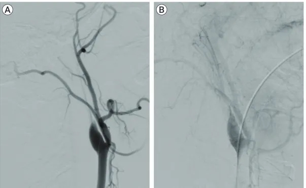
Admitted in the emergency room with left hemiplegia, right eyelid drop and swollen, chemosis, proptosis and ophthalmoplegia of the right eye. CTA revealed a direct CCF with dissection of the right ICA. DSA was indicat- ed to better understand the anatomy and to treat the le- sion. DSA showed a dissection in the right cervical ICA after the bulb with no flow beyond the dissection to the intracranial territory (Fig. 1). Right ECA injection with no communication to the cavernous sinus or anastomo-
Fig. 1. Right CCA DSA lateral view. (A) Early arterial phase of the right CCA. (B) Late arterial phase of the right CCA.
CCA, common carotid artery; DSA, digital subtraction angiography.
A B
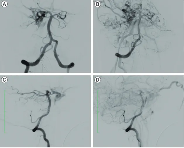
sis to the right ICA. Right vertebral injection was visual- ized and a good right posterior communicating (Pcom) artery that fills the fistula and the ICA bifurcation with a slow flow to the right middle cerebral artery (MCA) territory (Fig. 2). During the left ICA, a slight communi- cation through anterior communicating (Acom) artery and the right territory.
Patient under general anesthesia and a 6 F sheath was placed in the right femoral artery. A 6 F guiding catheter (Guider Soft tip [Boston Scientific, Fremont, CA, USA]) was positioned in the right vertebral artery.
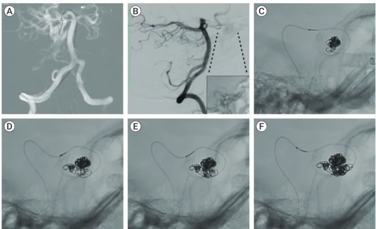
Through the guiding catheter a microcatheter Eche- lon 10 (Ev3, Irvine, CA, USA) with a Transend 0.014 Platinum guidewire (Stryker, CA, USA) was advanced through the Pcom artery to achieve the fistular point in the cavernous segment of the right ICA (Fig. 3). We
started to deploy 2 Target coils (Stryker, CA, USA) in the cavernous sinus and after that, we gently pullbacked the microcatheter into the fistula point and 8 Target Coils was detached to occlude the fistula point and the ICA beyond the ophthalmic artery (Fig. 3). Angiography demonstrated exclusion of the fistula and normal flow by the Pcom artery to the intracranial segments of the right ICA (Fig. 4). DSA control by the left ICA showed the same flow to the right territory.
DISCUSSION
An endovascular approach is the first treatment op- tion for direct CCF, with wide variety approach types:
transarterial, transvenous and retrograde. In this case,
Fig. 2. Right vertebral artery DSA. (A) Early arterial phase (frontal view) before treatment. (B) Late arterial phase (frontal view) before treatment. (C) Early arterial phase (lateral view) before treatment. (D) Late arterial phase (lateral view) be- fore treatment. DSA, digital subtraction angiography.
A B
C D
the most common arterial approach was not feasible due to the occlusion of the ipsilateral ICA. With a dissected carotid artery, the recanalization could be done to reach the cavernous sinus and this has already been described.
This approach is an option to avoid vasospasm during manipulation of Pcom or Acom arteries15) when retro- grade catheterization, mainly in young patients, can be dangerous. Because accessing the cavernous sinus and the fistula point through the venous route might be dif- ficult, the retrograde arterial access was chosen as treat- ment. Few reports in the literature using the retrograde approach to direct CCF4)14) are seen. All approaches may be acceptable in direct CCF, and the fastest but the least dangerous to the treatment was chosen.
Transarterial approach is the mainstay of endovascu- lar treatment. In some countries the detachable balloon is the first choice, but occasionally, technical problems
have been faced and the balloon had to be removed.
Thus, the use of coils or other material as the main en- dovascular approach is indicated. For indirect CCF, the transvenous approach became the preferred method, especially in cases with no arterial access. It is widely used with low rates of complications and high rates of treatment success (80%).12) Trans arterial approach is also possible in indirect CCF. The retrograde approach used in this case report should be the last option if some of the other approaches are feasible.
One of the most important tips-and-tricks for endo- vascular approach is to well recognize the fistula point and to close this hole with coils to avoid any minimal re- sidual fistula. We started the deployment of coils inside of the cavernous sinus and after that, with gentle manip- ulated the microcatheter and coils to deploy some coils inside of the fistula and then, we finished it with coils
Fig. 3. Right vertebral artery DSA during the treatment. (A) Right vertebral artery roadmap (frontal view) with the microcatheter navigation throw the Pcom artery to achieve the fistular point. (B) Right vertebral artery DSA (lateral view) after microcatheter navigation. Highlighted with dotted line: Unsubtraction image with the microcatheter position. (C) First coil deployment. (D) Second coil deployment. (E, F) Posi- tioned the microcatheter into the fistula point and Coils was detached to occlude the fistula point and the ICA beyond the ophthalmic artery.
DSA, digital subtraction angiography; ICA, internal carotid artery.
A B C
D E F
inside of the ICA beyond the ophthalmic artery.
Vertebral artery injections demonstrated completely occlusion of the fistula, with anterograde flow normal- ization into the right MCA and ACA. After that, the guiding catheter was withdrawn from the vertebral ar- tery and it was positioned in the left ICA to confirm the fistula occlusion and to have a better flow by the Acom artery to the right hemisphere.
CONCLUSIONS
We present a rare case of a combined traumatic cere- brovascular lesion, right carotid artery dissection and a right direct CCF treated by a retrograde endovascular
approach by the vertebral artery through the Pcom ar- tery to reach the fistula point and a complete cure of the CCF was achieved. The patient exhibited a minor neu- rological improvement of the left hemiplegia after the treatment.
Disclosure
The authors report no conflict of interest concerning the materials or methods used in this study or the find- ings specified in this paper.
REFERENCES
1. Barrow DL, Spector RH, Braun IF, Landman JA, Tindall Fig. 4. Right vertebral artery DSA after treatment. (A) Early arterial phase of the right vertebral artery (frontal view) after
endovascular treatment. (B) Late arterial phase of the right vertebral artery (frontal view) after endovascular treatment. (C) Early arterial phase (lateral view) after treatment. (D) Unsubtraction image of arterial phase (lateral view) after treatment.
DSA, digital subtraction angiography.
A B
C D
SC, Tindall GT. Classification and treatment of sponta- neous carotid-cavernous sinus fistulas. J Neurosurg. 1985 Feb;62(2):248-56.
2. Biffl WL, Moore EE, Ryu RK, Offner PJ, Novak Z, Coldwell DM, et al. The unrecognized epidemic of blunt carotid arte- rial injuries: early diagnosis improves neurologic outcome.
Ann Surg. 1998 Oct;228(4):462-70.
3. Chen CC, Chang PC, Shy CG, Chen WS, Hung HC. CT an- giography and MR angiography in the evaluation of carotid cavernous sinus fistula prior to embolization: a comparison of techniques. AJNR Am J Neuroradiol. 2005 Oct;26(9):2349-56.
4. Debrun GM, Ausman JI, Charbel FT, Aletich VA. Access to the cavernous sinus through the vertebral artery: technical case report. Neurosurgery. 1995 Jul;37(1):144-6; discussion 146-7.
5. De Campos JM, Ferro MO, Burzaco JA, Boixadós JR. Sponta- neous carotid-cavernous fistula in osteogenesis imperfecta. J Neurosurg. 1982 Apr;56(4):590-3.
6. Ducruet AF, Albuquerque FC, Crowley RW, McDougall CG.
The evolution of endovascular treatment of carotid cavernous fistulas: a single-center experience. World Neurosurg. 2013 Nov;80(5):538-48.
7. Fox R, Pope FM, Narcisi P, Nicholls AC, Kendall BE, Hou- rihan MD, et al. Spontaneous carotid cavernous fistula in Ehlers Danlos syndrome. J Neurol Neurosurg Psychiatry.
1988 Jul;51(7):984-6.
8. Gemmete JJ, Ansari SA, Gandhi D. Endovascular treatment of carotid cavernous fistulas. Neuroimaging Clin N Am. 2009
May;19(2):241-55.
9. Halbach VV, Hieshima GB, Higashida RT, Reicher M. Carot- id cavernous fistulae: indications for urgent treatment. AJR Am J Roentgenol. 1987 Sep;149(3):587-93.
10. Higashida RT, Halbach VV, Tsai FY, Norman D, Pribram HF, Mehringer CM, et al. Interventional neurovascular treatment of traumatic carotid and vertebral artery lesions: results in 234 cases. AJR Am J Roentgenol. 1989 Sep;153(3):577-82.
11. Lewis AI, Tomsick TA, Tew JM Jr. Management of 100 consecutive direct carotid- cavernous fistulas: Results of treatment with detachable balloons. Neurosurgery. 1995 Feb;36(2):239-44; discussion 244-5.
12. Meyers PM, Halbach VV, Dowd CF, Lempert TE, Malek AM, Phatouros CC, et al. Dural carotid cavernous fistula: defini- tive endovascular management and long-term follow-up. Am J Ophthalmol. 2002 Jul;134(1):85-92.
13. Ohlsson M, Consoli A, Rodesch G. Endovascular treatment of carotico-cavernous fistulas with acrylic glue: a series of nine cases. Neuroradiology. 2016 Dec;58(12):1181-8.
14. Oran I, Parildar M, Memis A, Dalbasti T. Posttraumatic intradural internal carotid artery-cavernous sinus fistula associated with ipsilateral carotid dissection. Transarterial embolization with detachable coils. Interv Neuroradiol. 2004 Mar;10(1):63-8.
15. Wilms G, Demaerel P, Lagae L, Casteels I, Mombaerts I. Di- rect caroticocavernous fistula and traumatic dissection of the ipsilateral internal carotid artery: endovascular treatment.
Neuroradiology. 2000 Jan;42(1):62-5.