773
Copyrights © 2019 The Korean Society of Radiology
Case Report
J Korean Soc Radiol 2019;80(4):773-776 https://doi.org/10.3348/jksr.2019.80.4.773 pISSN 1738-2637 / eISSN 2288-2928Unilateral Ureteric
Entrapment within the Sacroiliac Joint
Causing Unilateral
Hydroureteronephrosis
한쪽 수신 요관증을 유발한 요관의 천장관절 포착의 예
Jung Hwan Lee, MD1 , Bum Sang Cho, MD1,2*
1Department of Radiology, Chungbuk National University Hospital, Cheongju, Korea
2Department of Radiology, College of Medicine and Medical Research Institute, Chungbuk National University, Cheongju, Korea
Ureteric entrapment within the sacroiliac joint (SIJ) is very rare. However, we experienced a case of a 77-year-old woman who presented with right hydronephrosis during a routine health check. Computed tomography and retrograde pyelography showed entrapment of the right ureter at the level of the SIJ, with a right proximal hydroureter, which was confirmed during surgery. Herein, we described this case and present a review of the literature on similar cases.
Index terms Hydronephrosis; Sacroiliac Joint; Computed Tomography, X-Ray; Ureter; Urography
INTRODUCTION
Ureteric entrapment within the sacroiliac joint (SIJ) is a very rare condition, only 5 such cases have been reported to date (1-5). Almost all reported cases of ureteric en- trapment within the SIJ were associated with a history of trauma (2-4). However, we ex- perienced a case of unilateral ureteric entrapment within the SIJ in an elderly woman without a history of trauma. Here in we present this case along with a review of other similar cases.
CASE REPORT
A 77-year-old woman presented to our hospital with right hydronephrosis, discovered during a routine health check at outside hospital. She appeared healthy and had no his-
Received September 13, 2018 Revised October 5, 2018 Accepted October 23, 2018
*Corresponding author Bum Sang Cho, MD Department of Radiology, College of Medicine and Medical Research Institute, Chungbuk National University, 776 1sunhwan-ro, Seowon-gu, Cheongju 28644, Korea.
Tel 82-43-269-6365 Fax 82-43-269-6479
E-mail [email protected] This is an Open Access article distributed under the terms of the Creative Commons Attribu- tion Non-Commercial License (https://creativecommons.org/
licenses/by-nc/4.0) which permits unrestricted non-commercial use, distribution, and reproduc- tion in any medium, provided the original work is properly cited.
ORCID iDs Bum Sang Cho https://
orcid.org/0000-0002-3006-9207 Jung Hwan Lee
https://
orcid.org/0000-0002-8815-4092
jksronline.org
774
Ureteric Entrapment within Sacroiliac Joint
A
D E
B C
Post voiding
tory of trauma. No signs or symptoms of urinary tract infection were found on physical ex- amination. Laboratory evaluations, including urinalysis, blood urea nitrogen, creatinine, and electrolytes were within normal limits.
Outside ultrasonography had been performed 2 months earlier and had revealed hydrone- phrosis in the right kidney. Subsequently, intravenous pyelography (IVP) was performed. On IVP, the proximal ureter and renal pelvocalyx were shown to be dilated (Fig. 1A).
She was then referred to our hospital, where retrograde pyelography (RGP) and retrograde ureteric catheterization were attempted. RGP revealed kinking of the right mid-ureter at the SIJ level and dilatation of the proximal ureter (Fig. 1B). We next attempted retrograde ureter- ic catheterization during cystoscopy; a guide-wire could be inserted into the proximal ureter, and showed a kinking appearance, but a double-J catheter could not be inserted into the proximal ureter because the right mid ureter was entrapped at the SIJ (Fig. 1C).
Dynamic computed tomography (CT) was performed. On dynamic CT, the right kidney
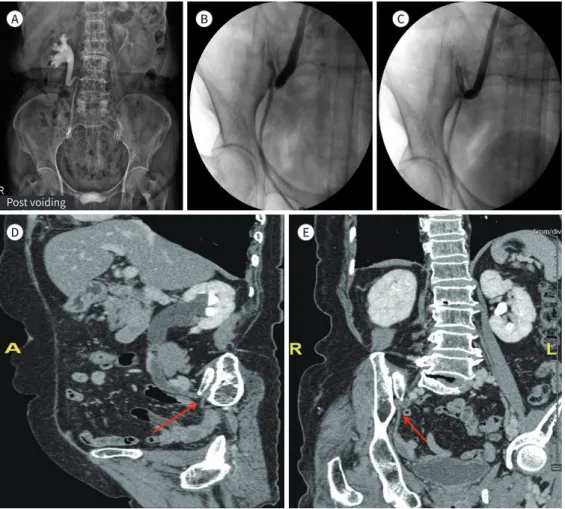
Fig. 1. A 77-year-old patient with unilateral hydronephrosis due to ureteric entrapment within the SIJ.
A. Intravenous pyelography shows that the proximal ureter and renal pelvocalyx are dilated.
B, C. Retrograde pyelogram shows dilatation of the right proximal ureter above the SIJ level and kinking and entrapment of the right mid-ureter at the SIJ.
D, E. Coronal (D) and oblique sagittal (E) images show that the right mid-ureter is seen to be entrapped in the right SIJ (arrow, D), and there is abrupt luminal narrowing of the ureter at the portion immediately distal to the SIJ (arrow, E).
SIJ = sacroiliac joint
https://doi.org/10.3348/jksr.2019.80.4.773 775
J Korean Soc Radiol 2019;80(4):773-776
was shown to have hydronephrosis accompanied by right proximal hydroureter and there was no evidence of a ureter stone or solid mass along the urinary tract. The size and shape of the right kidney were normal and the enhancement pattern was also shown to be normal. In coronal and oblique sagittal images, reconstructed with the 3D software TeraRecon (Aquari- us, Foster City, CA, USA; version 4.4.12), the right mid-ureter was shown to be entrapped in the right SIJ (Fig. 1D), and abrupt luminal narrowing of the ureter was seen at the portion im- mediately distal to the SIJ (Fig. 1E). Based on these imaging findings, our tentative diagnosis was ureter entrapment at the SIJ, and clinically differential diagnosis was ureter tuberculosis.
The patient underwent surgery in the urology department; it was confirmed that the upper ureter was severely dilated, that the mid-ureter was kinked, and that the ureter was en- trapped in the SIJ of the pelvic bone. Thus, ureter excision was performed at the entrapment site, and end to end anastomosis was performed. Microscopic examination revealed chronic inflammation with fibrosis at the entrapment point. The patient recovered well, and was dis- charged 1 week after surgery.
DISCUSSION
Ureteric entrapment within the SIJ is very rare, with only 5 cases reported to date; these in- clude one case reported by Otsuru et al. (1) from a cadaveric study, and there have been just 4 clinical cases reported. In 3 of the 4 clinical cases, there was a history of previous trauma; the exception is the case reported by Yeung et al. (5) In the first case, the ureter was entrapped during reduction of a pelvic fracture in a 6-year old boy (2). In the second case, the ureter was found entrapped in the SIJ during emergency exploration for ruptured bladder associated with pelvic fracture (3). In the third case, the ureter was found entrapped in the SIJ after emergency exploration for splenic injury and retroperitoneal hematoma associated with pel- vic fracture (4). These cases illustrate that significant pelvic trauma, especially pelvic bone fracture, may cause ureteric entrapment within the SIJ.
Yeung et al. (5) reported bilateral ureteral entrapment in an older woman who had no trau- matic or operative history, and they considered that her multiparous history or degenerative change of the SIJ predisposed her to ureteral entrapment at the SIJ.
In our case, the patient had no traumatic or operative history or multiparous history, but degenerative changes of osteophytes were seen, thus, we agree with Yeung et al. (5) that the widening caused by degenerative change may be a predisposing factor for ureteral entrap- ment at the SIJ.
In the case described by Yeung et al. (5), impaired renal function was observed, with ele- vated serum creatinine levels. Therefore, the patient was treated conservatively with bilateral percutaneous nephrostomy, and follow-up renal function tests showed normal creatinine level. Long-term management was discussed with the patient and her relatives. The option of bilateral uretero-ureterostomy was offered but declined in view of old age and fragility (5).
In contrast to the case by Yeung et al. (5), with a clinical renal problem, our patient did not show any clinical problem. We think that the hydronephrosis caused by ureteral entrapment in our case had occurred long ago; it had become a chronic problem, but did not result in complete obstruction of urine flow. We checked for signs of chronic inflammation and fibro-
jksronline.org
776
Ureteric Entrapment within Sacroiliac Joint
sis at the entrapment point, which supported our hypothesis. The imaging study revealed se- vere constriction of the lower ureter. Ureteric tuberculosis is characterized by a thickened ureteric wall and stricture. Stricture usually involves the distal one-third of the ureter and oc- curs at sites of normal anatomic narrowing, such as the uretero-pelvic junction, pelvic brim, and uretero-vesical junction (6). In our urologic department, ureter excision was performed in consideration of the possibility of tuberculous ureter. After excising the entrapped ureter, end-to-end anastomosis was performed.
In our case, imaging findings by RGP and multi-detector CT facilitated diagnosis. There- fore, advances in imaging techniques could make it possible to check for hidden cases of ureteral entrapment in the SIJ.
In conclusion, when encountering cases of hydronephrosis and hydroureter above the SIJ level, it is necessary to consider ureteral entrapment at the SIJ.
Conflicts of Interest
The authors have no potential conflicts of interest to disclose.
REFERENCES
1. Otsuru Y, Kondo C, Hara S, Takahashi H, Matsuno K. Ureteric entrapment in sacroiliac joint causing hydro- ureter and ipsilateral kidney hypertrophy. Anat Sci Int 2018;93:394-399
2. Noakes JE, Wese FX, Churchill BM. Ureteral entrapment injury in blunt pelvic trauma: a case report. J Urol 1982;127:764-765
3. Konety BR, Xerogeanes J, Corral DA, Gruen G, Hrebinko R. Ureteral entrapment in the sacroiliac joint in pel- vic trauma. J Trauma 1996;41:150-151
4. Shetty R, Thomas J, Rao S. Ureteral entrapment in traumatic sacroiliac joint dislocation - a rare injury. Indi- an J Urol 2001;18:75-77
5. Yeung JT, Ma JK, Yung AW, Cheng RL. Idiopathic bilateral ureteric entrapment within the sacroiliac joints causing bilateral hydroureteronephrosis. Clin Radiol 2010;65:662-665
6. Burrill J, Williams CJ, Bain G, Conder G, Hine AL, Misra RR. Tuberculosis: a radiologic review. Radiographics 2007;27:1255-1273
한쪽 수신 요관증을 유발한 요관의 천장관절 포착의 예
이정환1 · 조범상1,2*
요관의 천장관절 포착에 의한 수신증 발생은 매우 드문 것으로 알려져 있다. 저자는 건강검 진에서 우연히 발견된 수신증으로 내원한 77세 여자 환자의 전산화단층촬영과 역행성 요로 조영술에서 중부 요관이 천장관절에 포착된 것을 발견하였고, 이후 수술을 통해 확인하였다.
이에 문헌고찰과 함께 증례를 보고하는 바이다.
1충북대학교병원 영상의학과, 2충북대학교 의과대학 영상의학교실