Received December 19, 2008, Revised January 19, 2009, Accepted for publication January 29, 2009
Reprint request to: Nack-In Kim, M.D., Department of Dermatology, College of Medicine, Kyung Hee University, 1, Hoegi-dong, Dong- daemun-gu, Seoul 130-702, Korea. Tel: 82-2-958-8501, Fax: 82-2- 969-6538, E-mail: [email protected]
ORIGINAL ARTICLE
Expression of Toll-like Receptor 2 in Cultured Human Keratinocytes: The Effect of Bacterial Antigens,
Cytokines and Calcium Concentration
Bark-Lynn Lew, M.D., Woo-Young Sim, M.D., Nack-In Kim, M.D.
Department of Dermatology, College of Medicine, Kyung Hee University, Seoul, Korea
Background: Toll-like receptors (TLRs) are expressed by hu- man epidermal keratinocytes and are involved in immune responses. Objective: The goal of this was to investigate the expression of TLR2 in response to bacterial antigens, cyto- kines, and different calcium concentrations. Methods: The expression of TLR2 was assessed after stimulation by lip- oteichoic acid (LTA) and streptolysin O (SLO). In addition, TLR2 expression was evaluated after treatment with IFN-γ and TNF-α, and different concentrations of calcium. The ex- pression levels of TLR2 mRNA and protein were studied us- ing RT-PCR and Western blot analysis. Results: Cultured hu- man epidermal keratinocytes constitutively expressed TLR2 and the expression was stimulated by LTA and SLO; in addi- tion, IFN-γ and TNF-α upregulated TLR2 expression.
However, the changes in TLR2 expression associated with the calcium concentrations were insignificant. Conclusion:
TLR2 expression increased with the concentration and dura- tion of bacterial pathogens and this increase was amplified by several cytokines, from activated keratinocytes and other cells. (Ann Dermatol 21(4) 337∼344, 2009)
-Keywords-
Calcium, Cytokine, Keratinocyte, Microbacterial antigen, Toll-like receptor 2
INTRODUCTION
The immediate response by the host to microbial invasion
is mediated by the innate immune system which, in turn, alerts the adaptive immune response via cytokines/chemo- kines or co-stimulatory signals expressed by antigen-pre- senting cells. The innate immune system senses invading organisms via germ-line encoded, non-clonal pattern rec- ognition receptors that recognize molecular patterns shared by large groups of pathogens. One group of such receptors is the Toll-like receptors (TLRs); these receptors are the mammalian homologue of Toll, a type 1 trans-mem- brane receptor first described in Drosophila where, in adult flies, is involved in antifungal defence1. The TLR structure includes leucine-rich repeats in the extracellular domain and a cytoplasm domain that shares significant homology with the interleukin 1 receptor (IL-1R) signaling domain, termed the Toll/IL-1R (TIR) region2,3. Signaling pathways triggered via the TIR domain induce the activa- tion of the transcription factor NF-κB4, which translocates from the cytoplasm to the nucleus where it binds to the promoter regions of a wide variety of immune and in- flammatory genes.
The TLRs play a crucial role in the induction of anti- microbial responses in different cells. In the past few years, 10 different human TLRs have been identified. TLR2 has broad specificity and is involved in the recognition of yeast5, a wide variety of microbial compounds from Gram-positive bacteria such as peptidoglycan6 and lip- oproteins7,8, as well as mycobacterial cell wall compo- nents9. TLR2 is required for pro-inflammatory signaling to lipoteichoic acid, lipoproteins and lipoarabinomannan. By contrast, TLR4, together with CD14, recognizes lip- opolysaccharides (LPS) on Gram-negative bacteria10,11. The epidermis, the outermost skin layer, provides the first line of defense against the external environment. The ma- jor cell type of the epidermis is the keratinocyte. In addi- tion to forming a physical barrier, keratinocytes have been
shown to play an important regulatory role in the cuta- neous inflammatory and immune response by producing a variety of cytokines. Keratinocyte-derived cytokines are critical to mobilizing leukocytes from the blood and in sig- naling other cutaneous cells. In addition to regulating im- munological and inflammatory responses, the epidermal keratinocytes contribute to the protective barrier of the ep- ithelium and participate in the host defense by destroying invading microorganisms12,13. Human keratinocytes ex- press TLRs, which enable them to initiate the innate im- mune response to environmental microbiological chal- lenges. Several reports have demonstrated that human ker- atinocytes express TLR1, 2, 3, 4, 5, 6, 9 and 102-4,6-8,13,14; however, the expression of some of these compounds was not detected in all studies15.
Among them, TLR2 has been thought to have a more im- portant role in the keratinocytes than the others. This is because the main pathogens in the skin are predominantly gram positive bacteria. The molecules known to affect TLR2 activation include: TNF-α, IL-1β, IL-6, IL-8, IL-10, IL-12, NO, IL-4, IL-5, IL-6, and IL-1316-18. Baker et al.19 re- ported on the difference in TLR2 expression in psoriatic skin compared to normal skin and suggested that the dif- ference might be due to altered epidermal differentiation or the effects of proinflammatory cytokines. Therefore, TLR2 might be affected by TNF-α and IFN-γ, key cyto- kines in psoriasis, and the calcium level, which is asso- ciated with keratinocyte differentiation.
The aim of this study was to confirm the TLR-inducing ef- fects of bacterial antigens on keratinocytes, and to de- termine whether the expression of TLR2 is modulated by cytokines and calcium levels.
MATERIALS AND METHODS
Culture of human epidermal keratinocytes
Normal human keratinocytes were isolated from neonatal foreskin, and were cultured in 154 medium (Cascade Biologics, Portland, OR, USA) with human keratinocyte growth supplement (Cascade Biologics) and 1% pen- icillin-streptomycin-amphotericin B (10,000 U/ml, 10,000 μg/ml, and 25μg/ml, respectively; GIBCO BRL., Grand Island, NY, USA) in a humidified atmosphere containing 5% CO2 at 37oC. Isolated keratinocytes were cultivated at 37oC and 5% CO2 in Epilife (Cascade Biologics). Cells passed three times were used for the experiments and the media was renewed every second day.
Reagents
Lipoteichoic acid (LTA), derived from Staphylococcus aur- eus, and streptolysin O (SLO), derived from Streptococcus
pyogens, were purchased from sigma. LTA was used at a concentration of 0.1μg/ml, 1μg/ml, and 10μg/ml, and SLO was used at a concentration of 0.1 U/ml, 1 U/ml, and 10 U/ml. The duration of treatment with LTA and SLO was 3 and 24 hrs. Both IFN-γ and TNF-α (R&D System, Minneapolis, MN, USA) were used at a concentration of 10 ng/ml, 50 ng/ml, and 100 ng/ml. Culture medium with and without calcium (Medium 154CF) was purchased from Cascade Biologics. The concentrations of calcium used were: 0.05 mM, 0.1 mM, and 0.2 mM. The duration of treatment with IFN-γ, TNF-α and calcium was 6 and 24 hrs.
RNA isolation
Total cellular RNA was purified from cultured cells by the RNA-Bee solution (Tel-test Inc., Friendswood, TX, USA).
The cells were lysed with 1.0 ml RNA-Bee solution and extracted by adding 0.1 volume chloroform to the re- action tube. After the mixture was centrifuged at 12,000 g (4oC) for 15 min, the supernatant was transferred to a new 1.5 ml tube. After centrifugation, the cells were removed and an equal volume of isopropanol was added. The sam- ples were precipitated for 15 min at 4oC. After the mixture was centrifuged, the pellet was washed with 800μl of 75% ethanol and stored in diethylpyrocarbonate (DEPC) treated water. The total RNAs were measured at 260 nm with a spectrophotometer (Ultraspec 2000, Parmacia Biotech, Cambridge, England).
Reverse transcription-polymerase chain reaction (RT- PCR)
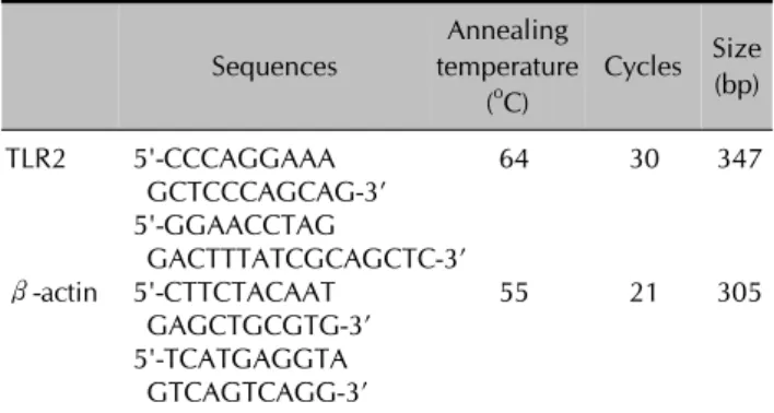
First-strand cDNA synthesis was performed by reverse transcription in a total volume of 20μl of reaction mixture containing 1μg of RNA, 1x reaction buffer, 1 mM each dNTP, 5μM random primers, 20 units RNase inhibitor, and 20 units AMV reverse transcriptase (Promega, Madison, WI, USA). The reaction mixture was incubated at 25°C for 10 min, 42°C for 1 hr, and terminated by heating at 95°C for 5 min. The polymerase chain reaction (PCR) was performed with 2μl of cDNA in a 50μl re- action mixture of 1x PCR buffer (10 mM Tris-HCl, pH 9.0, 50 mM KCl, 1.5 mM MgCl2), 200μM of each dNTP, 20 pmol of sense and antisense primer, and 1 unit of Ampli- Taq Gold DNA polymerase (Roche, Applied Biosystems, Foster City, CA, USA). The primer sequences are shown in Table 1. The amplification conditions were as follows: first denaturation at 95oC for 14 min, then denaturation at 95oC for 1 min, annealing at 53oC to 64oC for 1 min, and extension at 72oC for 1 min for 21 to 30 cycles, and final extension at 72oC for 5 min. Ten microliters of the PCR products were separated by electrophoresis on a 2% agar-
Table 1. Oligonucleotide sequences of PCR primers Sequences
Annealing temperature
(oC)
Cycles Size (bp)
TLR2 5'-CCCAGGAAA 64 30 347
GCTCCCAGCAG-3’
5'-GGAACCTAG
GACTTTATCGCAGCTC-3’
β-actin 5'-CTTCTACAAT 55 21 305
GAGCTGCGTG-3’
5'-TCATGAGGTA GTCAGTCAGG-3’
Fig. 1. (A) TLR2 mRNA expression levels at 0.1μg/ml, 1μg/ml and 10μg/ml of LTA compared to the control group for 3 hrs and 24 hrs (n=3, *p<0.05 when compared to the control). (B) TLR2 protein expression levels at 0.1μg/ml, 1μg/ml and 10μg/ml of LTA for 3 hrs and 24 hrs compared to the control group (n=3, *p<0.05 when compared to the control).
ose gel containing ethidium bromide and visualized by image analysis (Gel Doc 1000 gel documentation system, Bio-Rad, Hercules, CA, USA). The results are reported as the ratio of relative absorbance of TLR2/β-actin analyzed by densitometry. All experiments were performed in triplicate.
Western blot analysis
The cells were washed in PBS and solubilized in lysis buf- fer (20 mM Tris (pH 7.5), 150 mM NaCl, and 1 mM phe- nylmethylsulfonyl) for 15 min on ice and lysates were cen- trifuged at 13,000×g for 20 min. The protein concen- tration was determined by the BCA protein assay (Pierce,
Rockford, IL, USA). Fifty micrograms of protein was frac- tionated on 12.5% SDS-PAGE and transferred by electro- phoresis to a nitrocellulose membrane. The membranes were blocked with 5% non-fat dry milk in TBS-T buffer (20 mM Tris base, 137 mM NaCl, pH 7.6, 0.1%
Tween-20) at room temperature for 1 hr and incubated with primary antibodies (TLR2 and β-actin; Santa Cruz Biotechnology, CA, USA) at a 1:200 dilution with 5%
non-fat dry milk in TBS-T at room temperature for 1 hr.
After washing with TBS-T for 1 hr, the membranes were reacted with horseradish peroxidase-conjugated anti-rabbit IgG antibody diluted 1: 2,000 with TBS-T at room temper- ature for 1 hr. After washing the membranes with TBS-T for 1 hr, the membrane was reacted with ECL detection re- agents (ECL Western blotting detection reagents and analy- sis system, Amersham, Aylesbury, UK) according to the manufacture’s protocol and autoradiograms were exposed with Konica X-ray film (Konica corporation, Tokyo, Japan).
All experiments were performed in triplicate.
Statistical analysis
The results are expressed as the mean±standard devia- tion. Statistical significance was tested using the ANOVA with probabilities. A p value of less than 0.05 was consid- ered significant.
Fig. 2. (A) TLR2 mRNA expression levels at 0.1 U/ml, 1 U/ml and 10 U/ml of SLO compared to the control group at 3 hrs and 24 hrs (n=3, *p<0.05 when compared to the control). (B) TLR2 protein expression levels at 0.1 U/ml, 1 U/ml and 10 U/ml of SLO for 3 hrs and 24 hrs (n=3, *p<0.05 when compared to the control).
RESULTS
Expression of TLR2 mRNA and protein in cultured keratinocytes stimulated with LTA and SLO
After treatment with LTA for 3 hrs, the expression of TLR2 mRNA showed a tendency to increase as the LTA concen- tration increased. The increase of expression was statisti- cally significant compared to the control, only at the con- centration of 10μg/ml. After 24 hrs, the increase of ex- pression was more significant than at 3 hrs. At all concen- trations of LTA, the difference was statistically significant compared to the control (Fig. 1A). The results of the TLR2 protein expression were similar to those of the TLR2 mRNA. After treatment with LTA for 3 hrs and 24 hrs, the TLR2 protein expression levels increased. The difference was statistically significant at 10μg/ml, after 3 hrs, and 0.1μg/ml, 1μg/ml and 10μg/ml, after 24 hrs (Fig. 1B).
The expression of TLR2 mRNA and protein after treatment with SLO was also analyzed in the same manner. After treatment with SLO for 3 hrs, no significant change was detected. However, after treatment for 24 hrs, the TLR2 mRNA expression showed a significant increase asso- ciated with SLO stimulation. At all concentrations of SLO, the TLR2 mRNA expression increased significantly com- pared to the control (Fig. 2A). The results of the TLR2 pro- tein expression were the same as for the TLR2 mRNA.
After 24 hrs of treatment, it was observed that SLO in- creased the expression of TLR2 protein and the difference was more significant at greater SLO concentrations (Fig.
2B).
These findings provide evidence that both LTA and SLO stimulate the expression of TLR2 in human epidermal keratinocytes. The stimulative effects of both on TLR2 were distinct after 24 hrs of treatment and were propor- tional to the concentration levels.
Expression of TLR2 mRNA and protein in cultured keratinocytes stimulated with IFN-γ and TNF-α
The RT-PCR showed that IFN-γ was associated with an increase in the level of TLR2 mRNA after stimulation for 6 hrs. After treatment for 24 hrs, the increase in TLR2 mRNA expression was more significant. These effects were de- tected even at the lowest concentration of IFN-γ, 10 ng/ml; no difference in the concentrations was observed (Fig. 3A). The results of TLR2 protein expression after IFN- γ treatment were identical to the TLR2 mRNA results (Fig. 3B).
When TNF-α was added to the culture of epidermal kera- tinocytes, the TLR2 expression increased. This effect was confirmed in both the mRNA and protein levels (Fig. 4).
The TLR2-stimulating effect of TNF-α was more pro- nounced after treatment for 24 hrs compared to 6 hrs;
Fig. 3. (A) TLR2 mRNA expression levels after treatment with IFN-γ (10 ng/ml, 50 ng/ml and 100 ng/ml) for 6 hrs and 24 hrs compared to the control group (n=3, *p<0.05 when compared to the control). (B) TLR2 protein expression levels at 10 ng/ml, 50 ng/ml and 100 ng/ml of IFN-γ for 6 hrs and 24 hrs (n=3, *p<0.05 when compared to the control).
Fig. 4. (A) TLR2 mRNA expression levels after treatment with TNF-α (10 ng/ml, 50 ng/ml and 100 ng/ml) compared to the control group for 6 hrs and 24 hrs (n=3, *p<0.05 when compared to the control). (B) TLR2 protein expression levels at 10 ng/ml, 50 ng/ml and 100 ng/ml of TNF-α for 6 hrs and 24 hrs (n=3, *p<0.05 when compared to the control).
however, the differences were not statistically significant.
These results showed that IFN-γ and TNF-α significantly upregulated the expression of TLR2, regardless of their
concentration.
Fig. 5. (A) TLR2 mRNA expression levels according to the calcium concentration (0.05 mM, 0.1 mM, and 0.2 mM) in cultured human keratinocytes at 6 hrs and 24 hrs (n=3). (B) TLR2 protein expression levels according to the calcium concentration (0.05 mM, 0.1 mM, and 0.2 mM) in cultured human keratinocytes at 6 hrs and 24 hrs (n=3).
Expression of TLR2 mRNA and protein in cultured keratinocytes stimulated with calcium
Six hours after the addition of calcium, there was no sig- nificant change observed in the mRNA or protein, at all concentrations studied. Only the treatment with 0.2 mM of calcium for 24 hrs, was associated with an increased expression of TLR2 mRNA and protein; however, the dif- ference was not statistically significant (Fig. 5).
DISCUSSION
The results of this study showed that human keratinocytes constitutively express TLR2 mRNA and protein. Treatment of keratinocytes with LTA and SLO stimulated TLR2 ex- pression, especially after 24 hrs of treatment. IFN-γ and TNF-α both strongly induced TLR2 expression in kerati- nocytes, regardless of the concentration used. However, there were no significant changes in the expression of TLR2 associated with calcium concentrations.
Several prior studies reported that the expression of TLRs, including TLR2, in human keratinocytes was stimulated by yeast and bacteria. Most of these prior studies investigated the changes of TLR2 and/or TLR4 expression by the LPS of gram negative bacteria20-22; however, some investigated the effects of Staphylococcus aureus itself15 or, the LTA of Staphylococcus aureus23. In this study we demonstrated
that both SLO, a pathogenic toxin of Streptococcus pyo- genes, and LTA stimulated the expression of TLR2 in hu- man keratinocytes. Streptococcus pyogenes and Staphylo- coccus aureus are pathogens known to cause cutaneous infectious diseases. In addition, both are the main cause of secondary infection in atopic dermatitis, and contribute to the aggravation of atopic dermatitis. Streptococcus pyo- genes plays an important role in the development of psor- iasis and SLO has been associated with guttate psoriasis.
Staphylococcus aureus has been demonstrated in 20~50% of patients with psoriasis24,25. The results of this study showed that TLR2 expression was upregulated in keratinocytes as the concentration of LTA and SLO in- creased, although the increase was not directly propor- tional. These effects were more significant after 24 hrs of stimulation, which is consistent with the results of pre- vious reports21-23. The increase of TLR2 expression by LTA was previously reported to be statistically significant only at concentrations of 10μg/ml23; however, in the present study, a significant increase of TLR2 expression was ob- served starting at concentrations as low as 0.1μg/ml.
TLRs, pathogen-recognition receptors, transduce signals leading to the activation of NF-κB that subsequently drive the immune reaction by the transcriptional induction of several genes coding for cytokines, chemokines, and ad- hesion molecules26-28. Many investigators have focused mainly on the downstream pathway after activation of the
TLRs, such as the induction of cytokines or chemokines;
several molecules have been shown to affect the activa- tion of TLRs16-18,29-31. However, in the present study the goal was to identify the factors that regulate or influence the expression of TLR. In this study, it was confirmed that both IFN-γ and TNF-α stimulate TLR2 expression; the ef- fects were strong enough to increase TLR2 at 6 hrs, a rela- tively short duration of treatment. The stimulation by the two cytokines appears to occur regardless of their concentration. Pivarcsi et al.21 investigated co-treatment with LPS+IFN-γ, and found that 10 ng/ml increased the expression of TLR2 and the stimulation increased as the duration of treatment increased.
Therefore, induction of TLR2 by bacterial pathogens such as LTA and SLO might require a certain time interval for stimulation and this might have some correlation to their concentration. In the case of IFN-γ and TNF-α, both the duration of treatment and the concentration appeared to have little importance.
In addition, we studied whether TLR2 expression was in- fluenced by the calcium concentration. Prior to this study, we hypothesized that TLR2 activation would be influ- enced by the differentiation of epidermal keratinocytes, and expected that the expression of TLR2 would change with the calcium concentration. However, there was no significant effect of the calcium concentration on TLR2 ob- served in this study. It is well known that a culture me- dium with a high calcium concentration induces keratino- cytes to differentiate and proliferate. In addition, it has been reported that keratinocytes maintain a proliferative basal cell phenotype when they are cultured in media with a low calcium concentration (0.03 mM), and they are induced to differentiate by increasing the calcium concen- tration to above 0.1 mM32. A change of TLR2 expression was not detected at the low calcium concentration of 0.05 mM, in which, keratinocytes have a tendency to pro- liferate, or at the relatively high concentration of 0.2 mM, where keratinocytes tend to differentiate. Kawai et al.33 re- ported that TLR2 was expressed throughout the entire epidermis. By contrast, another study reported that TLR2 was highly expressed in proliferating basal keratinocytes19. Although it is difficult to interpret the present results, the calcium concentration itself might have little effect on TLR2 expression.
Baker et al.19 demonstrated a difference in TLR2 ex- pression in psoriatic epidermis compared to normal epi- dermis; TLR2 was more highly expressed in the upper epi- dermis of psoriatic skin, while, in normal skin, TLR2 was expressed throughout the epidermis, with higher staining in the basal keratinocytes. These findings might explain the different TLR2 expression patterns in psoriatic
epidermis. This study confirmed that LTA and SLO induce TLR2 expression, and that proinflammatory cytokines such as IFN-γ and TNF-α stimulate the expression of TLR2. In the presence of psoriasis, epidermal differentiation is al- tered, and the proliferation and differentiation balance is distorted34. In addition, IFN-γ and TNF-α are the main cytokines involved in the pathogenesis of psoriasis35. These results suggest that TLR2 is upregulated in response to the presence of Gram positive bacteria in the keratin layer24,25. In addition, TLR2 upregulation might not be sec- ondary to altered keratinocyte differentiation but result from the effects of proinflammatory cytokines such as TNF-α and IFN-γ present in the psoriatic lesions.
In conclusion, TLR2 plays a crucial role in the induction of antimicrobial responses in specific immune cells; it was found to be expressed in human keratinocytes even with- out stimulation. The expression of TLR2 increased with the concentration and duration of treatment with bacterial pathogens, and the increase was amplified by several cy- tokines, from activated keratinocytes and other cells.
Therefore, these results help us understand the expression of TLR2 in cutaneous infectious diseases as well as in- flammatory or immune-mediated skin diseases such as atopic dermatitis and psoriasis.
REFERENCES
1. Lemaitre B, Nicolas E, Michaut L, Reichhart JM, Hoffmann JA. The dorsoventral regulatory gene cassette spatzle/Toll/
cactus controls the potent antifungal response in Drosophila adults. Cell 1996;86:973-983.
2. Rock FL, Hardiman G, Timans JC, Kastelein RA, Bazan JF. A family of human receptors structurally related to Drosophila Toll. Proc Natl Acad Sci U S A 1998;95:588-593.
3. Kopp EB, Medzhitov R. The Toll-receptor family and control of innate immunity. Curr Opin Immunol 1999;11:13-18.
4. O’Neill LA, Greene C. Signal transduction pathways acti- vated by the IL-1 receptor family: ancient signaling machi- nery in mammals, insects, and plants. J Leukoc Biol 1998;63:650-657.
5. Underhill DM, Ozinsky A, Hajjar AM, Stevens A, Wilson CB, Bassetti M, et al. The Toll-like receptor 2 is recruited to macrophage phagosomes and discriminates between patho- gens. Nature 1999;401:811-815.
6. Yoshimura A, Lien E, Ingalls RR, Tuomanen E, Dziarski R, Golenbock D. Cutting edge: recognition of Gram-positive bacterial cell wall components by the innate immune system occurs via Toll-like receptor 2. J Immunol 1999;163:1-5.
7. Lien E, Sellati TJ, Yoshimura A, Flo TH, Rawadi G, Finberg RW, et al. Toll-like receptor 2 functions as a pattern recog- nition receptor for diverse bacterial products. J Biol Chem 1999;274:33419-33425.
8. Brightbill HD, Libraty DH, Krutzik SR, Yang RB, Belisle JT,
Bleharski JR, et al. Host defense mechanisms triggered by microbial lipoproteins through toll-like receptors. Science 1999;285:732-736.
9. Underhill DM, Ozinsky A, Smith KD, Aderem A. Toll-like re- ceptor-2 mediates mycobacteria-induced proinflammatory signaling in macrophages. Proc Natl Acad Sci U S A 1999;
96:14459-14463.
10. Poltorak A, He X, Smirnova I, Liu MY, Van Huffel C, Du X, et al. Defective LPS signaling in C3H/HeJ and C57BL/10ScCr mice: mutations in Tlr4 gene. Science 1998;282:2085-2088.
11. Qureshi ST, Lariviere L, Leveque G, Clermont S, Moore KJ, Gros P, et al. Endotoxin-tolerant mice have mutations in Toll-like receptor 4 (Tlr4). J Exp Med 1999;189:615-625.
12. Jung HC, Eckmann L, Yang SK, Panja A, Fierer J, Morzycka- Wroblewska E, et al. A distinct array of proinflammatory cy- tokines is expressed in human colon epithelial cells in re- sponse to bacterial invasion. J Clin Invest 1995;95:55-65.
13. Wang B, Ruiz N, Pentland A, Caparon M. Keratinocyte proinflammatory responses to adherent and nonadherent group A streptococci. Infect Immun 1997;65:2119-2126.
14. Lebre MC, van der Aar AM, van Baarsen L, van Capel TM, Schuitemaker JH, Kapsenberg ML, et al. Human keratino- cytes express functional Toll-like receptor 3, 4, 5, and 9. J Invest Dermatol 2007;127:331-341.
15. Mempel M, Voelcker V, Kollisch G, Plank C, Rad R, Gerhard M, et al. Toll-like receptor expression in human ker- atinocytes: nuclear factor kappaB controlled gene activation by Staphylococcus aureus is toll-like receptor 2 but not toll-like receptor 4 or platelet activating factor receptor dependent. J Invest Dermatol 2003;121:1389-1396.
16. Kang SS, Kauls LS, Gaspari AA. Toll-like receptors: applica- tions to dermatologic disease. J Am Acad Dermatol 2006;
54:951-983.
17. Thoma-Uszynski S, Stenger S, Takeuchi O, Ochoa MT, Engele M, Sieling PA, et al. Induction of direct antimicrobial activity through mammalian toll-like receptors. Science 2001;291:1544-1547.
18. Supajatura V, Ushio H, Nakao A, Akira S, Okumura K, Ra C, et al. Differential responses of mast cell Toll-like receptors 2 and 4 in allergy and innate immunity. J Clin Invest 2002;
109:1351-1359.
19. Baker BS, Ovigne JM, Powles AV, Corcoran S, Fry L. Normal keratinocytes express Toll-like receptors (TLRs) 1, 2 and 5:
modulation of TLR expression in chronic plaque psoriasis. Br J Dermatol 2003;148:670-679.
20. Song PI, Park YM, Abraham T, Harten B, Zivony A, Neparidze N, et al. Human keratinocytes express functional CD14 and toll-like receptor 4. J Invest Dermatol 2002;119:
424-432.
21. Pivarcsi A, Bodai L, Rethi B, Kenderessy-Szabo A, Koreck A,
Szell M, et al. Expression and function of Toll-like receptors 2 and 4 in human keratinocytes. Int Immunol 2003;15:721- 730.
22. Lebre MC, Antons JC, Kalinski P, Schuitemaker JH, van Capel TM, Kapsenberg ML, et al. Double-stranded RNA-ex- posed human keratinocytes promote Th1 responses by in- ducing a Type-1 polarized phenotype in dendritic cells: role of keratinocyte-derived tumor necrosis factor alpha, type I in- terferons, and interleukin-18. J Invest Dermatol 2003;120:
990-997.
23. Park YM, Kwon HJ, Kang YS, Koo JK, Kim MY, Kim HO, et al. Expression of Toll-like receptor 4 on human keratinocytes by lipoteichoic acid. Korean J Dermatol 2006;44:15-21.
24. Aly R, Maibach HE, Mandel A. Bacterial flora in psoriasis. Br J Dermatol 1976;95:603-606.
25. Leung DY, Harbeck R, Bina P, Reiser RF, Yang E, Norris DA, et al. Presence of IgE antibodies to staphylococcal exotoxins on the skin of patients with atopic dermatitis. Evidence for a new group of allergens. J Clin Invest 1993;92:1374-1380.
26. McInturff JE, Modlin RL, Kim J. The role of toll-like receptors in the pathogenesis and treatment of dermatological disease.
J Invest Dermatol 2005;125:1-8.
27. Akashi-Takamura S, Miyake K. Toll-like receptors (TLRs) and immune disorders. J Infect Chemother 2006;12:233-240.
28. Pandey S, Agrawal DK. Immunobiology of Toll-like re- ceptors: emerging trends. Immunol Cell Biol 2006;84:333- 341.
29. Mizel SB, Honko AN, Moors MA, Smith PS, West AP.
Induction of macrophage nitric oxide production by Gram-negative flagellin involves signaling via heteromeric Toll-like receptor 5/Toll-like receptor 4 complexes. J Immu- nol 2003;170:6217-6223.
30. Dahl MV. Imiquimod: a cytokine inducer. J Am Acad Dermatol 2002;47:S205-208.
31. Nagase H, Okugawa S, Ota Y, Yamaguchi M, Tomizawa H, Matsushima K, et al. Expression and function of Toll-like re- ceptors in eosinophils: activation by Toll-like receptor 7 ligand. J Immunol 2003;171:3977-3982.
32. Watt FM, Green H. Stratification and terminal differentiation of cultured epidermal cells. Nature 1982;295:434-436.
33. Kawai K, Shimura H, Minagawa M, Ito A, Tomiyama K, Ito M. Expression of functional Toll-like receptor 2 on human epidermal keratinocytes. J Dermatol Sci 2002;30:185-194.
34. Bernard BA, Asselineau D, Schaffar-Deshayes L, Darmon MY. Abnormal sequence of expression of differentiation markers in psoriatic epidermis: inversion of two steps in the differentiation program? J Invest Dermatol 1988;90:801-805.
35. Bonifati C, Ameglio F. Cytokines in psoriasis. Int J Dermatol 1999;38:241-251.