329
Copyrights © 2013 The Korean Society of Radiology
INTRODUCTION
Pseudoangiomatous stromal hyperplasia (PASH), a rare be- nign lesion, shows the proliferation of the breast stromal tissue mimicking the low grade angiosarcoma (1-7).
The most common mammographic and ultrasound finding of PASH is a circumscribed mass without calcification and it is difficult to distinguish from the phyllodes tumor and fibroade- noma (1-4, 8). Up to our knowledge, PASH presenting as rapid bilateral breast enlargement, as seen in our case, is very rare. In addition, several English medical literature were reported in this kind of manifestation of PASH (3, 4, 8).
We described imaging findings of diffuse, infiltrating, and bi- lateral manifectation of PASH.
CASE REPORT
A 19-year-old female had massive, progressive enlargement of both breasts for eight months. Although there was a history of
dull ache in both breasts, breast enlargement was not associated with nipple discharge, fever, weight loss or any other clinical symptoms. She was nulliparous and had no prior history of breast disease or trauma.
Physical examination revealed markedly enlarged breasts, measuring over 30 cm in the transverse and craniocaudal di- mension of each breast. Both breasts were firm in consistency and movable without any discrete palpable mass. The overlying skin appeared red, and tense with engorged superficial vein.
Skin ulceration was not present. There was no evidence of axil- lary or supraclavicular lymphadenopathy. Routine hematologic and biochemical examinations were within normal limits.
Due to huge size of both breasts, mammography could not be evaluated. On ultrasound, a diffuse, heterogeneously hypoecho- ic and infiltrating lesion in both breasts was present with diffi- culty in delineating the normal architecture. Ill-defined tubular shaped cystic areas were scattered within this lesion without vascular flow (Fig. 1).
The core needle biopsy was done to eliminate the possibility
Case Report
pISSN 1738-2637
J Korean Soc Radiol 2013;68(4):329-332 http://dx.doi.org/10.3348/jksr.2013.68.4.329
Received October 23, 2012; Accepted January 8, 2013 Corresponding author: Su Kyung Jeh, MD Department of Radiology, Kangnam Sacred Heart Hospital, College of Medicine, Hallym University, 1 Singil-ro, Yeongdeungpo-gu, Seoul 150-950, Korea.
Tel. 82-2-829-5241 Fax. 82-2-832-1845 E-mail: [email protected]
This is an Open Access article distributed under the terms of the Creative Commons Attribution Non-Commercial License (http://creativecommons.org/licenses/by-nc/3.0) which permits unrestricted non-commercial use, distri- bution, and reproduction in any medium, provided the original work is properly cited.
Pseudoangiomatous stromal hyperplasia (PASH), first described by Vuitch et al., is a rare benign lesion, shows the proliferation of the breast stromal tissue. We experi- enced a case of a 19-year-old woman with bilateral PASH manifesting as marked en- largement of the breasts for a duration of several months. Morphologic evaluation of imaging modalities was done, including ultrasound, computed tomography and mag- netic resonance images. Here, we describe bilateral PASH in a young girl presenting as bilateral breast gigantism from a main point of radiologic image finding.
Index terms
Pseudoangiomatous Stromal Hyperplasia Benign Breast Neoplasm
Breast
Radiologic Imaging Findings of Bilateral Infiltrating
Pseudoangiomatous Stromal Hyperplasia of the Breasts:
A Case Report
양측성 가성혈관종성 기질 증식증: 증례 보고
Hee Sun Go, MD, Su Kyung Jeh, MD
Department of Radiology, Kangnam Sacred Heart Hospital, College of Medicine, Hallym University, Seoul, Korea
Radiologic Imaging Findings of Bilateral Infiltrating Pseudoangiomatous Stromal Hyperplasia of the Breasts
submit.radiology.or.kr
J Korean Soc Radiol 2013;68(4):329-332
330
moplasty. The final right-sided excised lump measured 18 × 16
× 15 cm, and weighed 1200 grams. The left-sided lump mea- sured 22 × 17 × 11 cm and weighed 1200 grams. The micro- scopic examination showed proliferation of the stromal compo- nents. There was a proliferation of stroma with diffuse slit-like network of spaces, looking like vascular channels in the collagen stroma. No red blood cell was seen in this structure. Immuno- histochemistry was positive for CD34. Because no leaf-like growth pattern was noted, the phyllodes tumor has been ruled out. Also, cytological atypia or mitosis was absent. These fea- tures are regarded as being suitable for PASH (Fig. 1).
The patient is on regular follow up without evidence of recur- rence.
DISCUSSION
PASH is a benign stromal proliferation first described by Vuitch et al. Histologically, its most common feature is a complex pat- tern of the vascular channel, like a space lined by myofibro- blasts, without containing red blood cells, and within the stro- of malignancy. The result of biopsy suggested a possibility of
phyllodes tumor. Therefore, further work up was performed for surgical excision and detecting malignant features, such as inva- sion, or metastatic lesion.
CT and MRI scans revealed markedly enlarged breast parenchy- ma without discrete mass. Normal breast parenchyma couldn’t be delineated from this lesion. Both enlarged breasts appeared not to extend or invade into the chest wall muscles on CT and MRI, which suggested benign lesion rather than malignancy. No area of necrosis or hemorrhage was present on both CT scans and MR images. There was no evidence of enlarged lymph node in the axillary or supraclavicular area. On CT scans, both breasts showed homogeneous soft tissue density and a few fat densites were scattered throughout the whole breast. No calcification was detected on CT scans. On MR, both breasts showed homoge- neously low intensity on T1 as compared with the muscle and heterogeneously high signal intensity on T2 weighted images, respectively. Both breasts showed a mild, heterogeneous en- hancement on fat suppressed and enhanced images (Fig. 1).
The patient underwent bilateral simple mastectomy and mam-
F B
G C
H D
E A
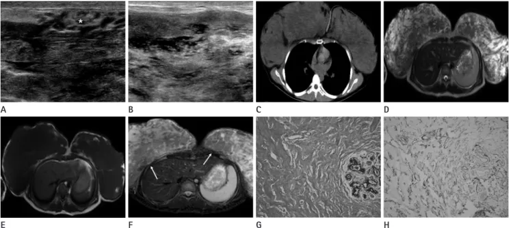
Fig. 1. A 19-year-old female has massive enlargement of both breasts.
A. On ultrasound, normal parenchymal structure (*) is mixed with heterogenously hypoechoic and infiltrating lesion.
B. Ill-defined cystic structures are scattered with the lesion.
C. On non-enhanced CT scan, the lesions on both breasts show relatively homogeneous density with focal fatty nodules. The retromammary fat is preserved well.
D, E. These reveal heterogeneously high signal intensity on T2 weighted images (D) and homogeneously low signal intensity on T1 weighted images (E).
F. On fat saturated, contrast-enhanced T1 weighted images, heterogeneous enhancement (arrows) is noted.
G. The microscopic image (hematoxylin and eosin stained section, × 100) reveals that proliferation of stromal components with diffuse network of spaces in the collagen stroma and vascular channel-like space.
H. In immunohistochemistry (× 100), it shows the positive stain of CD34.
Hee Sun Go, et al
submit.radiology.or.kr J Korean Soc Radiol 2013;68(4):329-332
331
enhancement pattern (4, 8, 9).
Although it is a very rare manifestation of PASH, it should be considered to be one of the differential diagnoses of massive bi- lateral breast enlargement in young women.
REFERENCES
1. Goel NB, Knight TE, Pandey S, Riddick-Young M, de Pare- des ES, Trivedi A. Fibrous lesions of the breast: imaging- pathologic correlation. Radiographics 2005;25:1547-1559 2. Celliers L, Wong DD, Bourke A. Pseudoangiomatous stro- mal hyperplasia: a study of the mammographic and sono- graphic features. Clin Radiol 2010;65:145-149
3. Choi YJ, Ko EY, Kook S. Diagnosis of pseudoangiomatous stromal hyperplasia of the breast: ultrasonography find- ings and different biopsy methods. Yonsei Med J 2008;49:
757-764
4. Ryu EM, Whang IY, Chang ED. Rapidly growing bilateral pseudoangiomatous stromal hyperplasia of the breast. Ko- rean J Radiol 2010;11:355-358
5. Hargaden GC, Yeh ED, Georgian-Smith D, Moore RH, Raf- ferty EA, Halpern EF, et al. Analysis of the mammographic and sonographic features of pseudoangiomatous stromal hyperplasia. AJR Am J Roentgenol 2008;191:359-363 6. Singh KA, Lewis MM, Runge RL, Carlson GW. Pseudoangi-
omatous stromal hyperplasia. A case for bilateral mastec- tomy in a 12-year-old girl. Breast J 2007;13:603-606 7. Sng KK, Tan SM, Mancer JF, Tay KH. The contrasting pre-
sentation and management of pseudoangiomatous stro- mal hyperplasia of the breast. Singapore Med J 2008;49:
e82-e85
8. Teh HS, Chiang SH, Leung JW, Tan SM, Mancer JF. Rapidly enlarging tumoral pseudoangiomatous stromal hyperpla- sia in a 15-year-old patient: distinguishing sonographic and magnetic resonance imaging findings and correlation with histologic findings. J Ultrasound Med 2007;26:1101- 1106
9. Baskin H, Layfield L, Morrell G. MRI appearance of pseu- doangiomatous stromal hyperplasia causing asymmetric breast enlargement. Breast J 2007;13:209-210
ma. Therefore, it resembled the low grade angiosarcoma, except for absence of red blood cells (1-7).
Immunohistochemically, PASH are known to be positive for CD34 and negative for vascular markers CD31 and factor VIII- related antigen, so it can be distinguished from angiosarcoma (4-6, 8). PASH mainly occurred in young premenopausal wom- en; in some cases, it also occurred in postmenonpausal women in hormonal replacement therapy. This suggests that the devel- opment of PASH may be related with hormonal stimulation (1, 3, 4, 7-9). It is found as an incidental finding of breast biopsy specimens, up to 25% and can occur as mass forming tumor.
PASH is known not to progress to malignancy, but it can be pres- ent within malignant lesions. However, some reports also had produced the possibility of recurrence. As such, final diagnosis by core needle biopsy alone was not recommended (1-6, 8).
Imaging findings of PASH are non specific. The most com- mon mammographic finding of PASH is a circumscribed mass without calcification, followed by increased stromal density.
Some reports were suggesting that PASH could show as slow- glowing asymmetry on mammography, although, some PASH lesions cannot be detected on mammography. The case is shown as a well-defined mass, can mimick the phyllodes tumor and the fibroadenoma. The size of PASH lesions varies, ranged from a few millimeters to a huge mass (1, 3, 4, 8).
On ultrasound, it is mainly presented as a hypoechoic mass, also mimicking fibroadenoma or phyllodes tumor. It usually are associated with tubular or round shaped cystic space (4, 8). Cel- liers et al. (2) reported the normal ultrasound as a second com- mon finding. During pregnancy, PASH lesions can occur as a breast mass, causing breast enlargement with skin necrosis (1).
Manifestation of PASH woman as rapid bilateral breast enlarge- ment is very rare. Further, a few English medical literature were reported in this kind of manifestation of PASH (3, 4, 8).
On MRI, which had been reported less frequently, PASH showed intermediate or low signal intensity on T1 weighted im- ages in reference of the muscle, as our case. Low signal intensity mixed with high signal intensity is a common finding on T2 weighted images. High signal intensity area is thought to be a cystic space on ultrasound images. After contrast injection, PASH was known to show avid enhancement with benign, persistent
Radiologic Imaging Findings of Bilateral Infiltrating Pseudoangiomatous Stromal Hyperplasia of the Breasts
submit.radiology.or.kr
J Korean Soc Radiol 2013;68(4):329-332
332
양측성 가성혈관종성 기질 증식증: 증례 보고
고희선 · 제수경
양측성 유방통과 유방 확대를 주소로 내원한 19세 여자 환자에서, 유방 초음파, 전산화단층촬영, 자기공명영상을 시행하 였다. 시행한 영상소견상 경계가 불분명한 종괴양의 병변이 정상 유방 조직과 서로 섞여 있는 양상을 보였으며 수술 후 병 리조직 검사상, 양측성 가성혈관종성 기질 증식증으로 확인되어 증례 보고를 하고자 한다.
한림대학교 의과대학 강남성심병원 영상의학과