Received: 18 April, 2013 Revised: 20 May, 2013 Accepted: 10 June, 2013 Corresponding author: Young Kim
Department of Physical Therapy, The Graduate School, Sahmyook University, 815 Hwarang-ro, Nowon-gu, Seoul 139-742, Republic of Korea Tel: 82-2-3399-1637 Fax: 82-2-3399-1639 E-mail: saleeva@naver.com
This is an Open-Access article distributed under the terms of the Creative Commons Attribution Non-Commercial License (http://creativecommons.org/licens es/by-nc/3.0) which permits unrestricted non-commercial use, distribution, and reproduction in any medium, provided the original work is properly cited.
Copyright © 2013 Korean Academy of Physical Therapy Rehabilitation Science
pISSN 2287-7576 Phys Ther Rehabil Sci
eISSN 2287-7584 2013, 2 (1), 1-6
www.jptrs.org
The effects of EMG-triggered functional electrical stimulation on upper extremity function in stroke patients
Young Kim
Department of Physical Therapy, The Graduate School, Sahmyook University, Seoul, Republic of Korea
Objective: The aim of this review is to explore the latest intervention trends and effects of EMG-triggered functional electrical stimulation on the upper extremity functions in stroke patients.
Design: Systematic review on clinical trials.
Methods: A systematic literature search was performed to identify clinical trials evaluating the effects of EMG-triggered func- tional electrical stimulation (EMG-FES) and task-oriented EMG-triggered FES on the hand functions in stroke patients. Literature review was conducted with the following key words: hand function, functional electrical stimulation, task-oriented, stroke.
Results: Ten clinical trials were included; 8 of them were randomized controlled trial, 1 was block-randomized, and 1 was a pre-post comparison study. A positive effect of electrical stimulation was reported in the patient groups that were treated with func- tional electrical stimulation combined with specific tasks, and volitional muscle contraction-triggered stimulation that was synchronized with tasks. Motor capabilities of the hand and arm were improved after the rehabilitation.
Conclusions: EMG-triggered electrical stimulation may be more effective than non-triggered electrical stimulation in facilitat- ing the hand functions in stroke patients in terms of muscle strength and voluntary muscle contraction of the paretic hand and arm.
Triggered electrical stimulation can be even more effective when it is combined with specific tasks.
Key Words: Electrical stimulation, Hand, Stroke, Task
Introduction
Of all stroke-induced impairments, arm hemiparesis may be the most disabling, considering its impact on the ability to perform activities of daily living (ADL) [1]. Recovery of up- per extremity function is most rapid during the first months after stroke, but only 20% of the stroke survivors who are 3 months post stroke have normal upper extremity function [2,3].
There is growing evidence that electrical stimulation (ES) has a positive effect on upper extremity motor recovery fol- lowing stroke [4-6]. ES might be a useful therapy in the re- habilitation of patients with stroke, but research reports demonstrate a wide variety of stimulation paradigms in
terms of stimulation parameters, method of stimulations, and duration of treatment. This raises the question of how ES should be applied in order to achieve the optimum outcome.
Various devices are available for the application of ES,
and different adjustments of stimulation parameters includ-
ing amplitude, pulse duration, and pulse frequency are
provided. With regard to motor stimulation, several methods
of application have been reported [6]. Cyclic neuromuscular
electrical stimulation (NMES) or functional electrical stim-
ulation (FES) is applied by a pre-programmed scheme,
which causes repetitive muscle contraction without active
participation of the patient [5]. This passive neuromuscular
stimulation has been reported to produce increased muscle
strength, but the evidence is less convincing for more com-
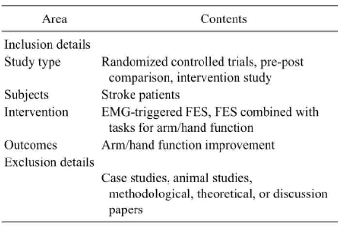
Table 1. List of inclusion and exclusion details
Area Contents
Inclusion details
Study type Randomized controlled trials, pre-post comparison, intervention study Subjects Stroke patients
Intervention EMG-triggered FES, FES combined with tasks for arm/hand function
Outcomes Arm/hand function improvement Exclusion details
Case studies, animal studies,
methodological, theoretical, or discussion papers
EMG: electromyogram, FES: functional electrical stimulation.
plicated hand manipulation tasks [7-9]. In contrast, electro- myogram (EMG)-triggered functional electrical stimula- tion stimulation (EMG-FES) involves initiating a voluntary contraction for a specific movement until the muscle activity reaches a pre-set threshold level, and then an assisting elec- trical stimulus begins [7,10-12]. Compared to passive ES, voluntary initiating motor actions is known to be more effec- tive in strengthening the muscles. Moreover, EMG-FES re- quires cognitive involvement by the motor cortex; improve- ment following EMG-FES was reported to be accompanied by changes in somatosensory cortex activation as measured by functional magnetic resonance imaging (fMRI) [13].
Other rehabilitative methods such as task-oriented training, bilateral movement training, and intensive tracking were as- sociated with changes in size and location of motor output areas [10,14-17]. Therefore, task-oriented EMG-triggered FES can be a beneficial therapeutic intervention for hand function recovery in stroke patients.
The purpose of the present systematic review was to in- vestigate the effects of task-oriented EMG-triggered FES on the arm/hand functions of stroke patients; mainly the wrist and finger extensors, which are known to be essential in re- gaining functional movement of the upper limb for ADL [18].
Methods
A computer-aided literature search up to March 2013 was performed in the following electronic databases: Pubmed (MEDLINE), Cochrane Central register of Controlled Trials, and CINAHL. The following medical subject head- ings and key words were used: stroke, hemiplegia, upper ex- tremity, wrist extensors, hand function, task oriented, neuro- muscular stimulation, EMG-triggered, and FES. References in relevant studies were examined, and the ones that were published after the year 2000 were included in this review.
Inclusion criteria for the present review were as follows.
First, the studies involved patients diagnosed with a stroke.
Second, the study investigated the effects of EMG-NMES or EMG-triggered FES by means of surface electrodes as the experimental intervention. Third, the EMG-NMES applied was targeted to activating the extensor muscles of the forearm. Fourth, the study was classified as a randomized controlled trial, involving at least one test treatment and one control treatment. Fifth, the application of EMG-NMES was the experimental treatment in the randomized controlled trial,
not the control treatment. Sixth, the study analyzed the func- tional measures for the hemiparetic arm/hand functions.
Case studies, animal studies, methodological, and theoret- ical discussions were exempted (Table 1).
Results
After searching the latest EMG-triggered NMES research studies, 10 clinical intervention studies were selected for re- view on the efficacy of the intervention. Clinical character- istics and results of the included studies are summarized in Table 2 and 3. Among the 10 studies, 8 of them were randomized controlled studies. One study was block-rando- mized [19], and one other study was a pre-post comparison study [20]. Except 2 studies [21], chronic stroke patients were recruited for the research.
For outcome measures, clinical tests, kinematic meas- ures, and electromyographic measures were used. As a clin- ical test, Box and Block test was the most commonly used measure, and then the upper-extremity portion of the Fugl-Meyer assessment was the second most commonly used measure.
Four studies by Cauraugh and Kim [10,11] and Cauraugh
et al. [22,23] were included in this study. The results of theirstudies showed significantly decreased motor dysfunction
and improved motor capabilities of the wrist and finger ex-
tensors after intervention. When different durations of stim-
ulation was applied, longer active stimulation decreased re-
sidual motor dysfunctions more than the shorter stimulation
duration (10 s>5 s>0 s). Longer stimulation group (10 s)
displayed improvements on all outcome measures com-
pared to the control group. The two coupled motor recovery
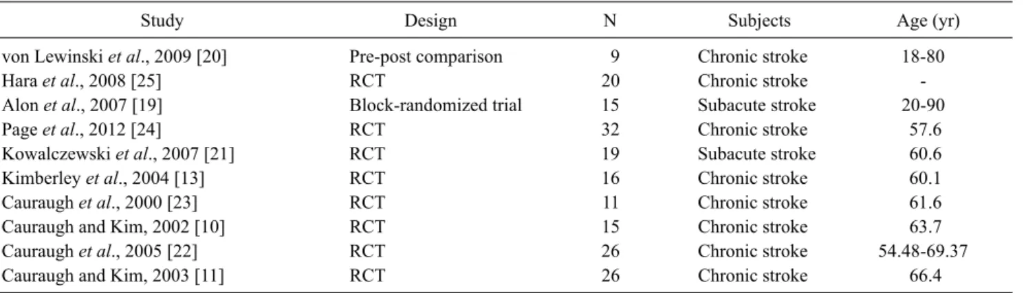
Table 2. Clinical characteristics of included trials
Study Design N Subjects Age (yr)
von Lewinski et al., 2009 [20] Pre-post comparison 9 Chronic stroke 18-80
Hara et al., 2008 [25] RCT 20 Chronic stroke -
Alon et al., 2007 [19] Block-randomized trial 15 Subacute stroke 20-90
Page et al., 2012 [24] RCT 32 Chronic stroke 57.6
Kowalczewski et al., 2007 [21] RCT 19 Subacute stroke 60.6
Kimberley et al., 2004 [13] RCT 16 Chronic stroke 60.1
Cauraugh et al., 2000 [23] RCT 11 Chronic stroke 61.6
Cauraugh and Kim, 2002 [10] RCT 15 Chronic stroke 63.7
Cauraugh et al., 2005 [22] RCT 26 Chronic stroke 54.48-69.37
Cauraugh and Kim, 2003 [11] RCT 26 Chronic stroke 66.4
RCT: randomized controlled trial.
protocols increased movement capabilities in patients with stroke. The coupled bilateral protocol involved concurrent wrist/finger movements on the unimpaired limb that were coupled with active stimulation on the impaired limb. The group that was treated with unilateral EMG-FES on the im- paired wrist/fingers exceeded the control group in Box &
Block test and rapid onset of muscle contraction.
Kimberley et al. [13] compared the EMG-FES training group and the sham group in the aspect of arm and hand functions. Significant improvements were found in Box and Block test, isometric finger-extension strength, motor activ- ity log (MAL), Jebsen-Taylor hand function test (JTHFT) for small objects, stacking, and heavy ans. The sham group improved isometric finger-extension strength, but no other test.
Kowalczewski et al. [21] used the scores from sensor readings on the workstation, which was used for task-ori- ented training for the high-intensity FES group. Kinematic scores were obtained from the 3 tasks given to each subject, and the maximal displacement was divided by the time taken. This score was normalized to that of control subjects, and the mean of 3 normalized task scores was calculated.
This is called combined kinematic score (CKS). The CKS provided quantitative information on improvement in motor performance of the specific tasks on the workstation. The high-intensity group had more than tripled CKS, sig- nificantly increased Wolf motor function test, MAL (amount of use) and MAL (quality of movement) (p<0.05) whereas the low-intensity group only increased about 20% in CKS.
Page et al. [24] compared different durations of task-ori- ented FES, and analyzed the effects on improving hand functions of stroke patients. Tasks were tailored for each subject based on their lifestyle and preference, but mainly
included grasping, releasing, and pinching actions for ADL-related movements. The 'repetitive task-specific prac- tice+FES 120 minute' group, which repetitively performed specific tasks with FES for 120 minutes a day, exhibited the most consistent pattern of motor change, and the largest number of significant changes of all of the groups, on the assessments. The Fugl-Meyer assessment subsets showed the most increase in shoulder/forearm and wrist, although hand and coordination/speed was increased. The group sig- nificantly increased functional movement ability on the arm motor ability test functional ability scale, and exhibited sig- nificantly increased quality of movement (p<0.05).
Alon et al. [19] applied a microprocessor based FES to concurrently synchronize ES with specific tasks. The Box and Block test and JTHFT quantified the recovery of upper extremity function. The mean number of blocks transferred increased more in the FES group compared to the control, and JTHFT task was 6.7±2.9 seconds faster that the 11.8±5.4 seconds of the control group. Mean Fugl-Meyer score at 12 weeks was 49.3±5.1 points out of 54 compared to the control group that scored 40.6±8.2 points (p<0.05). All patients re- gained hand function.
Hara et al. [25] also used instrumental tasks that were con- sisted of reaching, grasping, moving (pulling, rotating) and releasing an object using the hemiplegic upper extremity.
Objects were chosen on the basis of each patient's ability to
grasp the object with FES assistance at the beginning of the
training period. ADL activity was also trained, and it in-
cluded washing, drying dishes, and folding clothes accord-
ing to individual ability. All of the tasks were performed
with power-assisted FES. The stimulator induces greater
muscle contraction by ES in proportion to the integrated
EMG signal picked up from the target muscles as they ac-
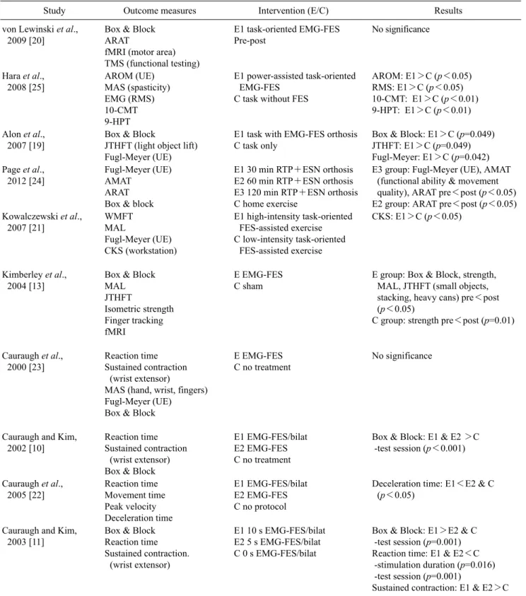
Table 3. Outcome measures, intervention, and results of the studies included
Study Outcome measures Intervention (E/C) Results
von Lewinski et al., 2009 [20]
Box & Block ARAT
fMRI (motor area) TMS (functional testing)
E1 task-oriented EMG-FES Pre-post
No significance
Hara et al., 2008 [25]
AROM (UE) MAS (spasticity) EMG (RMS) 10-CMT 9-HPT
E1 power-assisted task-oriented EMG-FES
C task without FES
AROM: E1>C (p<0.05) RMS: E1>C (p<0.05) 10-CMT: E1>C (p<0.01) 9-HPT: E1>C (p<0.01)
Alon et al., 2007 [19]
Box & Block
JTHFT (light object lift) Fugl-Meyer (UE)
E1 task with EMG-FES orthosis C task only
Box & Block: E1>C (p=0.049) JTHFT: E1>C (p=0.049) Fugl-Meyer: E1>C (p=0.042) Page et al.,
2012 [24]
Fugl-Meyer (UE) AMAT
ARAT Box & block
E1 30 min RTP+ESN orthosis E2 60 min RTP+ESN orthosis E3 120 min RTP+ESN orthosis C home exercise
E3 group: Fugl-Meyer (UE), AMAT (functional ability & movement quality), ARAT pre<post (p<0.05) E2 group: ARAT pre<post (p<0.05) Kowalczewski et al.,
2007 [21]
WMFT MAL
Fugl-Meyer (UE) CKS (workstation)
E1 high-intensity task-oriented FES-assisted exercise C low-intensity task-oriented
FES-assisted exercise
CKS: E1>C (p<0.05)
Kimberley et al., 2004 [13]
Box & Block MAL JTHFT
Isometric strength Finger tracking fMRI
E EMG-FES C sham
E group: Box & Block, strength, MAL, JTHFT (small objects, stacking, heavy cans) pre<post (p<0.05)
C group: strength pre<post (p=0.01)
Cauraugh et al., 2000 [23]
Reaction time Sustained contraction
(wrist extensor)
MAS (hand, wrist, fingers) Fugl-Meyer (UE)
Box & Block
E EMG-FES C no treatment
No significance
Cauraugh and Kim, 2002 [10]
Reaction time Sustained contraction
(wrist extensor) Box & Block
E1 EMG-FES/bilat E2 EMG-FES C no treatment
Box & Block: E1 & E2 >C -test session (p<0.001)
Cauraugh et al., 2005 [22]
Reaction time Movement time Peak velocity Deceleration time
E1 EMG-FES/bilat E2 EMG-FES C no protocol
Deceleration time: E1<E2 & C (p<0.05)
Cauraugh and Kim, 2003 [11]
Box & Block Reaction time Sustained contraction.
(wrist extensor)
E1 10 s EMG-FES/bilat E2 5 s EMG-FES/bilat C 0 s EMG-FES/bilat
Box & Block: E1>E2 & C -test session (p=0.001) Reaction time: E1 & E2<C -stimulation duration (p=0.016) -test session (p=0.001)
Sustained contraction: E1 & E2>C E: experimental group, C: control group, EMG: electromyogram, EMG-FES: EMG-triggered functional electrical stimulation, bilat: bilateral, MAS (hand, wrist, fingers): motor assessment scale, UE: upper extremity, MAL: motor activity log, JTHFT: Jebsen-Taylor hand function test, fMRI: functional MRI, WMFT: Wolf motor function test, CKS: combined kinematic score from the workstation, RTP+ESN: repetitive task-specific practice (RTP) combined with functional electrical stimulation neuroprosthesis (ESN), FM: Fugl-Meyer assessment, AMAT: arm motor ability test, ARAT: action research arm test, AROM: active range of motion, MAS (spasticity): modified Ashworth scale, RMS: root mean square, ROM: range of motion, 10-CMT: ten-cup-moving test, 9-HPT: nine-hole-peg test, TMS: transcranial magnetic stimulation.
tively contract. The electrodes detect electromyography in the affected muscles, and then provide amplified stimulation to these muscles. A computer inside the device evaluates the amount of activity present in the muscle, and the stimulator does not work when there is no volitional muscle contraction detected. After participating in the FES program, subjects exhibited improved active range of motion, spasticity, hand performance according to ten-cup-moving test (F=18.72, p
<0.01) and nine-hole-peg test (F=12.27, p<0.01) com- pared to the control group. The subjects in FES group also exhibited a significantly increased root mean square (RMS) of target muscle EMG, with values of 102.8±78.9 μv in ex- tensor carpi radialis longus, 111.2±95.2 μv in extensor dig- itorum communis and 120.0±81.2 μv in deltoid compared to preintervention levels (p<0.05).
von Lewinski et al. [20] studied the effects of task-ori- ented EMG-triggered FES training, and analyzed the activa- tion patterns in the motor areas of the brain by using fMRI and transcranial magnetic stimulation. The subjects were asked to put plastic cups of different sizes into each other, and voluntary activity in at least one UE muscle group trig- gered the FES. fMRI showed an increase in the spatial extent of activation in the sensorimotor cortex (SMC) in 4 of 7 pa- tients after EMG-FES therapy. The findings supported the notion that intensified EMG-FES may improve the arm function in individual chronic stroke patients but not in more severely impaired individuals. Functional improvements were paralleled by increased ipsilesional SMC activation and intracortical facilitation, supporting neuroplasticity.
Discussion
The present review was conducted to evaluate the effects of EMG-triggered FES on the arm/hand function after stroke. The important finding of this study was that EMG- FES, when performed in an intensive manner with higher voluntary muscle contraction, produced significant im- provements in functional activities in the patients who were in the experimental groups. The control groups, or the sham treatment groups only showed gains in strength without functional improvements from pre-test to post-test.
The application of EMG-triggered FES was proved to be effective in post stroke hand function recovery in terms of grasping, releasing, and pinching; degree of improvements depending on the severity of impairment.
In this review, the studies that conducted various func-
tional tests that would focus on finger and wrist movements for the hand function were included in order to understand the effective and accurate outcome measures for the hand function tests after EMG-FES training. Most of the studies used Box and Block and some JTHFT for dexterity function assessment. The changes in EMG amplitudes were analyzed by using the mean increase in RMS of target muscles during sustained contraction and for rapid onset of the target mus- cles [10,11,22,23,25]. The improved results were reflected on enhanced muscle strength and motor performance.
Most of the studies in this review were conducted with chronic stroke patients, but according to Meilink et al. [26], the return of finger extension is critical for improvement of dexterity after stroke, and because the return of dexterity is not fully defined within the initial five weeks post stroke, the effects of EMG-FES should be applied within this critical time window. More studies need to be conducted to find the effects of EMG-FES on the hand function recovery in sub- acute stroke patients.
References
1. Hill-Hermann V, Strasser A, Albers B, Schofield K, Dunning K, Levine P, et al. Task-specific, patient-driven neuroprosthesis training in chronic stroke: results of a 3-week clinical study. Am J Occup Ther 2008;62:466-72.
2. Parker VM, Wade DT, Langton Hewer R. Loss of arm function after stroke: measurement, frequency, and recovery. Int Rehabil Med 1986;8:69-73.
3. Nakayama H, Jørgensen HS, Raaschou HO, Olsen TS. Recovery of upper extremity function in stroke patients: the Copenhagen stroke study. Arch Phys Med Rehabil 1994;75:394-8.
4. Barreca S, Wolf SL, Fasoli S, Bohannon R. Treatment inter- ventions for the paretic upper limb of stroke survivors: a critical review. Neurorehabil Neural Repair 2003;17:220-6.
5. Chae J, Yu D. Neuromuscular stimulation for motor relearning in hemiplegia. Crit Rev Phys Rehabil Med 1999;11:279-97.
6. de Kroon JR, van der Lee JH, IJzerman MJ, Lankhorst GJ.
Therapeutic electrical stimulation to improve motor control and functional abilities of the upper extremity after stroke: a system- atic review. Clin Rehabil 2002;16:350-60.
7. Burridge JH, Ladouceur M. Clinical and therapeutic applications of neuromuscular stimulation: a review of current use and speculation into future developments. Neuromodulation 2001;4:147-54.
8. Chae J, Bethoux F, Bohine T, Dobos L, Davis T, Friedl A.
Neuromuscular stimulation for upper extremity motor and func- tional recovery in acute hemiplegia. Stroke 1998;29:975-9.
9. Powell J, Pandyan AD, Granat M, Cameron M, Stott DJ.
Electrical stimulation of wrist extensors in poststroke hemiple- gia. Stroke 1999;30:1384-9.
10. Cauraugh JH, Kim S. Two coupled motor recovery protocols are better than one: electromyogram-triggered neuromuscular stim-
ulation and bilateral movements. Stroke 2002;33:1589-94.
11. Cauraugh JH, Kim SB. Chronic stroke motor recovery: duration of active neuromuscular stimulation. J Neurol Sci 2003;215:
13-9.
12. Woldag H, Hummelsheim H. Evidence-based physiotherapeutic concepts for improving arm and hand function in stroke patients:
a review. J Neurol 2002;249:518-28.
13. Kimberley TJ, Lewis SM, Auerbach EJ, Dorsey LL, Lojovich JM, Carey JR. Electrical stimulation driving functional improve- ments and cortical changes in subjects with stroke. Exp Brain Res 2004;154:450-60.
14. Carey JR, Kimberley TJ, Lewis SM, Auerbach EJ, Dorsey L, Rundquist P, et al. Analysis of fMRI and finger tracking training in subjects with chronic stroke. Brain 2002;125:773-88.
15. Jang SH, Kim YH, Cho SH, Lee JH, Park JW, Kwon YH.
Cortical reorganization induced by task-oriented training in chronic hemiplegic stroke patients. Neuroreport 2003;14:137- 41.
16. Liepert J, Bauder H, Wolfgang HR, Miltner WH, Taub E, Weiller C. Treatment-induced cortical reorganization after stroke in humans. Stroke 2000;31:1210-6.
17. Richards LG, Stewart KC, Woodbury ML, Senesac C, Cauraugh JH. Movement-dependent stroke recovery: a systematic review and meta-analysis of TMS and fMRI evidence. Neuropsychol- ogia 2008;46:3-11.
18. Fritz SL, Light KE, Patterson TS, Behrman AL, Davis SB.
Active finger extension predicts outcomes after constraint- in- duced movement therapy for individuals with hemiparesis after stroke. Stroke 2005;36:1172-7.
19. Alon G, Levitt AF, McCarthy PA. Functional electrical stim-
ulation enhancement of upper extremity functional recovery dur- ing stroke rehabilitation: a pilot study. Neurorehabil Neural Repair 2007;21:207-15.
20. von Lewinski F, Hofer S, Kaus J, Merboldt KD, Rothkegel H, Schweizer R, et al. Efficacy of EMG-triggered electrical arm stimulation in chronic hemiparetic stroke patients. Restor Neurol Neurosci 2009;27:189-97.
21. Kowalczewski J, Gritsenko V, Ashworth N, Ellaway P, Prochazka A. Upper-extremity functional electric stimulation- assisted exercises on a workstation in the subacute phase of stroke recovery. Arch Phys Med Rehabil 2007;88:833-9.
22. Cauraugh JH, Kim SB, Duley A. Coupled bilateral movements and active neuromuscular stimulation: intralimb transfer evi- dence during bimanual aiming. Neurosci Lett 2005;382:39-44.
23. Cauraugh J, Light K, Kim S, Thigpen M, Behrman A. Chronic motor dysfunction after stroke: recovering wrist and finger ex- tension by electromyography-triggered neuromuscular stimula- tion. Stroke 2000;31:1360-4.
24. Page SJ, Levin L, Hermann V, Dunning K, Levine P. Longer ver- sus shorter daily durations of electrical stimulation during task- specific practice in moderately impaired stroke. Arch Phys Med Rehabil 2012;93:200-6.
25. Hara Y, Ogawa S, Tsujiuchi K, Muraoka Y. A home-based re- habilitation program for the hemiplegic upper extremity by pow- er-assisted functional electrical stimulation. Disabil Rehabil 2008;30:296-304.
26. Meilink A, Hemmen B, Seelen HA, Kwakkel G. Impact of EMG-triggered neuromuscular stimulation of the wrist and fin- ger extensors of the paretic hand after stroke: a systematic review of the literature. Clin Rehabil 2008;22:291-305.